
 Worksheets
Eye - Structure
Total questions: 74
Worksheet time: 37mins
Prevent sweat from getting into the eyes and shades the eye.
Eyebrows
Eyelids
Nose
Hair
Protect the Anterior part of the eye.
Eyebrows
Eyelids
Nair
Nose
Levator palpabrae - ______ the eye (CN-III)
Orbicularis Oculi - ______ the eye (CN-VII)
Closes
Open
Opens
Closes
Closes
Closes
Opens
Opens
Thin transparent membrane covers the cornea. When infected called CONJUNCTIVITIS or pink-eye
Retina
Lens
Sclera
Conjunctiva
Consists of the Lacrimal gland (which produces tears) & Nasolacrimal duct system.
Tears are made of a dilute saline solution.
The composition of tears differs between happy and sad tears.
We produce tears not just for emotions; every time we BLINK, we make tears.
Tears are emptied into the NOSE via the NASOLACRIMAL DUCT.
Conjunctiva
Eyelids
Lacrimal Apparatus
Aqueous Humor
The OUTER TUNIC
Aka: Fibrous Layer is made up of _____ & ______
Sclera & Cornea
Choroid, ciliary body
Iris
Retina
The MIDDLE TUNIC
Aka: Vascular Layer is made up of _______, ________ & ________
Retina
Sclera, Cornea, Iris
Choroid, Ciliary body, Iris
Chroid
The INNER TUNIC
Aka: Nervous Layer is made up of ________
Sclera
Cornea
Choroid
Retina
Extrinsic Eye Muscles:
Laterally innervation (CN)
Oculomotor (CN-III)
Optic (CN-II)
Abducens (CN-VI)
Trochlear (CN-IV)
Extrinsic Eye Muscles:
Medially innervation (CN)
Trochlear (CN-IV)
Oculomotor (CN-III)
Optic (CN-II)
Abducens (CN-VI)
Extrinsic Eye Muscles:
Elevates/Medially innervation (CN)
Oculomotor (CN-III)
Optic (CN-II)
Abducens (CN-VI)
Trochlear (CN-IV)
Extrinsic Eye Muscles:
Depress/Medially innervation (CN)
Trochlear (CN-IV)
Abducens (CN-VI)
Oculomotor (CN-III)
Optic (CN-II)
Extrinsic Eye Muscles:
Elevates & turns Laterally innervation (CN)
Oculomotor (CN-III)
Trochlear (CN-IV)
Optic (CN-II)
Abducens (CN-VI)
Extrinsic Eye Muscles:
Depresses & turns Medially innervation (CN)
Oculomotor (CN-III)
Abducens (CN-VI)
Trochlear (CN-IV)
Optic (CN-II)
The outermost fibrous layer of the eye.
The WHITE of the eye.
Protects & shapes the eye.
Site of INSERTION of ALL the extrinsic muscles of the eye.
Lens
Sclera
Cornea
Retina
The transparent central portion of the Sclera.
Outermost Fibrous layer of the eye.
Aids in REFRACTION.
It contains 3 disorders of Refraction (Myopia, Hyperopia, Astigmatism)
Retina
Sclera
Cornea
Choroid
"NEARSIGHTEDNESS" the inability to see DISTANT IMAGES. The eyeball is too long.
MYOPIA
HYPEROPIA
ASTIGMATISM
"FARSIGHTEDNESS" the inability to see NEAR IMAGES.
MYOPIA
HYPEROPIA
ASTIGMATISM
Distorted images unequal curvature of LENS or CORNEA lead to this. Results in distorted images since light rays are not bale to all meet at a common focus.
MYOPIA
HYPEROPIA
ASTIGMATISM
Plasma-like fluid.
Supplies NUTRIENTS to lens & cornea
CONTINOUSLY CIRCULATES!!
It gets reabsorbed into venous blood through the Canal of Schlemm.
Aqueous Humor
Canal of Schlemm
Ciliary Body
Vitreous Humor
The site where Aqueous Humor gets reabsorbed and taken into the venous blood.
Canal of Schlemm
Cornea
Fovea Centralis
Optic Disc
The opening in the IRIS that regulates the amount to light entering the eye.
Light adaptation occurs when moving from bright light into darkness.
Cornea
Pupil
Iris
Lens
Moving from Bright Light to Darkness --> The pupil will _______ to let more light in!!!
CONSTRICT
DILATE
Moving from Darkness to Bright Light --> Pupil will ________ to reduce the amount of light going in!!
DILATE
CONSTRICT
The visible color part of the eye around the pupil.
Can be blue, green, brown, hazel.
Made up of 2 SMOOTH MUSCLES layers that change shape depending on the environmental light.
Divides the eye into the Anterior & Posterior Chamber.
Iris
Lens
Retina
Cornea
Which structure of the eye is made up of 2 SMOOTH MUSCLES layers that change shape depending on the environmental light.
Sclera
Cornea
Lens
Iris
Focuses (refracts) light on the retina.
Has a flexible biconvex crystal-like structure.
Is held in position by suspensory fibers called Ciliary Zonule.
Divides the intraocular cavity into a posterior segment that contains the Vitreous Humor & an anterior segment that contains the Aqueous Humor.
With accommodation the lens bulges, becomes flatter when holding an object close to the eye.
Retina
Iris
Lens
Cornea
Secretes Aqueous Humor into the Intraocular Cavity.
Choroid
Ciliary body
Fovea Centralis
Optic Disc
Gel-like fluid that reinforces the eyeball & prevents it from collapsing inward.
In the Posterior segment of the eye.
Contributes to the Intraocular pressure & DOES NOT CIRCULATE!!
Vitreous Humor
Ciliary Body
Aqueous Humor
Canal if Schlemm
The innermost sensory layer of the eye.
The ONLY area of the body we can directly examine blood vessels & get information regarding cardiovascular health!!!!
Has 2 Layer: Pigmented Layer & Neural Layer.
Also contains Photoreceptors: RODS & CONES
Choroid
Retina
Cornea
Optic Nerve
The retina has 2 layers!
_______ - Absorbs light & stores Vitamin A
Pigmented Layer
Neural Layer
The retina has 2 layers!
_______ - Reacts to light energy via PHOTORECEPTORS
Pigmented Layer
Neural Layer
The retina has Photoreceptors:
_____ - Operates in DIM LIGHT!
RODS
CONES
The retina has Photoreceptors:
_____ - Operates in BRIGHT LIGHT!
RODS
CONES
The retina has Photoreceptors:
Night-blindness is a problem with the RODS due to lack of Vitamin A.
CAN BE CURED by Vitamin A.
RODS
CONES
The retina has Photoreceptors:
Colorblindness is a problem of the _____ due to the lack of one or more _____.
CAN NOT be cured.
RODS
CONES
3 types of CONES named for colors of light:
Blue, Green, Red
Red, Yellow, Blue
Green, Orange, Blue
Aka: Macula Lutea
The GREATEST VISUAL ACUITY is found here due to having ALL CONES
Optic Disc
Optic Nerve
Choroid
Fovea Centralis
Aka: BLIND SPOT
No photoreceptors there!!!
CN-II leaves the eye from the retina.
Retina
Optic Disc
Optic Nerve
Choroid
CN-II
Light Travels via the Optic Nerve --> Optic Tract --> Primary Visual Cortex in the OCCIPITAL LOBE
If damaged, it will result in blindness!!
Optic Disc
Choroid
Optic Nerve
Lens
Part of the MIDDLE REGION of the eye.
Contains PIGMENT & prevents light from scattering in the eyeball.
Optic Disc
Choroid
Fovea Centralis
Retina
Sensory Receptors:
Mechanoreceptors
temp
pressure, vibration, stretch, & itch
light
pain (extreme heat/cold, excessive pressure, inflammation) --> warn of possible tissue damage
& blocked by inhibitory endogenous opioids
chemicals
Sensory Receptors:
Thermoreceptors
Light
Temp
Chemicals
pressure, vibration, stretch, & itch
pain (extreme heat/cold, excessive pressure, inflammation) --> warn of possible tissue damage
& blocked by inhibitory endogenous opioids
Sensory Receptors:
Photoreceptors
Light
Temp
Chemicals
pressure, vibration, stretch, & itch
pain (extreme heat/cold, excessive pressure, inflammation) --> warn of possible tissue damage
& blocked by inhibitory endogenous opioids
Sensory Receptors:
Chemoreceptors
Chemicals
Light
Temp
pressure, vibration, stretch, & itch
pain (extreme heat/cold, excessive pressure, inflammation) --> warn of possible tissue damage
& blocked by inhibitory endogenous opioids
Sensory Receptors:
Nociceptors
pain (extreme heat/cold, excessive pressure, inflammation)
--> warn of possible tissue damage
& blocked by inhibitory endogenous opioids
light
pressure, vibration, stretch, & itch
chemicals
temp
General Senses by Location:
Stimulus OUTSIDE body --> receptors in skin for touch, pressure, pain, special sense organs
Interoreceptors (Visceroreceptors)
Exteroceptors
Proprioceptors
General Senses by Location:
stimulus in internal viscera (organs) & blood vessels --> chemical
changes in body, tissue stretch, temp changes
Interoreceptors (Visceroreceptors)
Proprioceptors
Exteroceptors
General Senses by Location:
skeletal muscles, tendons, joints, ligaments, & connective tissue --> tells brain
about one’s movement + position
Interoreceptors (Visceroreceptors)
Exteroceptors
Proprioceptors
AWARENESS of changes in internal + external environment
Occurs in the brain
Sensation
Perception
Conscious INTERPRETATION of stimuli
Occurs in the brain
Sensation
Perception
70% of all sensory receptors are in the eye
Nearly half of the cerebral cortex is involved in processing visual information!
True
False
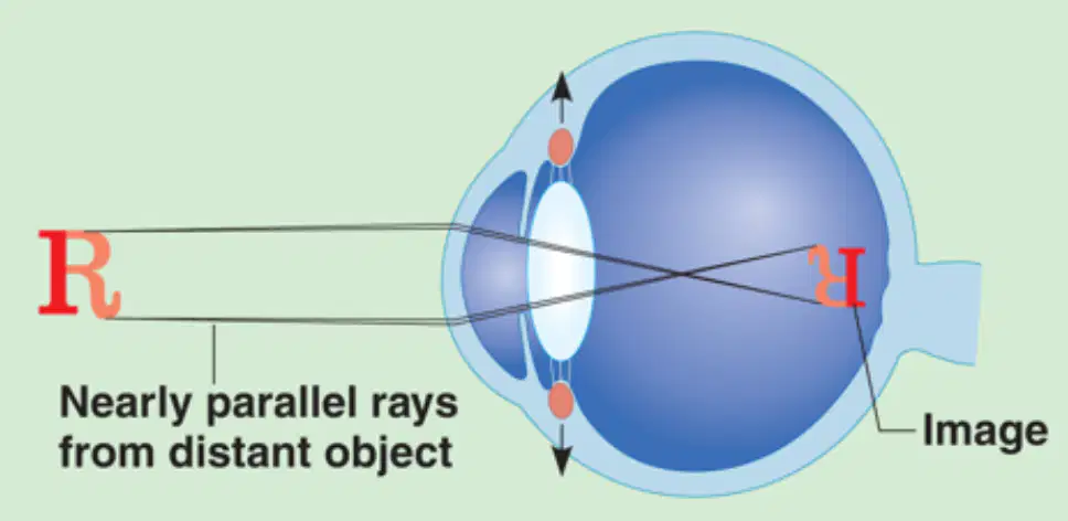
What is the term for the bending of a light ray due to a change in speed as it passes from one transparent medium to another—a process that occurs at the cornea and lens, allowing light to converge at a focal point for image formation?
Reflection
Absorption
Refraction
Dispersion
Which process involves changing the shape of the lens using the ciliary muscles to increase the eye’s refractory power, allowing you to focus on close objects?
Convergence
Accommodation
Dilation
Adaptation
Which process occurs when the pupils constrict to prevent the most divergent light rays from entering the eye during close vision?
Constriction (accommodation pupillary reflex)
Dilation
Convergence
Refraction
Which close-vision process involves the medial rotation of the eyeballs toward the object being viewed?
Accommodation
Divergence
Convergence
Rotation reflex
What condition occurs when the lens loses its ability to accommodate—typically after age 50—resulting in difficulty focusing on close objects and often requiring reading glasses?
Myopia
Hyperopia
Presbyopia
Astigmatism

Which sequence correctly describes the pathway of light as it enters the eye and is processed by the brain?
Cornea → Aqueous humor → Pupil → Lens → Vitreous humor → Retina → Photoreceptors → Optic disc → Optic nerve → Optic chiasm → Optic tract → Occipital lobe → Primary visual cortex
Lens → Cornea → Vitreous humor → Pupil → Optic nerve → Retina → Occipital lobe
Pupil → Lens → Cornea → Retina → Optic tract → Primary visual cortex
Retina → Cornea → Optic nerve → Lens → Occipital lobe

#1
Ciliary Zonule
Cornea
Iris
Ciliary Body

#2
Ciliary Body
Ciliary Zonule
Cornea
Iris

#3
Cornea
Iris
Pupil
Lens

#4
Lens
Iris
Pupil
Aqueous Humor

#5
Cornea
Iris
Pupil
Lens

#6
Vitreous Humor
Canal of Schlemm
Lens
Aqueous Humor

#7
Iris
Lens
Aqueous Humor
Cornea

#8
Retina
Sclera
Sclera Venous Sinus (Canal of Schlemm)
Cornea

#9
Vitreous Humor
Aqueous Humor
Optic Disc
Fovea Centralis

#10
Cornea
Retina
Sclera
Choroid

#11
Choroid
Retina
Cornea
Sclera

#12
Iris
Retina
Choroid
Cornea

#13
Optic Disc
Vitreous Humor
Optic Nerve
Fovea Centralis

#14
Central artery and vein of the retina
Optic Nerve
Optic Disc
Fovea Centralis

#15
Optic Nerve
Central artery and vein of the retina
Retina
Optic Disc

#16
Vitreous Humor
Optic Disc
Optic Nerve
Fovea Centralis

