
 Worksheets
Because Siena made the request: Integumentary System Review
Total questions: 150
Worksheet time: 1hrs 15mins
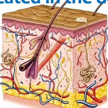
According to the overview, the skin includes two distinct layers. Which pair names them?
epidermis and dermis
dermis and hypodermis
epidermis and subcutaneous layer
hypodermis and subcutaneous layer

Based on the slide, the skin is connected to underlying tissue by which structure?
subcutaneous layer (hypodermis)
epidermis
dermis
stratum corneum
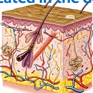
Where are the skin accessory structures located, according to the slide?
in the dermis
in the epidermis
in the hypodermis
on the skin surface
Which of the following is listed as a skin accessory structure?
Nails
Melanin
Stratum basale
Keratinocytes
Which set best lists the glands identified as skin accessory structures in this overview?
sweat and oil
salivary and endocrine
mucous and lacrimal
sebaceous and parathyroid

Skin structure includes 2 distinct layers. Which pair lists the true layers of the skin?
Epidermis and dermis
Dermis and hypodermis
Epidermis and hypodermis
Basement membrane and dermis

What type of tissue composes the epidermis, the outer layer of the skin?
Stratified squamous epithelium
Dense irregular connective tissue
Adipose tissue
Smooth muscle
Which skin layer is the inner, thicker layer that includes connective tissue?
Epidermis
Dermis
Hypodermis
Basement membrane
What is the primary role of the basement membrane in skin?
Anchors epidermis to dermis
Produces melanin for the epidermis
Stores adipose beneath the dermis
Provides blood supply to the epidermis
Which statement best describes the subcutaneous layer (hypodermis)?
It lies beneath the dermis and is predominantly loose connective tissue with adipose
It is the outer layer of the skin composed of stratified squamous epithelium
It is a dense connective tissue layer within the dermis
It forms the basement membrane between epidermis and dermis
Is the subcutaneous layer (hypodermis) considered a true layer of the skin?
No, it is not a true layer of skin
Yes, it is the third true layer of skin
Yes, it is part of the dermis
Yes, it forms the epidermal surface
Composed of what tissue type is the epidermis
Stratified squamous epithelium
Simple cuboidal epithelium
Pseudostratified columnar epithelium
Transitional epithelium
Which structure is absent in the epidermis
Blood vessels
Keratinocytes
Melanocytes
Desmosomes
In healthy skin, what is the relationship between production of epidermal cells and the loss of dead cells
They are equal
Production exceeds loss
Loss exceeds production
They vary unpredictably
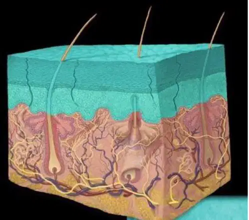
Refer to the cross-sectional skin diagram. Which statement correctly identifies the epidermis
It is the outermost layer
It is the middle vascular layer
It is the deep adipose layer
It is the subcutaneous fascia
Based on the slide titled "Epidermis – Functions", which item is listed as a protective role of the epidermis?
Protection from excess water loss
Blood oxygenation
Nutrient absorption from the environment
Endocrine hormone secretion
According to the slide titled "Epidermis – Functions", select all items the epidermis protects the body from.
Excess water loss
Injury
Chemicals
Disease-causing microorganisms
UV radiation
Which of the following is not included among the protective functions listed for the epidermis on the slide?
UV radiation
Chemicals
Injury
Nutrient absorption
According to the epidermis structure list, which layer is on top of the epidermis?
Stratum corneum
Stratum lucidum
Stratum granulosum
Stratum spinosum
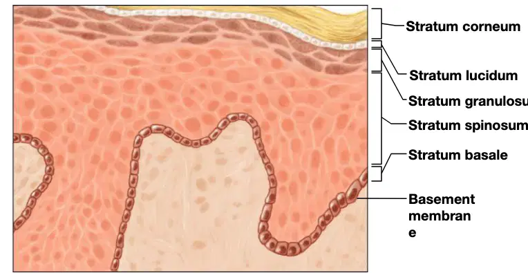
Which epidermal layer lies directly above the basement membrane as shown in the labeled skin diagram?
Stratum basale
Stratum spinosum
Stratum granulosum
Stratum corneum
Which layer is immediately superficial to the stratum basale in the epidermis structure list?
Stratum spinosum
Stratum granulosum
Stratum lucidum
Stratum corneum
Choose the correct arrangement of the epidermal layers from superficial to deep as presented in the list.
Stratum corneum, Stratum lucidum, Stratum granulosum, Stratum spinosum, Stratum basale
Stratum basale, Stratum spinosum, Stratum granulosum, Stratum lucidum, Stratum corneum
Stratum lucidum, Stratum corneum, Stratum granulosum, Stratum spinosum, Stratum basale
Stratum granulosum, Stratum corneum, Stratum lucidum, Stratum spinosum, Stratum basale
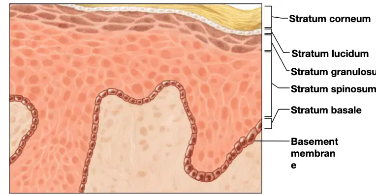
In the labeled diagram, what structure is indicated beneath the epidermal layers and supports them?
Basement membrane
Dermal papillae
Hair follicle
Sebaceous gland

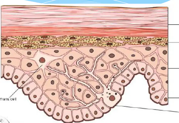
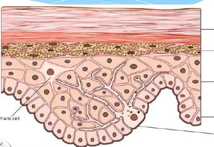
Refer to the epidermis diagram showing labeled layers and cells. Which epidermal layer consists of dead, flattened cells filled with keratin, with glycolipids present in the extracellular space?
Stratum corneum
Stratum granulosum
Stratum spinosum
Stratum basale

In the epidermis diagram, which layer contains flattened cells with deteriorating organelles and cytoplasm full of lamellated granules that release lipids, along with keratohyaline?
Stratum corneum
Stratum granulosum
Stratum spinosum
Stratum basale
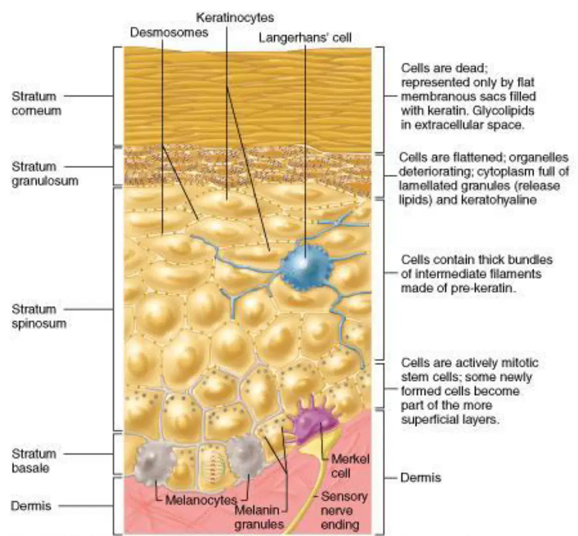
According to the diagram, cells in which epidermal layer contain thick bundles of intermediate filaments made of pre-keratin?
Stratum basale
Stratum spinosum
Stratum granulosum
Stratum corneum

Based on the diagram text, which epidermal layer is composed of actively mitotic stem cells, some of which become part of the more superficial layers?
Stratum corneum
Stratum spinosum
Stratum basale
Stratum granulosum

Which cell type labeled in the diagram functions in immune surveillance within the epidermis?
Keratinocyte
Langerhans’ cell
Melanocyte
Merkel cell

Which labeled cell in the diagram produces the pigment that forms melanin granules?
Keratinocyte
Langerhans’ cell
Melanocyte
Merkel cell

In the diagram, which structure connects adjacent keratinocytes to each other, contributing to epidermal cohesion?
Desmosomes
Lamellated granules
Sensory nerve ending
Keratohyaline

According to the diagram, which labeled cell is associated with a sensory nerve ending in the epidermis?
Keratinocyte
Langerhans’ cell
Melanocyte
Merkel cell
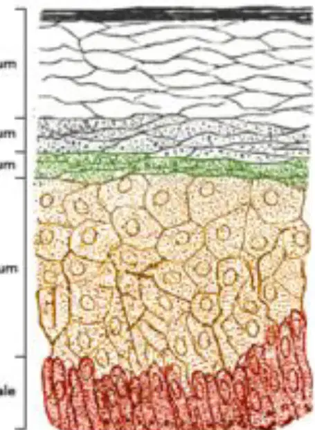
Identify the deepest layer of the epidermis.
Stratum basale
Stratum granulosum
Stratum lucidum
Stratum corneum
Cells in the stratum basale are close to the dermis and are nourished primarily by which source?
Dermal blood vessels
Sebaceous gland secretions
Sweat duct fluid
Ambient oxygen diffusion
Which processes occur in the stratum basale that drive cells toward the skin surface? Select all that apply.
Cell division
Cell growth
Active pushing of older cells upward
Immediate keratinization at origin
As epidermal cells move farther from the dermis, how does their nutrient supply change?
It becomes poorer
It remains constant
It increases moderately
It fluctuates unpredictably
What is the ultimate direction of cell migration from the stratum basale?
Toward the top of the epidermis
Into the dermis
Laterally across the basement membrane
Toward hair follicles
During upward migration through the epidermis, what happens to the cytoplasm of the cells?
It is replaced by keratin
It expands to store water
It is converted to melanin
It is expelled as sweat
What is the eventual fate of epidermal cells as they reach the surface after migrating from the stratum basale?
They die
They become nerve cells
They fuse into multinucleated muscle fibers
They revert to stem cells
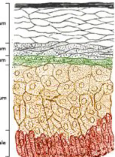
Based on the labeled diagram of epidermal layers, which layer is most superficial?
Stratum corneum
Stratum spinosum
Stratum basale
Stratum granulosum
Define keratinization in the epidermis.
Process by which older cells harden
Process of rapid cell division in the dermis
Secretion of melanin by melanocytes
Formation of collagen fibers in connective tissue
During keratinization, the cytoplasm fills with strands of which tough, fibrous, water-proof protein?
Keratin
Actin
Elastin
Myosin
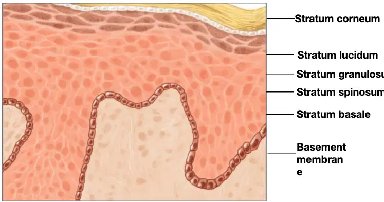
Identify the outermost epidermal layer shown in the labeled cross‑section diagram of the skin.
Stratum corneum
Stratum basale
Stratum spinosum
Stratum granulosum

Which epidermal layer lies directly above the basement membrane in the labeled skin diagram?
Stratum basale
Stratum spinosum
Stratum granulosum
Stratum lucidum
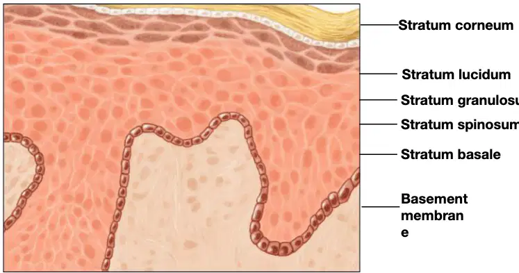
Which epidermal layer, shown between the stratum corneum and stratum granulosum in the diagram, is characteristic of thick skin?
Stratum lucidum
Stratum spinosum
Stratum basale
Stratum corneum

According to the diagram and notes on the epidermis, which layer is on top of the skin surface?
Stratum corneum
Stratum basale
Stratum spinosum
Dermis
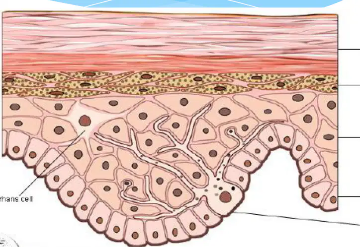
What best describes the cells in the stratum corneum as presented in the notes?
Many layers of tough, tightly packed dead cells formed from keratinization
Single layer of living cells actively dividing
Loose connective tissue with abundant blood vessels
Clusters of melanocytes producing pigment
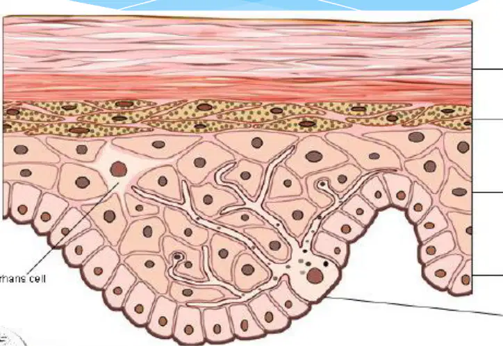
In the stratum corneum, what is present instead of cytoplasm?
Keratin
Melanin
Collagen
Hemoglobin
What ultimately happens to the dead cells of the stratum corneum?
They are eventually shed
They divide and form new epidermal layers
They migrate into the dermis
They transform into melanocytes
Based on the notes, which functions are attributed to keratin in the stratum corneum? Select all that apply.
Acts as a barrier
Provides waterproofing
Stores energy
Catalyzes metabolic reactions
Identify the epidermal layer that is present in thick, hairless skin of the palms and soles but is missing where the epidermis is thin.
Stratum lucidum
Stratum granulosum
Stratum spinosum
Stratum basale
Which epidermal layer forms the outermost surface of the skin?
Stratum corneum
Stratum lucidum
Stratum granulosum
Stratum basale
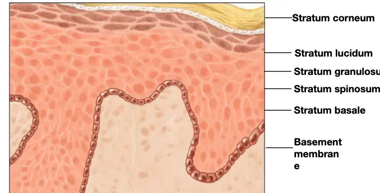
In the cross‑section image of the epidermis labeled with layers and a basement membrane, which labeled layer lies directly above the basement membrane?
Stratum basale
Stratum spinosum
Stratum granulosum
Stratum corneum
Choose the correct order of epidermal layers from deep to superficial in thin skin (where the layer found only in thick, hairless skin is absent).
Stratum basale → Stratum spinosum → Stratum granulosum → Stratum corneum
Stratum basale → Stratum granulosum → Stratum spinosum → Stratum corneum
Stratum corneum → Stratum granulosum → Stratum spinosum → Stratum basale
Stratum granulosum → Stratum lucidum → Stratum corneum → Stratum basale
For thick, hairless skin of the palms and soles, which additional epidermal layer appears between the stratum granulosum and the stratum corneum?
Stratum lucidum
Stratum basale
Stratum spinosum
Basement membrane

According to the slide, melanocytes are melanin-producing cells in the epidermis.
immune cells that fight pathogens
melanin-producing cells
sweat gland cells that secrete perspiration
sensory nerve cells that detect touch

Where are melanocytes located within the epidermis, as stated on the slide?
in the deepest layer of the epidermis
uniformly throughout the dermis
in the hypodermis beneath the skin
only in hair follicles
What is the primary function of melanin described on the slide?
waterproofing the skin
protecting from the sun’s UV rays
regulating body temperature
producing vitamin D

Which factors determine the amount of melanin produced, as listed on the slide? Select all that apply.
genetics (DNA)
direct exposure to sunlight
dietary protein intake
chronological age
Which substance do melanocytes produce?
keratin
collagen
melanin
hemoglobin
Skin color is due largely to which pigment in the epidermis?
Melanin
Hemoglobin
Carotene
Keratin
Variations in human skin color result from which factors? Select all that apply.
Amount of melanin produced
Distribution of pigment granules
Size of pigment granules
Density of sweat glands
According to the epidermis information, skin color is mostly what?
Genetically determined
Entirely environmentally induced
Solely due to diet
Random and unregulated
Which statement best reflects the number of genes that affect pigmentation of skin, hair, and irises?
More than a hundred genes affect pigmentation
Exactly ten genes control pigmentation
Fewer than five genes influence pigmentation
No genes are involved in pigmentation
What effect can sunlight have on pigmentation?
Stimulates production of additional pigment
Permanently removes pigment
Has no effect on pigment production
Only affects hair pigmentation but not skin
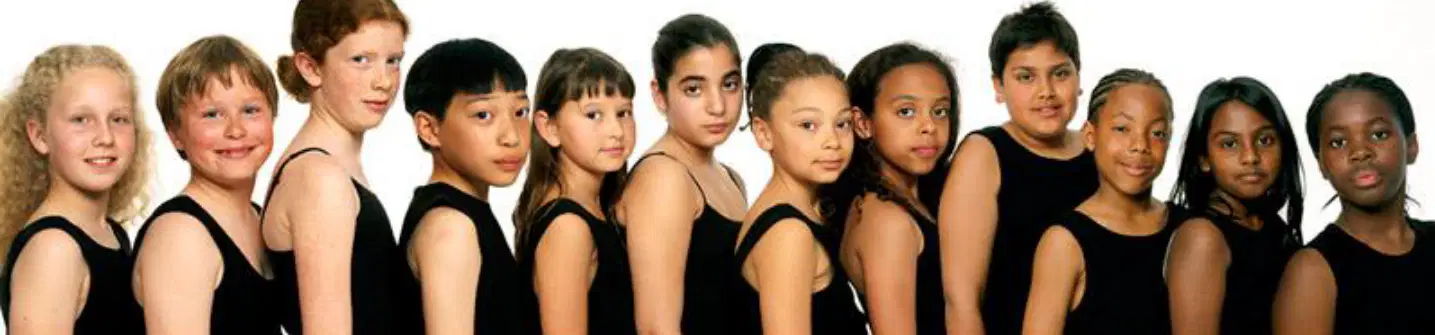
Based on the photo of individuals with varying skin tones, which concept from the epidermis content is most directly illustrated?
Variation due to amount, distribution, and size of melanin pigment granules
Role of sweat glands in thermoregulation
Structure and function of subcutaneous fat
Effect of cartilage on joint movement
In the epidermis, what is the thin outer layer of dead skin cells called?
Cuticle
Dermis
Melanocyte
Basal lamina
Which skin layer is the one you see on the surface?
Epidermis
Dermis
Hypodermis
Subcutaneous tissue
According to the worksheet, most of the “dust” around your house is primarily composed of what?
Dead skin cells
Pollen grains
Fabric fibers
Soil particles
What lies beneath the cuticle within the epidermis?
Layers of living epithelial cells
Adipose tissue
Keratinized hair shafts
Skeletal muscle
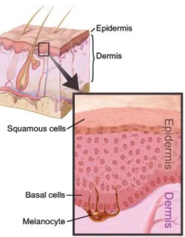
In the labeled skin diagram, which cell type is found in the basal layer of the epidermis?
Basal cells
Squamous cells
Neurons
Chondrocytes
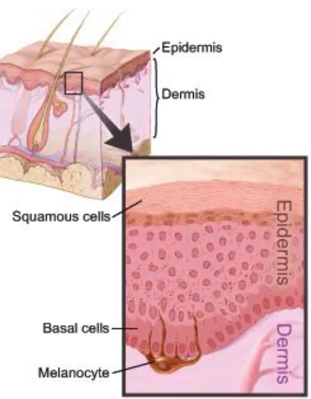
In the labeled skin diagram, which pigment-producing cell is indicated?
Melanocyte
Macrophage
Fibroblast
Lymphocyte
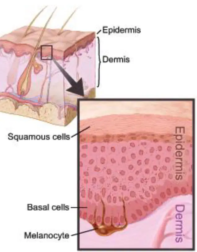
Based on the diagram, which statement best describes the position of the epidermis relative to the dermis?
Epidermis lies superficial to the dermis
Epidermis lies deep to the dermis
They are the same layer
Dermis is part of the epidermis
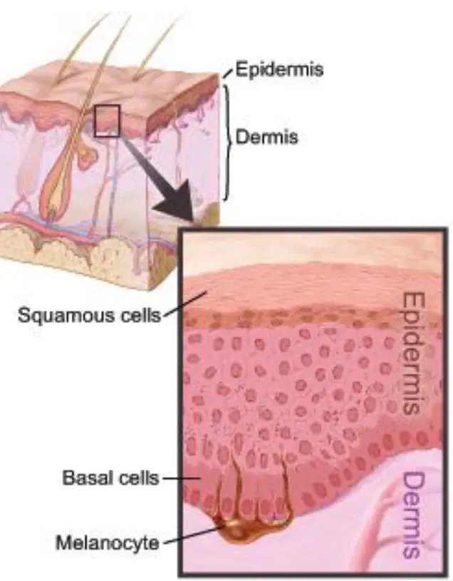
Where are squamous cells located within the epidermis as shown in the diagram?
Upper layers of the epidermis
Basal layer next to the dermis
Within the dermis
In the hypodermis
Keratinization is described as the process by which what occurs in the epidermis?
older cells harden
new blood vessels form in the dermis
melanin pigments break down
collagen is replaced with elastin
During keratinization, the cytoplasm fills with which material?
strands of tough, fibrous, water-proof keratin protein
globules of lipid hormones
vesicles containing hemoglobin
calcium carbonate crystals
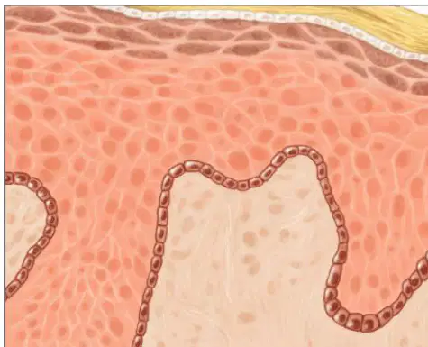
Refer to a labeled diagram of the epidermis showing Stratum corneum, Stratum lucidum, Stratum granulosum, Stratum spinosum, Stratum basale, and the basement membrane. Which layer is the outermost?
stratum corneum
stratum basale
stratum spinosum
basement membrane
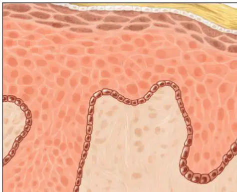
In the labeled epidermis diagram, which layer lies directly above the basement membrane?
stratum basale
stratum corneum
stratum granulosum
stratum lucidum
Which properties are attributed to the keratin protein that fills the cytoplasm during keratinization? Select all that apply.
tough
fibrous
water-proof
highly soluble
primarily elastic

Refer to the labeled cross-section of the skin. Which labeled structure separates the epidermis from the dermis?
Stratum corneum
Basement membrane
Stratum germantivum (basale)
Subcutaneous layer

In the diagram of the skin, which labeled epidermal stratum forms the outermost surface layer?
Stratum germantivum (basale)
Dermis
Stratum corneum
Basement membrane

According to the labeled skin diagram, which epidermal stratum is the basal layer where new cells originate?
Stratum corneum
Basement membrane
Stratum germantivum (basale)
Subcutaneous layer
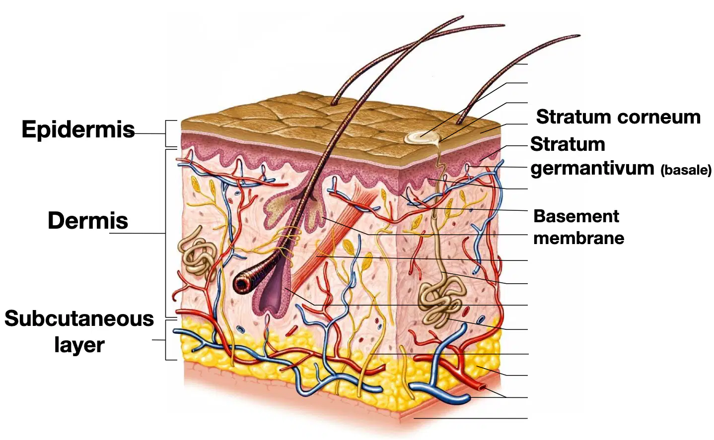
Based on the diagram, which major skin region is deepest and shown as the yellow layer beneath the dermis?
Epidermis
Dermis
Subcutaneous layer
Basement membrane

Using the labeled cross-section, which major skin region contains most of the depicted blood vessels and nerves?
Epidermis
Dermis
Subcutaneous layer
Stratum corneum
The dermis is what type of tissue?
Connective tissue
Epithelial tissue
Nervous tissue
Muscle tissue
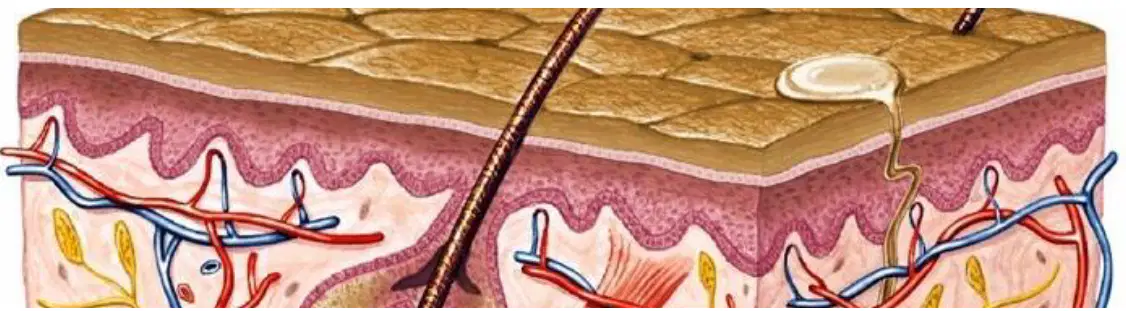
Accessory structures are located in which layer of the skin?
Dermis
Epidermis
Hypodermis
Stratum corneum
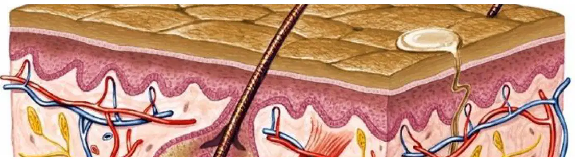
The wavy, bumpy top of the dermis is called what?
Dermal papillae
Stratum basale
Hair follicle
Sebaceous gland
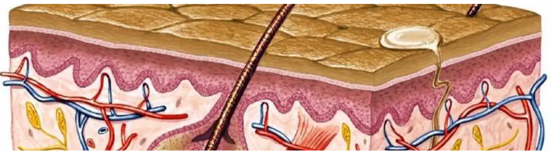
Which description best defines dermal papillae?
Projections of the dermis that extend into spaces between ridges of the epidermis
Coiled ducts of sweat glands that open to the skin surface
Clusters of sensory receptors embedded in the hypodermis
Networks of blood vessels that run parallel to the epidermal surface
Dermal papillae are best described as projections of the dermis that extend into spaces between ridges of the epidermis.
Projections of the dermis into spaces between epidermal ridges
Folds of the epidermis that cover hair follicles
Openings of sweat glands in the dermis
Bundles of collagen that anchor the hypodermis
Dermal papillae mark the juncture point between which two tissue types?
Epithelial and connective tissue
Muscle and nervous tissue
Adipose and epithelial tissue
Cartilage and muscle tissue
Where are dermal papillae most abundant in the body?
Hands and feet
Scalp
Abdomen
Back
The ridges associated with dermal papillae primarily help to do what?
Secure the dermis to the epidermis
Increase melanin production
Reduce heat loss through the skin
Lubricate the skin surface
Which features are attributed to dermal papillae? Select all that apply.
Fingerprints
Palm contours
Sweat gland secretion
Hair shaft formation
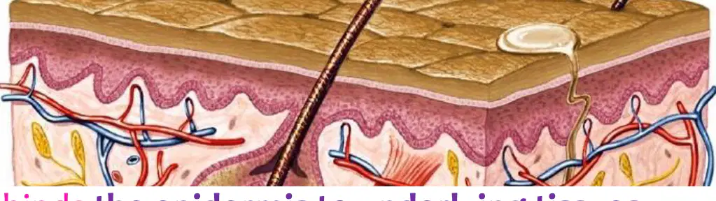
The dermis binds what to the underlying tissues?
The epidermis
The hypodermis
The hair follicles
The sweat glands
Based on the list "In the dermis is:", which of the following are located in the dermis? Select all that apply.
Blood vessels
Nerve receptors (motor and sensory)
Muscles
Fibers
According to the list "In the dermis is:", which of the following are located in the dermis? Select all that apply.
Hair
Nails
Sweat glands
Sebaceous glands (oil)
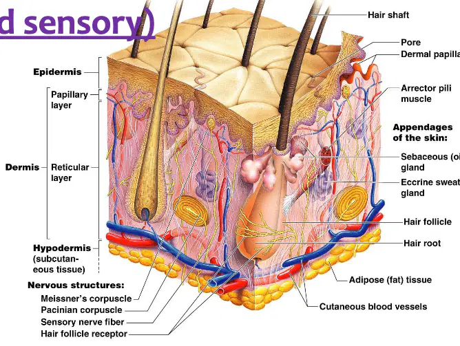
In the labeled cross-section of skin, which appendage is the oil-producing gland located in the dermis?
Sebaceous gland
Eccrine sweat gland
Meissner's corpuscle
Arrector pili muscle
Which item from the slide refers to receptors in the dermis that can be motor and sensory?
Nerve receptors
Hair follicle
Cutaneous blood vessels
Adipose tissue
According to the slide subtitle "Where Accessory Structures are Located", in which layer of the skin are these accessory structures located?
Dermis
Epidermis
Hypodermis
Stratum corneum
Select all locations where hair is most visible.
Scalp
Eyebrows
Eyelashes
Palms
Soles
Select all skin surfaces that lack hair.
Palms
Soles
Lips
Nipples
Parts of external reproductive organs
Which statement best describes hair at birth and later life?
Humans are born with hair, which changes as they grow
Humans are born without any hair
Hair present at birth is permanent and does not change
Hair first appears only at puberty
What happens after hair follicles develop before birth?
Baby hair is later replaced
Follicles disappear at birth
Hair never changes after birth
Hair develops only on the scalp
Which type of hair develops at puberty according to the slide?
Axillary and pubic hair
Scalp hair only
Eyebrow hair only
Lash hair only
What triggers the development of coarse hair at puberty?
Hormones
Dietary protein
Sunlight exposure
Regular exercise
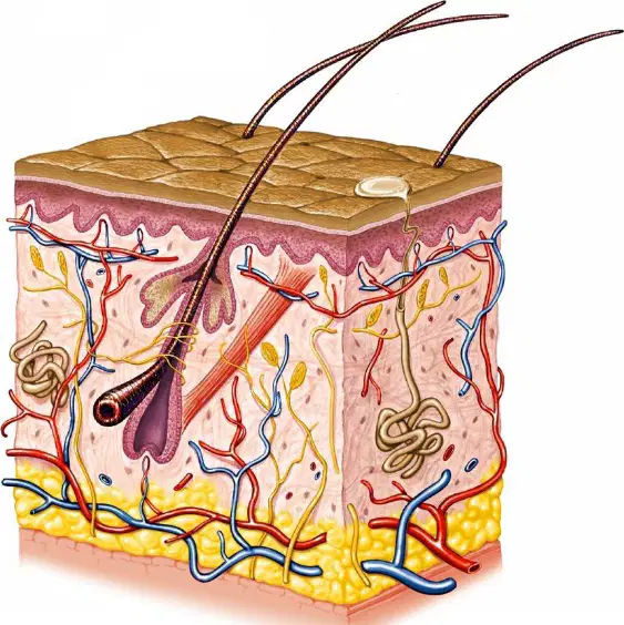
According to the slide, each hair develops from a group of epidermal cells at the base of a tube-like depression. What is this structure called?
Hair follicle
Sebaceous gland
Sweat pore
Arrector pili muscle
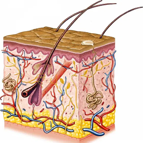
Where is the hair root located as described on the slide?
At the skin surface within the epidermis
In the dermis at the base, connected to blood vessels
Deep in the hypodermis embedded in adipose tissue
Within the stratum corneum near the outer surface
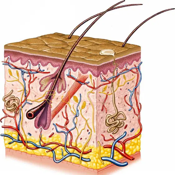
What nourishes the base of the hair root according to the slide?
Blood vessels supplying the dermis
Sebum secreted by sebaceous glands
Sweat from eccrine glands
Lymphatic fluid from dermal capillaries
Based on the slide, glands are considered appendages of which organ?
skin
liver
endocrine system
bone
Which pair lists the two kinds of skin glands identified on the slide?
sudoriferous and sebaceous
eccrine and apocrine
endocrine and exocrine
ceruminous and mammary
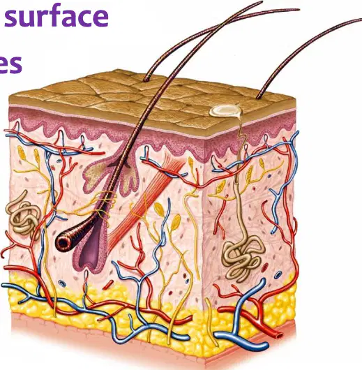
What is the duct opening target of eccrine sweat glands?
pore on skin surface
hair follicles
sebaceous ducts
lymphatic vessels
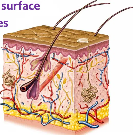
Where do apocrine sweat gland ducts empty?
hair follicles
directly to a pore on the skin surface
blood capillaries
sebaceous glands
Which gland type produces oil in the skin?
sebaceous glands
sudoriferous glands
eccrine glands
apocrine glands
Sudoriferous (sweat) glands are classified as which type of glands and are described as widespread in skin.
Endocrine glands
Exocrine glands
Paracrine glands
Sebaceous glands
According to the worksheet, sudoriferous (sweat) glands are the most numerous in the body, with how many present.
Fewer than 1 million
About 300,000
More than 3 million
About 30 million
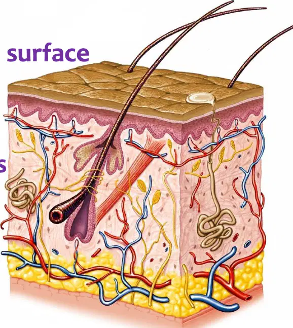
Eccrine sweat glands open via a duct to which location.
Hair follicles
Sebaceous glands
Blood vessels
A pore on the skin surface
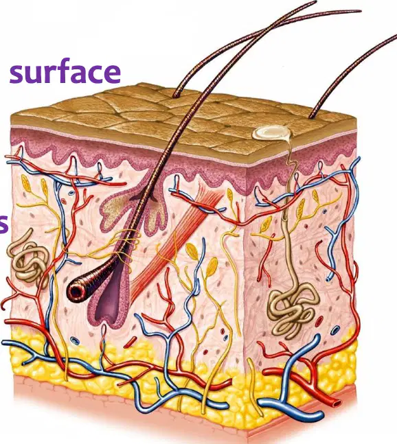
Apocrine sweat gland ducts empty into which structure.
Hair follicles
A pore on the skin surface
Sebaceous glands
Blood vessels
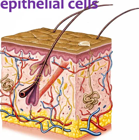
Sudoriferous glands each consist of a tiny tube that originates as what structure in the deeper dermis?
a ball-shaped coil
a straight duct
an alveolar sac
a branched acinus
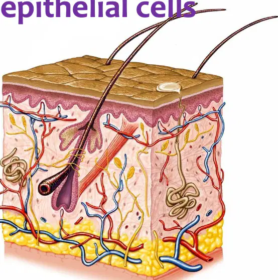
The coiled portion of a sudoriferous gland is closed at which end?
the deep end
the superficial end
both ends
neither end
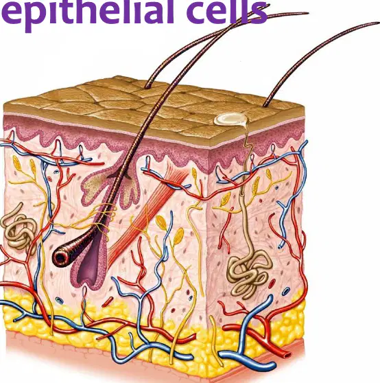
What type of cells line the coiled portion of the sudoriferous gland?
sweat-secreting epithelial cells
keratinocytes only
fibroblasts
adipocytes
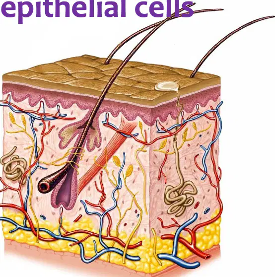
In the illustrated skin cross-section, which structure is responsible for producing sweat?
the coiled sudoriferous gland
the sebaceous gland
the hair papilla
a Meissner corpuscle
Which type of sweat gland is identified as the most numerous?
Eccrine glands
Apocrine glands
Sebaceous glands
Ceruminous glands
What best describes the composition of secretion from eccrine glands?
Strong odor with volatile compounds
Mostly water with some ions
Rich in lipids and proteins
Primarily sebum
What is the primary physiological role of eccrine glands?
Cool the body down
Lubricate hair shafts
Produce earwax
Secrete hormones into blood
Where are eccrine glands commonly found according to the material?
Forehead, neck, back
Axilla and groin around hair follicles
External auditory canal
Eyelids
Under which conditions do eccrine glands produce profuse sweat?
On hot days or during exercise
In cold environments at rest
During sleep only
After meals
During emotional stress, where does moisture from eccrine glands typically appear?
Palms and soles
Scalp and eyelids
Chest and abdomen
Forearms
Which statement about the smell of eccrine gland perspiration is supported by the material?
No smell; produces perspiration
Strong odor intrinsic to secretion
Always aromatic due to pheromones
Smell increases because it is lipid-rich
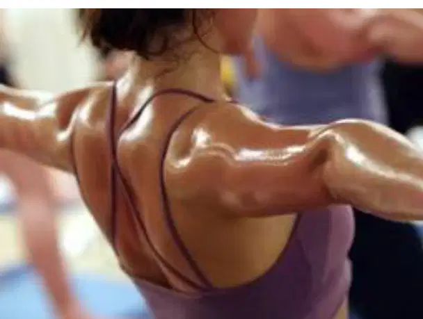
Sudoriferous glands: Sweat—fluid that eccrine glands secrete. Which gland type produces this fluid?
Eccrine glands
Sebaceous glands
Ceruminous glands
Mammary glands
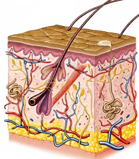
Sudoriferous glands carry sweat away by a tube (duct) that opens at the surface as what?
A pore
A hair follicle
An alveolus
A synapse
Sweat from eccrine glands is mostly water with small amounts of salts and wastes (urea and uric acid). Which components are typically present? Select all that apply.
Water
Salts
Urea
Uric acid
Proteins
Sweat—mostly water with small amounts of salts and wastes—serves which additional function noted for sudoriferous glands?
Excretory function
Endocrine function
Digestive function
Reproductive function
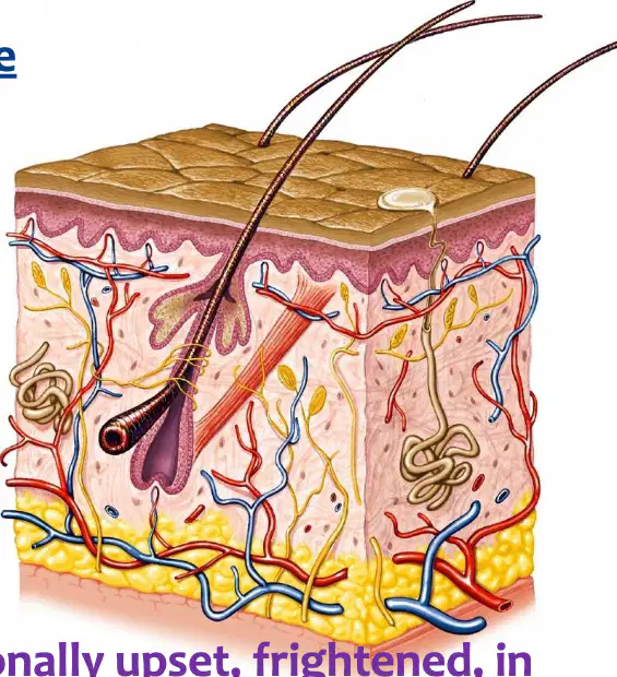
Apocrine glands are other sweat glands that become active at puberty. Which type of sweat gland is described as becoming active at puberty?
Apocrine glands
Eccrine glands
Sebaceous glands
Ceruminous glands
According to the slide, apocrine glands are not as many compared to other sweat glands. Which statement best describes their abundance?
They are fewer in number than other sweat glands
They are more numerous than other sweat glands
They are equal in number to other sweat glands
They occur only in infants
Apocrine gland secretions are associated with sex scents/sex attractants. What is the stated idea behind these secretions?
To attract the opposite sex
To regulate body temperature
To lubricate the skin surface
To neutralize skin pH
Where are apocrine glands localized, as indicated on the slide?
Genitals, nipples, face
Palms, soles, eyelids
Scalp only
Abdomen and back only
When are apocrine glands usually secreted, according to the slide?
When a person is emotionally upset, frightened, in pain, or during sexual arousal
During deep sleep
After eating a large meal
During dehydration
Who uses apocrine glands enormously, as stated on the slide?
Other animals
Plants
Bacteria
Fungi
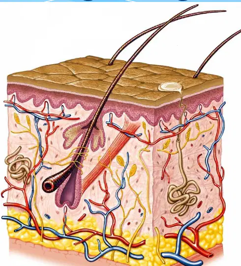
In adults, apocrine glands are most numerous in which body regions?
Palms and soles
Axillary regions and groin
Scalp and forehead
Back and chest
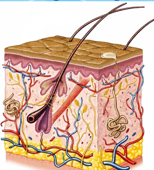
The ducts of apocrine glands open into which structures of the skin?
Sweat pores on the epidermal surface
Sebaceous glands
Hair follicles
Dermal capillaries
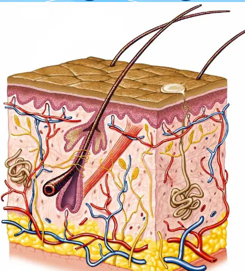
Secretions of apocrine glands develop a scent primarily because they are metabolized by what?
Melanocytes
Skin bacteria
Keratinocytes
Langerhans cells
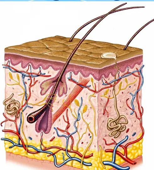
Which sudoriferous gland type is characterized by ducts opening into hair follicles and increased presence in axillary and groin regions?
Eccrine glands
Apocrine glands
Sebaceous glands
Ceruminous glands
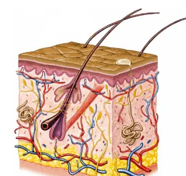
Identify the modified sweat gland of the external ear canal that secretes ear wax.
Ceruminous glands
Mammary glands
Identify the modified sweat gland that secretes milk.
Ceruminous glands
Mammary glands
According to the summary, the average square inch of skin holds how many sweat glands?
650
20
60,000
More than 1,000
According to the summary, the average square inch of skin holds how many blood vessels?
650
20
60,000
More than 1,000
According to the summary, the average square inch of skin holds how many melanocytes?
650
20
60,000
More than 1,000
According to the summary, the average square inch of skin holds more than how many nerve endings?
650
20
60,000
1,000
Groups of specialized epithelial cells are usually associated with which structure in the skin?
Hair follicles
Sweat pores
Nerve endings
Blood vessels
Sebaceous glands secrete an oily mixture of fatty material and cellular debris. What is this secretion called?
Sebum
Melanin
Keratin
Sweat
Through which pathway is the secretion of sebaceous glands released?
Into hair follicles through small ducts
Directly onto the epidermal surface
Into nearby sweat glands
Into the bloodstream

