
 Worksheets
Types of Epithelial Tissue
Total questions: 96
Worksheet time: 48mins
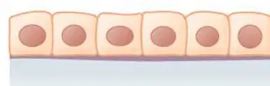
Identify the type of epithelium
Simple squamous
Cuboidal
Stratified squamous
Pseudo stratified
Esophagus epithelium is:
Simple squamous
Cuboidal
Stratified squamous non keratinized
Pseudo stratified
What is the key function of Simple Squamous epithelium?
Secretion
Absorption
Allows for rapid passage (diffusion and filtration) of substances; forms frictionless lining
Protection
Fill in the blank: The cells of Simple Cuboidal epithelium are _________?
cube-shaped or spherical; central, large nucleus
flat and thin; elongated nucleus
column-shaped; nucleus near the base
irregularly shaped; multiple nuclei
Where is Simple Columnar epithelium typically found?
Lining of the digestive tract
Alveoli of lungs
Skin surface
Lining of blood vessels
Which type of epithelial tissue has the thickest layers with deepest cells that are cuboidal/columnar and surface cells that are flattened (squamous)?
Stratified Squamous
Stratified Cuboidal
Stratified Columnar
Pseudostratified Columnar
What is the main function of stratified cuboidal epithelium?
Absorption
Protection and some limited secretion
Filtration
Sensation
Stratified columnar epithelium is found in small areas of the pharynx, male urethra, and lining of some large glandular ducts.
True
False
Fill in the blank: The keratinized type of stratified squamous epithelium is found in the _________.
epidermis of the skin
lining of the esophagus
inner surface of the cheek
lining of the vagina
Which specialized epithelial tissue appears multilayered but is actually a single layer and is often ciliated in the trachea?
Stratified Squamous
Stratified Cuboidal
Stratified Columnar
Pseudostratified Columnar
Identify the type of cartilage shown in diagram:
Hyaline
Fibrous
Elastic
Cornea
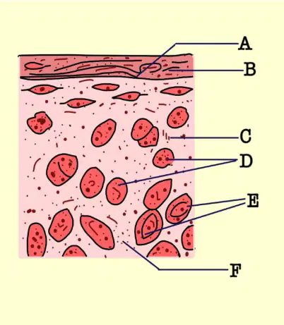
Refer to the diagram of cartilage. Which type of cartilage is labeled as 'A'?
Hyaline cartilage
Elastic cartilage
Fibrocartilage
None of the above
What is the appearance (gross) of Hyaline Cartilage?
Glassy, smooth, translucent, bluish-white
Opaque, rough, yellowish-brown
Fibrous, dull, grayish-white
Spongy, porous, reddish-pink
Which type of cartilage contains thick bundles of Type I Collagen fibers?
Fibrocartilage
Hyaline cartilage
Elastic cartilage
Articular cartilage
Which type of cartilage is the most flexible and easily bends and recoils?
Elastic cartilage
Hyaline cartilage
Fibrocartilage
Articular cartilage
What is the main fiber type found in Elastic Cartilage?
Type II Collagen and abundant Elastic Fibers (Elastin)
Type I Collagen only
Reticular fibers only
Type III Collagen and minimal Elastic Fibers
Which type of cartilage usually has perichondrium present (except on articular surfaces)?
A) Hyaline cartilage
B) Elastic cartilage
C) Fibrocartilage
Where are chondrocytes located in elastic cartilage?
Scattered in groups (isogenous groups)
Located in lacunae between elastic fibers
Arranged in rows/columns between collagen bundles
Fill in the blank: The main function of fibrocartilage is _________.
Shock absorption; resists heavy pressure and stretching forces
Providing insulation and energy storage
Facilitating rapid nerve impulse transmission
Secreting hormones into the bloodstream
Which type of cartilage is always present with perichondrium?
Hyaline cartilage
Elastic cartilage
Fibrocartilage
Chondrocytes in fibrocartilage are arranged in rows or columns between collagen bundles.
True
False
Which type of cartilage is found in the external ear (auricle)?
Hyaline cartilage
Elastic cartilage
Fibrocartilage
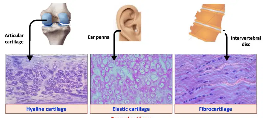
Identify the type of cartilage shown in the image labeled 'Articular cartilage'.
Hyaline cartilage
Elastic cartilage
Fibrocartilage
Calcified cartilage
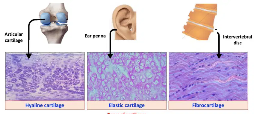
Identify the type of cartilage shown in the image labeled 'Ear pinna'.
Elastic cartilage
Hyaline cartilage
Fibrocartilage
Articular cartilage
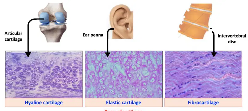
Identify the type of cartilage shown in the image labeled 'Intervertebral disc'.
Fibrocartilage
Hyaline cartilage
Elastic cartilage
Articular cartilage
Dilator pupillae & constrictor pupillae muscles of eye are derived from which germ layer?
Neuroectoderm
Mesoderm
Endoderm
Surface ectoderm
Which germ layer gives rise to the endothelium of blood and lymph vessels?
Mesoderm
Endoderm
Neuroectoderm
Ectoderm
Red and white blood cells are derived from which germ layer?
Mesoderm
Endoderm
Neuroectoderm
Ectoderm
The spleen, kidney, and adrenal cortex are derivatives of the ____ germ layer.
Mesoderm
Ectoderm
Endoderm
Neural crest
Testes, epididymis, ductus deferens, seminal vesicle, and ejaculatory duct are derived from which germ layer?
Mesoderm
Endoderm
Neuroectoderm
Ectoderm
Ovary, uterus, uterine tubes, and superior 1/3 of vagina are derived from the ____ germ layer.
Mesoderm
Ectoderm
Endoderm
Neural crest
Microglia and Kupffer cells are derived from which germ layer?
Mesoderm
Endoderm
Neuroectoderm
Ectoderm
Hepatocytes are derived from the ____ germ layer.
Endoderm
Mesoderm
Ectoderm
Mesothelium
Principal and oxyphil cells of parathyroid are derivatives of which germ layer?
Mesoderm
Endoderm
Neuroectoderm
Ectoderm
Parafollicular cells are derived from the ____ germ layer.
Endoderm
Mesoderm
Ectoderm
Neural crest
Epithelial reticular cells of thymus are derived from which germ layer?
Mesoderm
Endoderm
Neuroectoderm
Ectoderm
Acinar and islet cells of pancreas are derived from the ____ germ layer.
Endoderm
Mesoderm
Ectoderm
Periderm
Acinar cells of submandibular and sublingual glands are derivatives of which germ layer?
Mesoderm
Endoderm
Neuroectoderm
Ectoderm
Epithelial lining of gastrointestinal tract, trachea, bronchi, and lungs are derived from the ____ germ layer.
Endoderm
Mesoderm
Ectoderm
Neural crest
Biliary apparatus is derived from which germ layer?
Mesoderm
Endoderm
Neuroectoderm
Ectoderm
Urinary bladder, female urethra, and male urethra are derivatives of the ____ germ layer.
Endoderm
Mesoderm
Ectoderm
Neural crest
Auditory tube and middle ear cavity are derived from which germ layer?
Mesoderm
Endoderm
Neuroectoderm
Ectoderm
Crypts of palatine tonsils are derived from the ____ germ layer.
Endoderm
Mesoderm
Ectoderm
Neural crest
All neurons of the brain and spinal cord are derived from which germ layer?
Mesoderm
Endoderm
Neuroectoderm
Ectoderm
Retina, iris, epithelium, and ciliary body epithelium are derivatives of the ____ germ layer.
Neuroectoderm
Endoderm
Mesoderm
Surface ectoderm
Optic nerve (CN II), optic chiasma, and optic tract are derived from which germ layer?
Mesoderm
Endoderm
Neuroectoderm
Ectoderm
Dilator and sphincter pupillae muscles are derived from the ____ germ layer.
Neuroectoderm
Mesoderm
Endoderm
Surface ectoderm
Astrocytes, oligodendrocytes, ependymocytes, tanycytes, and choroid plexus cells are derivatives of which germ layer?
Mesoderm
Endoderm
Neuroectoderm
Ectoderm
Neurohypophysis and pineal gland are derived from the ____ germ layer.
Neuroectoderm
Endoderm
Mesoderm
Surface ectoderm
A patient report with sudden complete loss of vision on the left side field of both the eyes. What is the site of lesion?
Left optic nerve
Right optic tract
Optic chiasma decussating fibers
Visual cortex lesion
Fill in the blank: The central area of each retina is called the _______.
macula
cornea
iris
sclera
The image in the retina is inverted (upside down) which is converted to the upright image in the ________.
cerebral visual cortex
retina
optic nerve
lens
The projection of the exterior light on the retina is _______ in each eye, i.e., structures on the temporal field are sensed by the nasal part of the retina, and structures on the nasal field are sensed by the temporal part of the retina.
contralateral
bilateral
unilateral
parallel
After retinal stimulation, the fibers of the optic nerve (basically axons of the retinal ganglion cells) carry the signal from retina to the _______.
optic chiasm
lateral geniculate nucleus
primary visual cortex
superior colliculus
The optic nerve is carrying 2 sets of fibers: those from the nasal side, and those from the temporal side of each retina, respectively.
True
False
Within the optic chiasm, the fibers of the 2 sides decussate and nasal fibres of each optic nerve cross over to the opposite side while the temporal fibres retain their side. The nasal fibres of the left retina will project to the ______ cerebral cortex.
right
left
frontal
occipital
A person with section of the right optic tract will not be able to see things on the temporal side of the left eye and the nasal side of the right eye.
True
False
After the optic tract ends in the lateral geniculate body in the Thalamus, optic radiations originate from there which project to the ______ cortex of each cerebral hemisphere.
visual
auditory
motor
olfactory
There are two geniculocalcarine radiations on each hemisphere: the upper and the lower. Each one of them carries information of the opposite visual field in the tract. The upper tract will carry information of the ______ visual field, while the lower tract will carry information of the upper field.
lower
right
central
peripheral
According to the passage, which tract carries information of the upper visual field?
Upper tract
Lower tract
Refer to the diagram of the visual pathway and the visual field deficit illustrations. Which visual field defect is represented by a central scotoma?
Central scotoma
Monocular vision loss
Bitemporal hemianopia
Contralateral homonymous hemianopia
Contralateral superior quadrantanopia
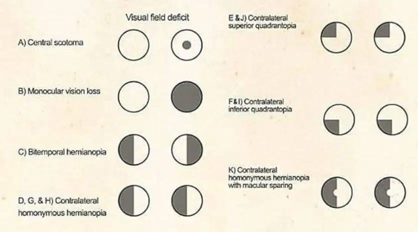
Refer to the diagram of the visual pathway and the visual field deficit illustrations. Which visual field defect is represented by complete vision loss in one eye?
D) Contralateral homonymous hemianopia
C) Bitemporal hemianopia
A) Central scotoma
B) Monocular vision loss
E) Contralateral superior quadrantanopia
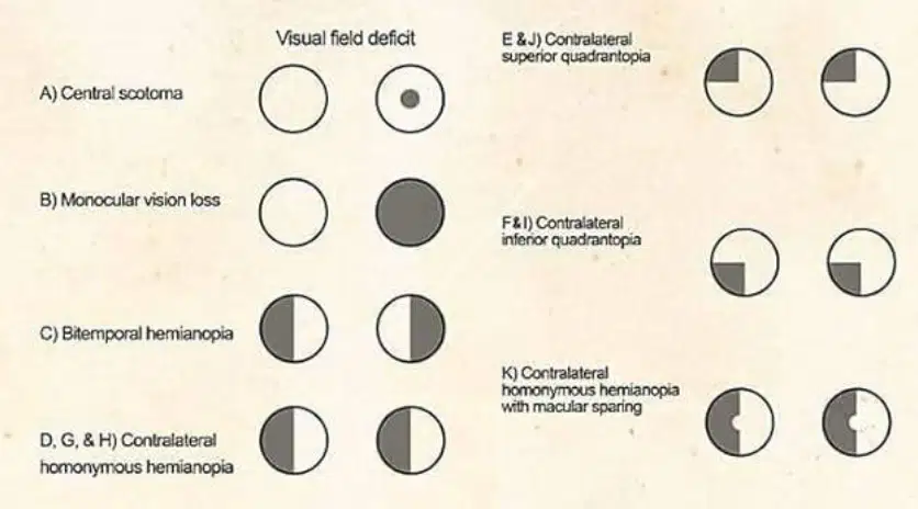
Refer to the diagram of the visual pathway and the visual field deficit illustrations. Which visual field defect is represented by loss of vision in the temporal half of both visual fields?
Monocular vision loss
Central scotoma
Contralateral superior quadrantanopia
Contralateral homonymous hemianopia
Bitemporal hemianopia
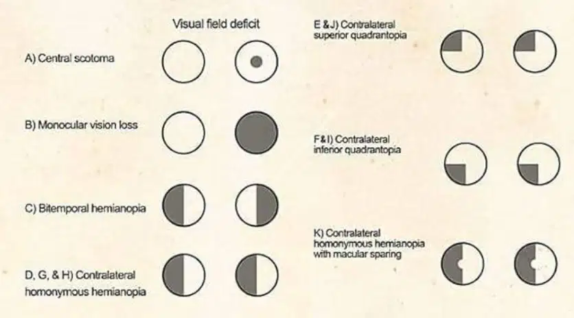
Refer to the diagram of the visual pathway and the visual field deficit illustrations. Which visual field defect is represented by loss of vision in the same half of the visual field in both eyes?
Contralateral homonymous hemianopia
Central scotoma
Monocular vision loss
Bitemporal hemianopia
Contralateral superior quadrantanopia
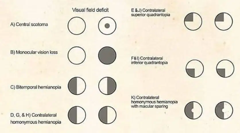
Which visual field defect is represented by loss of vision in the upper quadrant of the contralateral visual field?
E) Contralateral superior quadrantanopia
F) Contralateral inferior quadrantanopia
K) Contralateral homonymous hemianopia with macular sparing.
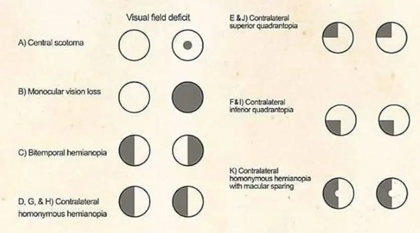
Refer to the diagram of the visual pathway and the visual field deficit illustrations. Which visual field defect is represented by loss of vision in the lower quadrant of the contralateral visual field?
E) Contralateral superior quadrantanopia
F) Contralateral inferior quadrantanopia
K) Contralateral homonymous hemianopia with macular sparing.
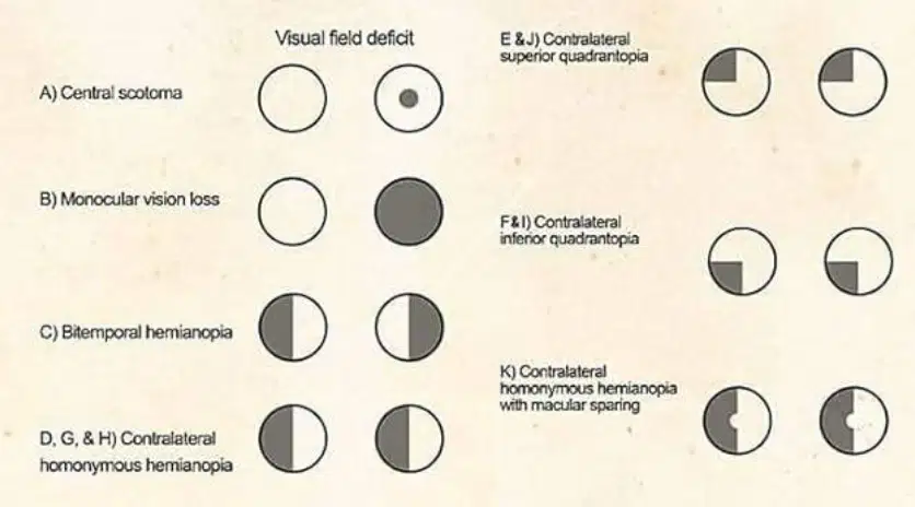
Refer to the diagram of the visual pathway and the visual field deficit illustrations. Which visual field defect is represented by contralateral homonymous hemianopia with macular sparing?
Contralateral superior quadrantanopia
Contralateral inferior quadrantanopia
Contralateral homonymous hemianopia with macular sparing
Purkinje cells are found in:
Cerebrum
Midbrain
Medulla
Cerebellum
Loss of vision in one eye is due to a defect in the _________
Ipsilateral Optic Nerve
Contralateral Optic Tract
Optic Chiasma
Lateral Geniculate Body
Bitemporal Hemianopia is caused by a lesion at the _________
Optic chiasm
Optic nerve
Lateral geniculate body
Occipital cortex
Binassal hemianopia is associated with a defect in the _________
Optic chiasm
Optic nerve
Lateral geniculate body
Occipital cortex
Left homonymous hemianopia is due to a lesion in the _________.
Right optic tract / radiation
Left optic nerve
Right occipital lobe
Left optic tract
Right homonymous hemianopia is due to a lesion in the _________
Left optic tract / radiation
Right optic nerve
Left retina
Right occipital pole
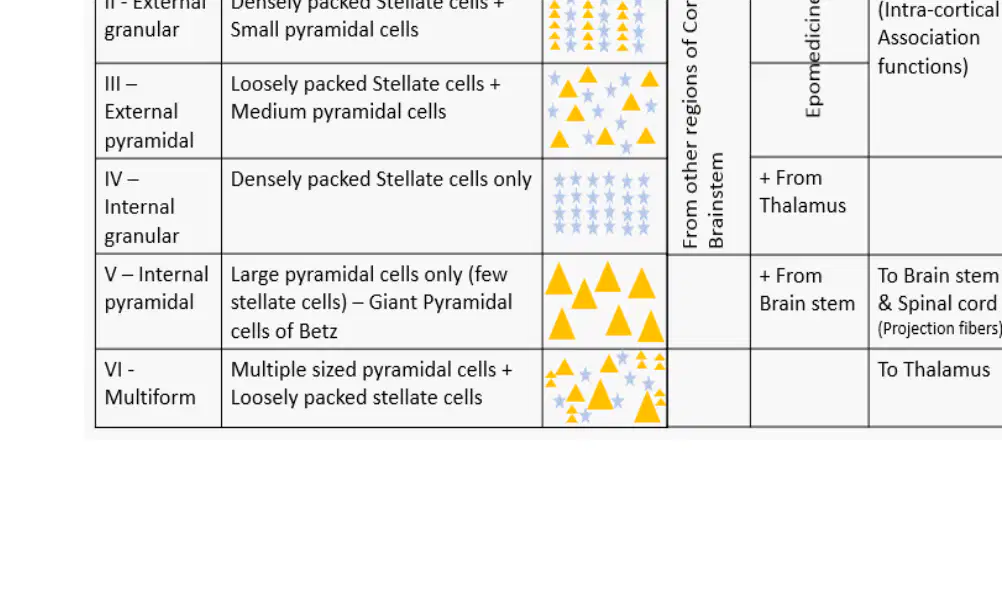
Which layer of the cortex contains axons and dendrites (cell processes)?
I – Molecular
II – External granular
IV – Internal granular
VI – Multiform
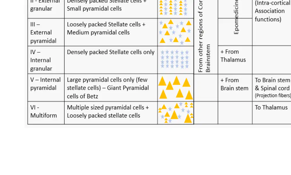
Which layer of the cortex contains densely packed stellate cells and small pyramidal cells?
II – External granular
I – Molecular
IV – Internal granular
VI – Multiform
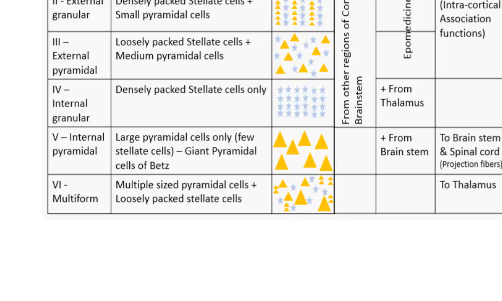
Which layer of the cortex contains loosely packed stellate cells and medium pyramidal cells?
III – External pyramidal
I – Molecular
IV – Internal granular
V – Internal pyramidal
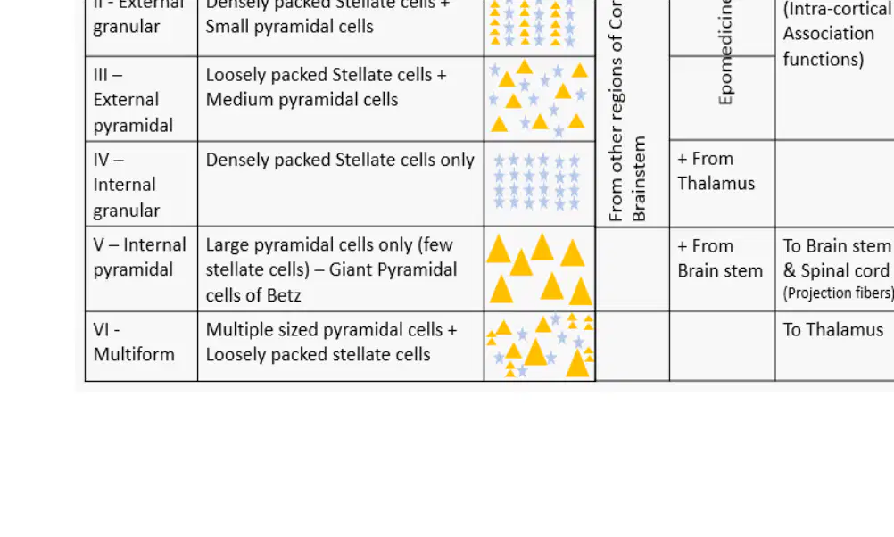
Which layer of the cortex contains densely packed stellate cells only?
IV – Internal granular
I – Molecular
V – Internal pyramidal
VI – Multiform
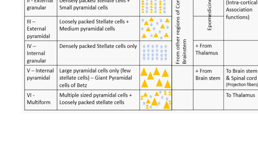
Which layer of the cortex contains large pyramidal cells (Giant Pyramidal cells of Betz)?
V – Internal pyramidal
I – Molecular
II – External granular
IV – Internal granular
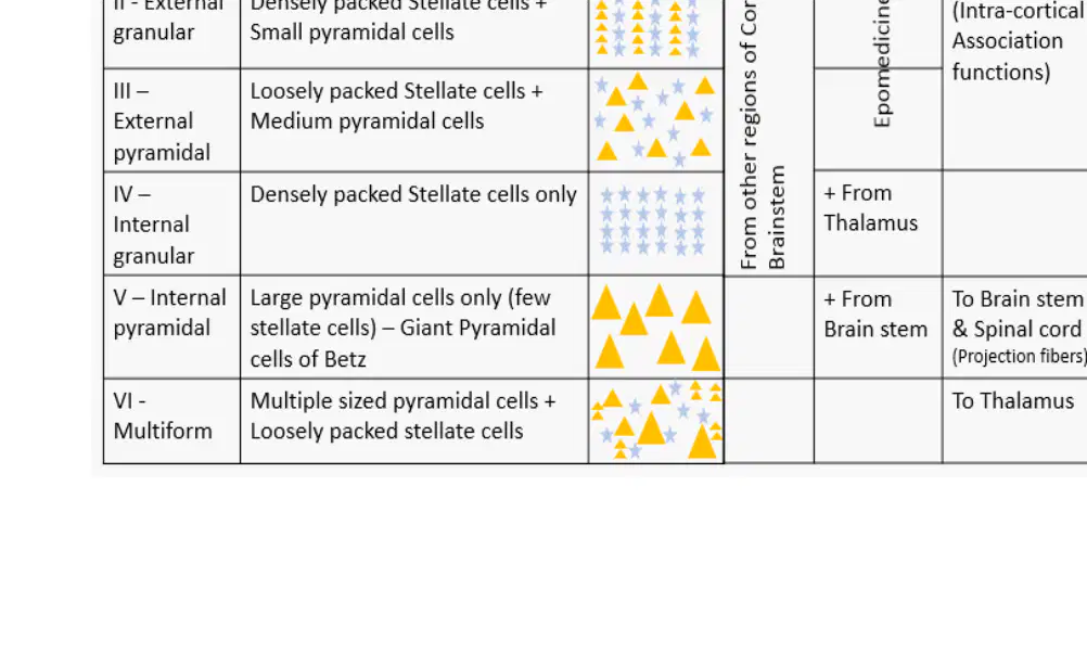
Which layer of the cortex contains multiple sized pyramidal cells and loosely packed stellate cells?
VI – Multiform
I – Molecular
II – External granular
IV – Internal granular
Which artery supplies the superior surface of the cerebellum?
SCA
AICA
PICA
The AICA originates from which artery?
Basilar Artery
Vertebral Artery
Internal Carotid Artery
The PICA supplies which surface of the cerebellum?
Superior
Anteroinferior
Posteroinferior
What is the main function of the cerebrocerebellum (lateral hemispheres)?
Planning, timing, and initiation of skilled movements.
Regulation of heart rate and respiration.
Processing visual information.
Control of basic reflexes.
Which nucleus is key for the cerebrocerebellum (lateral hemispheres)?
Dentate Nucleus
Fastigial Nucleus
Interposed Nucleus
Vestibular Nucleus
What is the main deficit seen in spinocerebellum (vermis & intermediate zones) lesions?
Truncal/Gait Ataxia (Unsteady, wide-based walking, balance issues).
Intention tremor (tremor during voluntary movement).
Resting tremor (tremor at rest).
Hemiballismus (violent, involuntary limb movements).
The output of the spinocerebellum is to which structures?
Brainstem/Spinal Cord
Cerebral Cortex
Basal Ganglia
Thalamus
The artery marked with arrow is palpated at which point:
Petrous temporal
Condyle
Zygoma
Ramus of mandible
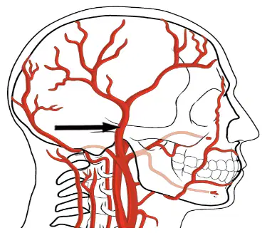
Which bone does the superficial temporal artery pass over, as shown in the diagram?
Mandible
Maxilla
Zygoma
Parietal bone
The superficial temporal artery is a branch of which artery?
Internal carotid artery
External carotid artery
Maxillary artery
Parietal artery
Fill in the blank: The STA is one of the two terminal branches of the ________, the other being the Maxillary Artery.
External Carotid Artery (ECA)
Internal Carotid Artery (ICA)
Subclavian Artery
Common Carotid Artery
Name the two main terminal branches of the superficial temporal artery after its bifurcation above the zygomatic arch.
Frontal Branch (Anterior) and Parietal Branch (Posterior)
Maxillary Branch and Occipital Branch
Temporal Branch and Mandibular Branch
Zygomatic Branch and Auricular Branch
Transverse Facial Artery: ________ the parotid gland, parotid duct, and the Masseter muscle.
Supplies
Drains
Innervates
Protects
Middle Temporal Artery: ________ the Temporalis muscle.
Supplies
Innervates
Originates from
Attaches to
Zygomatico-orbital Artery: ________ the Orbicularis Oculi muscle (around the eye).
Supplies
Innervates
Drains
Contracts
Anterior Auricular Branches: ________ the anterior aspect of the external ear.
Supply
Drain
Protect
Compress
The Fontanelle between Coronal & Sagittal sutures is:
Anterior fontanelle and fuses at 18 months
Posterior fontanelle & fuses at 12 months
Lambdoid fontanelle & fuses at 2-3 months

