
 Worksheets
Sonographic Evaluation of Ovarian Neoplasms
Total questions: 73
Worksheet time: 37mins
At what cyst size do doctors usually recommend surgery?
Greater than 10 cm
Greater than 8 cm
Greater than 3 cm
Greater than 5 cm
In postmenopausal ovarian sonograms, which mass feature is the most suspicious?
Mixed texture to solid with papillae
Thin-walled simple anechoic cyst
Stable size with multiple follicles
Uniform low echogenicity throughout
Which ultrasound lesion description is most likely benign?
Well-defined anechoic lesion
Thick, irregular septations
Mural nodules present
Solid echogenic elements
Which Doppler pattern in ovarian masses suggests possible malignancy?
Low-resistive arterial flow
High-resistive venous flow
Triphasic high-impedance flow
Absent detectable Doppler signal
Which statement about septation and locularity in ovarian cysts is most accurate?
Multiloculated thin septations ensure benignity
No septations always indicate malignancy
Unilocular, thinly septated cysts tend to be benign
Any septations confirm malignancy
Which finding suggests extra-ovarian spread and requires further evaluation?
Extension to omentum or peritoneum
Presence of small physiologic cysts
Normal ovarian vascular resistive index
Mobile free fluid without echoes
Which type of mass is most likely to be malignant?
Multilocular, thickly septated mass
Smooth-walled, avascular lesion
Simple, unilocular, thin-walled cyst
Uniform, anechoic fluid collection
Which change in ovarian volume is considered suspicious during screening?
More than 20 ml
More than 40 ml
More than 10 ml
More than 30 ml
Which ovarian feature is seen in postmenopausal women not on HRT?
Atrophic ovaries without follicles
Enlarged ovaries with many follicles
Normal-sized ovaries with cysts
Hypertrophic ovaries with papillae
Which sonographic pattern in postmenopausal ovaries suggests malignancy?
Isoechoic ovaries with smooth walls
Uniform anechoic follicles
Small hypoechoic ovaries
Enlarged echogenic ovaries
Which statement best describes the mortality rate of ovarian carcinoma among gynecologic cancers?
Lowest mortality due to early detection
Highest mortality from late-stage diagnosis
Moderate mortality with effective screening
Declining mortality from widespread vaccination
At what ages is ovarian carcinoma most commonly seen?
40 to 60 years
70 to 90 years
20 to 30 years
10 to 20 years
Which is the most important risk factor for ovarian carcinoma?
Use of combined oral contraceptives
Nulliparity without infertility
Early menarche with obesity
Family history of ovarian cancer
Why is ovarian carcinoma often diagnosed at an advanced stage?
Routine screening detects precancerous lesions.
Symptoms are prominent early.
Asymptomatic and undetectable early.
Patients present with dramatic bleeding.
Which clinical finding is commonly seen in advanced ovarian carcinoma?
Ascites with abdominal distension
Productive cough with hemoptysis
Severe pruritus with excoriations
Hematemesis with jaundice
Which serum marker is commonly elevated in epithelial ovarian cancer?
CA-125
AFP
CA 19-9
CEA
Which patient presentation is most suggestive of ovarian carcinoma?
Acute diarrhea with high fever and chills
Vague abdominal pain, bloating, constipation
Right upper quadrant colicky pain after eating
Sharp localized pain with rebound tenderness
On physical examination, what adnexal findings might be observed in ovarian carcinoma?
Normal ovaries to slightly enlarged, irregular ovaries
Large, tender uterus with homogeneous texture
Smooth cystic mass, always unilateral
Firm, mobile mass resolving with rest
Which imaging findings suggest malignant spread of ovarian carcinoma?
Ascites, peritoneal implants, lymphadenopathy
Single mural nodule without other features
Isolated ovarian cyst without ascites
Normal ovaries with mild free fluid only
Which description best matches Stage I ovarian cancer?
Confined to one or both ovaries only
Spread to pelvic organs and tissues
Peritoneal implants on bowel and omentum
Metastasis to liver parenchyma or beyond
Which type of epithelial ovarian tumor accounts for approximately 30% of all ovarian neoplasms?
Serous neoplasms
Mucinous neoplasms
Brenner neoplasms
Endometrioid neoplasms
Which statement best describes the origin of epithelial ovarian tumors?
Sex cord–stromal cells
Surface epithelium covering the ovary
Peritoneal mesothelial lining
Germ cells within follicles
Cystadenoma and cystadenocarcinoma are types of which category of epithelial tumors?
Serous and mucinous types
Endometrioid only type
Clear cell only type
Brenner only type
Which epithelial ovarian tumor subtype accounts for about 20–25% of all ovarian neoplasms?
Clear cell tumors
Endometrioid tumors
Serous tumors
Mucinous tumors
What is the typical size range of giant mucinous cystadenomas of the ovary?
5–15 cm diameter
15–50 cm diameter
3–8 cm diameter
10–30 cm diameter
A patient presents with a large multilocular cystic ovarian mass featuring papillary projections, calcifications, and ascites. Which malignant tumor is most likely?
Mucinous cystadenocarcinoma
Serous cystadenocarcinoma
Clear cell carcinoma
Brenner tumor
Which clinical feature is most consistently associated with mucinous cystadenocarcinoma of the ovary?
Krukenberg metastasis
Pseudomyxoma peritonei
Meigs syndrome triad
History of endometriosis
A patient presents with pelvic fullness and weight loss. In which age range does serous cystadenocarcinoma most commonly present with these symptoms?
Age 30–50, dysuria and hematuria
Age >60, acute pain and fever
Age 20–40, amenorrhea and infertility
Age 40–60, pelvic fullness and weight loss
Abigail and Kai discover a solid, hypoechoic ovarian mass with calcification, possibly associated with Meig’s syndrome. Which benign epithelial tumor is this?
Brenner tumor
Serous cystadenoma
Mucinous cystadenoma
Endometrioid tumor
A patient presents with a large, unilateral abdominal mass on ultrasound, showing thick, irregular walls, septations, papillary projections, and internal echoes. What is the most likely diagnosis?
Serous cystadenocarcinoma
Mucinous cystadenocarcinoma
Mucinous cystadenoma
Endometrioid carcinoma
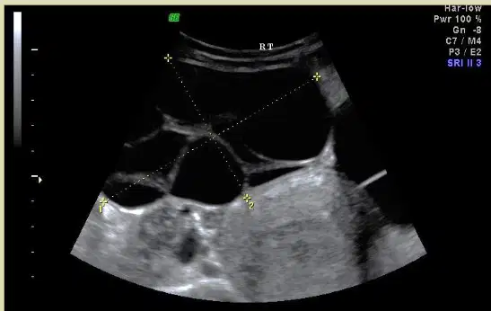
Using the image given, what ovarian neoplasm should be considered?
Fibroma
Serous cystadenoma
Endometrioma
Dermoid cyst
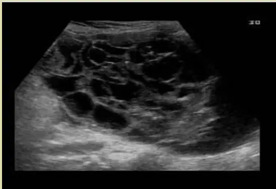
Using the image given, which ultrasound feature makes you most suspicious of a mucinous cystadenoma rather than a simple ovarian cyst?
Posterior acoustic shadowing throughout
Uniform anechoic single cavity
Circumferential calcified wall appearance
Multiple compartments with thin septa
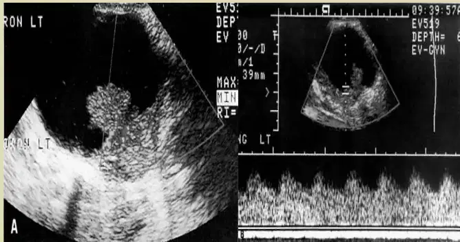
Spectral Doppler was obtained from the ovarian mass shown. The waveform and the sonographic image are consistent with which of the following?
Paraovarian cyst with no internal flow
Benign serous cystadenoma, avascular
Hemorrhagic cyst with increase vascularity
Serous cystadenocarcinoma with vascularity
Which statement best describes ovarian germ cell tumors?
Develop from secondary Müllerian structures
Arise from mature stromal thecal cells
Derived from primitive embryonic germ cells
Originate from surface epithelial mesothelium
What proportion of ovarian neoplasms are germ cell tumors?
Roughly thirty to forty percent
Approximately five to ten percent
About fifteen to twenty-five percent
Nearly fifty to sixty percent
Which is the most common ovarian neoplasm?
Dermoid cystic teratoma
Immature teratoma
Dysgerminoma
Endodermal sinus tumor
A patient with a germ cell tumor presents with pelvic or abdominal pain and a mass. Which markers are often elevated?
TSH, prolactin, cortisol
CEA, CA-19-9, PSA
AFP, LDH, hCG
AST, ALT, alkaline phosphatase
Which sonographic feature is most characteristic of a dermoid (cystic teratoma)?
Hypervascular solid lesion with central scar
Multiloculated cyst with thick septations and ascites
Uniform hypoechoic solid mass without calcification
Calcifications with shadowing, floating hair strands
Which feature best describes an immature teratoma?
Rapidly growing unilateral malignant mass
Slow-growing bilateral benign lesion
Hormone-secreting tumor causing virilization
Cystic benign lesion with teeth and bone
Which marker pattern is most commonly associated with immature teratoma?
High inhibin and estradiol levels
Isolated elevation of CA-125 only
Marked increase in CA-19-9 alone
Elevated AFP, hCG, LDH, and CEA
Which clinical scenario is most commonly associated with dysgerminoma?
Postmenopausal bleeding episodes
Infertility with oligomenorrhea
Children with precocious puberty
Adult males with gynecomastia
What is the typical ultrasound appearance of a dysgerminoma?
Predominantly cystic lesion with septations
Complex mass with fat-fluid levels
Solid mass with central scar and calcified rim
Solid homogeneous mass with irregular borders
Which serum marker is most strongly associated with an endodermal sinus tumor?
High hCG with normal AFP
Alpha-fetoprotein elevation
Raised CA-125 without AFP change
Marked increase in LDH only
Which ovarian germ cell tumor often shows calcifications such as teeth or bone on imaging?
Dysgerminoma
Endodermal sinus tumor
Choriocarcinoma
Dermoid cystic teratoma
Avery says dermoid cystic teratomas are always bilateral. Ethan says only 15% are bilateral. Who is correct about their bilateral occurrence?
More common in midline extragonadal sites
Exclusively unilateral involvement
Predominantly always bilateral involvement
Fifteen percent bilateral involvement
Which statement best describes the sonographic appearance of germ cell tumors?
The appearance varies from cystic to complex to solid.
They always present as homogeneous hypoechoic masses.
They consistently show multiloculated cystic morphology.
They uniformly demonstrate central necrotic cavities.
In which age range is dysgerminoma most common?
Adolescence only
Above seventy years
Under five years of age
Twenty to thirty years
Which marker is associated with dysgerminoma?
Consistent rise in CEA
Strong increase in hCG
Markedly high AFP levels
Elevated LDH and sometimes CA-125
Which set of symptoms best fits a dermoid cystic teratoma?
Ascites and weight loss
Fever and leukocytosis
Vaginal bleeding and anemia
Asymptomatic mass or torsion pain
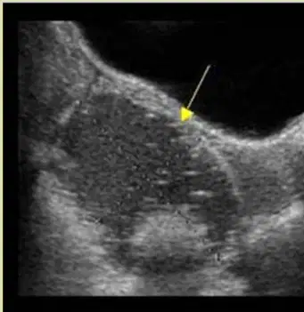
What is the 'dermoid plug' in a cystic dermoid?
Homogeneous hypoechoic solid mass
Peripheral calcified rim around the lesion
Echogenic mural nodule attached to the cyst wall
Thin avascular septation within the cyst
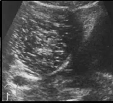
Which ultrasound finding is seen with the classic 'dermoid mesh' pattern in a mature cystic teratoma?
Multiple linear echogenic interfaces floating in a cyst
Uniform hypoechoic solid mass without cystic areas
Layered fluid-debris level with posterior enhancement
Single calcified tooth creating strong shadowing
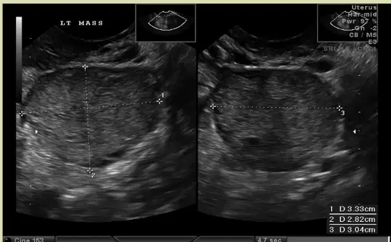
The left ovary shows a solid mass with hypoechoic irregular septa and a 'lobulated' pattern on ultrasound. What is the most likely diagnosis?
Endometrioma with ground-glass echoes
Immature solid teratoma of the ovary
Hemorrhagic cyst of the ovary
Serous cystadenoma with thin septa
Which ovarian tumor category arises from the sex cords of the embryonic gonad or the ovarian stroma?
Metastatic gastrointestinal lesions
Sex cord–stromal tumor category
Germ cell tumor derivatives
Epithelial neoplasms of surface lining
Which ovarian stromal tumor is known for producing estrogen?
Fibroma produces progesterone.
Thecoma produces estrogen.
Granulosa cell tumor produces androgens.
Sertoli-Leydig cell tumor produces cortisol.
Which ovarian stromal tumor is classically associated with Meigs syndrome?
Thecoma with ascites and hydrothorax
Sertoli-Leydig with pleural effusions
Fibroma with ascites and hydrothorax
Granulosa with endocrine hypertension
In which age group are thecomas most commonly found?
Any age
Adolescents (10–19 years)
Postmenopausal women
Women aged 20–30 years
Maya and Avery are studying sonography. They encounter a case of thecoma. Which sonographic feature are they most likely to find?
Cystic lesion with strong enhancement
Anechoic simple cyst without walls
Hypoechoic solid mass with attenuation
Mixed echogenicity with calcified septa
Which statement best describes granulosa cell tumors?
Androgen-producing, causes rapid virilization
Most common estrogenic tumor, occurs at varied ages
Always bilateral, uniformly malignant
Predominantly cystic, does not produce hormones
Which scenario best suggests a granulosa cell tumor?
Amenorrhea with deepening of the voice
Postmenopausal bleeding and pain
Precocious puberty, oligomenorrhea
Asymptomatic pressure without bleeding
Grace says fibromas can be found on both sides. James asks how often that occurs. What is the usual location pattern for fibromas?
Always bilateral in presentation
Ten percent bilateral occurrence
Uniformly unilateral occurrence
Predominantly midline pelvic location
Which age range is most commonly associated with Sertoli-Leydig cell tumors?
Children under ten years
Ages twenty to thirty years
Postmenopausal, over sixty years
Any age without limit
What hormonal effect is seen with Sertoli-Leydig cell tumors?
Cortisol excess with Cushing's features
Progesterone excess with lactation
Androgen excess with signs of virilization
Estrogen excess with endometrial growth
What is the typical sonographic appearance of a granulosa cell tumor?
Solid, complex, and heterogeneous
Purely anechoic, simple, thin-walled
Calcified, multilocular, with septations
Uniformly hyperechoic, fat-containing
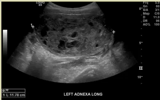
Which ultrasound pattern best helps to identify a granulosa cell tumor?
Multiloculated cyst with thin, avascular septations
Solid lesion with dense, shadowing calcifications
Predominantly solid with few cystic spaces
Large, complex mass with solid and cystic components
Purely cystic lesion without internal echoes
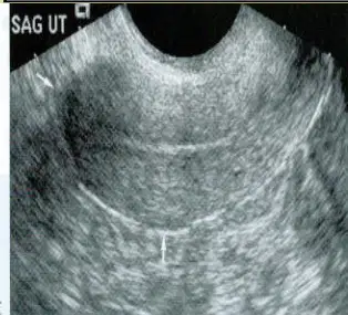
Lily and Michael observe an ovarian fibroma on ultrasound. Which echotexture best describes it?
Solid mass with coarse calcifications
Predominantly cystic with mural nodule
Complex solid-cystic with septations
Heterogeneous solid with necrotic areas
Homogeneous solid mass echotexture
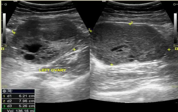
What vascular and cystic features are typically seen in Sertoli-Leydig cell tumors?
Avascular solid mass without cysts
Predominantly solid mass with few cystic areas and vascularity
Solid mass with extensive calcifications and no flow
Multiloculated cyst with high-resistance flow
Purely cystic lesion with no vascular flow
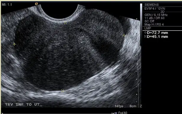
This transvaginal image shows a solid ovarian mass with small cystic areas. Which stromal tumor diagnosis best matches this pattern?
Sertoli-Leydig tumor due to calcified solid mass.
Granulosa cell tumor due to mixed solid-cystic appearance.
Dysgerminoma due to purely cystic presentation.
Fibroma due to homogeneous solid echotexture.
Theca cell tumor due to multiloculated cystic nature.
Which primary site most commonly causes Krukenberg tumor in the ovaries?
Pancreatic ductal adenocarcinoma
Cutaneous melanoma
Gastric or colon origin
Endometrial carcinoma
Breast carcinoma
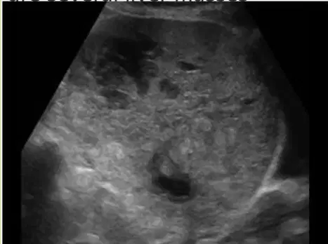
Which option best describes the classic ultrasound appearance of a Krukenberg tumor in the ovary?
Uniform, solid, echogenic oval mass
Predominantly calcified, hyperechoic lesion
Simple anechoic cyst without septations
Multiloculated cyst with thin, clear fluid only
Moth-eaten solid mass with cystic or complex areas
What is the most common laterality pattern seen in Krukenberg tumors of the ovaries?
Bilateral ovarian involvement is common
Alternating side involvement over time
Midline extraovarian involvement exclusively
Pelvic sidewall involvement without ovarian disease
Strictly unilateral involvement only
Isla, James, and Mia are studying for their clinical exams. They encounter a case of secondary ovarian metastases, such as Krukenberg tumors. Which combination of symptoms would help them identify it?
Pruritus with urticarial eruptions
Abdominal or pelvic pain with bloating
Vertigo with tinnitus
Polyuria with predominant nocturia
Fever and productive cough
Who is most commonly associated with metastatic ovarian disease of gastrointestinal origin?
Postoperative patients
Pregnant patients
Young males
Middle-aged and elderly females
Adolescent females
Lily, Zoe, and Jackson observe an encapsulated solid Krukenberg mass in the ovary on an ultrasound. What other finding might be seen on the scan?
Hydronephrosis in both kidneys
Ascites in the peritoneal cavity
Gallbladder sludge and polyps
Pleural effusions in both lungs
Pericardial effusion around the heart

