Search Header Logo
Powers Microscope Notes

Powers Microscope Notes

Assessment

Presentation

Biology

9th Grade

Hard

NGSS
MS-LS1-1, MS-LS1-2

Standards-aligned

Created by

Christopher Powers

Used 2+ times

FREE Resource

16 Slides • 34 Questions

1
media

2

media

3

media

4

media

5

media

6

media

7

media

8

media

9

media

10

media

11

media

12

Multiple Choice

Question image
Which part of the microscope controls the amount of light available?
1

coarse focus

2
diaphragm
3
objective lens
4
revolving nosepiece

13

Multiple Choice

Question image

A thin flat piece of glass used to hold objects for examination is called a ___

1

sheet

2

slide

3

cover slip

4

objective

14

media

15

Multiple Choice

Question image

What is circled here?

1

Magnification

2

Scanner

3

Coarse Adjustment knob

4

Fine adjustment knob

16

Multiple Choice

Question image

When do we use the coarse adjustment knob

1

Medium and high power

2

Low power only

3

High power only

4

Medium power only

17

Multiple Choice

Question image
A microscope is an instrument that.
1
makes faraway objects look closer.
2
makes small objects appear larger.
3
decreases the size of small objects.
4
increases the size of small objects.

18

media

19

Multiple Choice

Question image

The table compares four types of microscopes and their functional abilities. Which of the following represents and Scanning Electron Microscope?

1

Microscope A

2

Microscope B

3

Microscope C

4

Microscope D

20

Multiple Choice

Question image

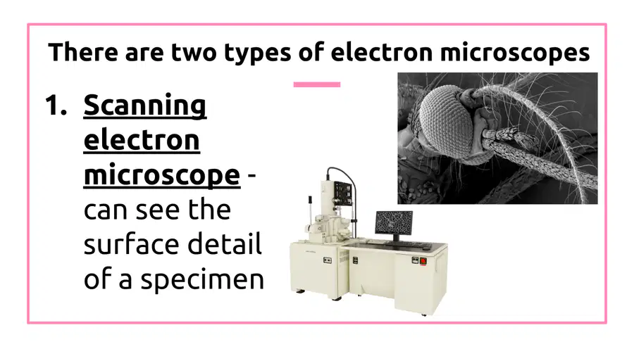
Which microscope took a 3D image of this fly's head?

1

Compound Light

2

Transmission Electron

3

Dissecting

4

Scanning Electron

21

media

22

Multiple Choice

Lets light pass through an object and then through two or more lenses
1
Binocular Microscope
2
Compound Microscope
3
Electron Microscope
4
Simple Microscope

23

media

24

Multiple Choice

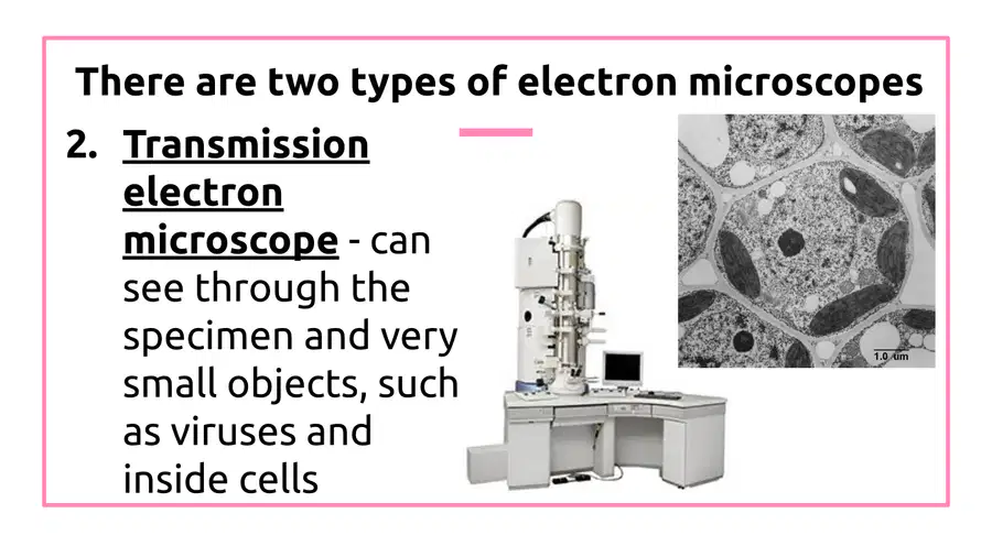
What is the function of a Transmission electron microscope?

1

To see through specimens

2

To magnify objects

3

To illuminate samples

4

To capture images in color

25

Multiple Choice

Question image

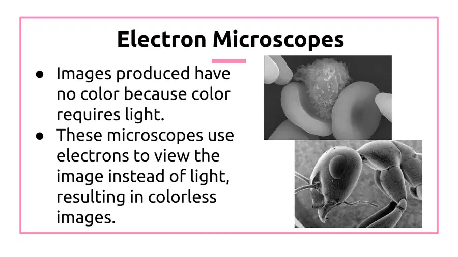
Jon viewed the surface of red blood cells while researching a new medication. What type of microscope did he use?

1

Light Microscope

2

Dissecting Microscope

3

Scanning Electron Microscope

4

Transmission Electron Microscope

26

Multiple Choice

Question image

This image shows the detailed structure of the tiny mitochondria organelle.  Which microscope most likely took this image?

1

Compound Light

2

Scanning Electron

3

Dissecting

4

Transmission Electron

27

media

28

Multiple Choice

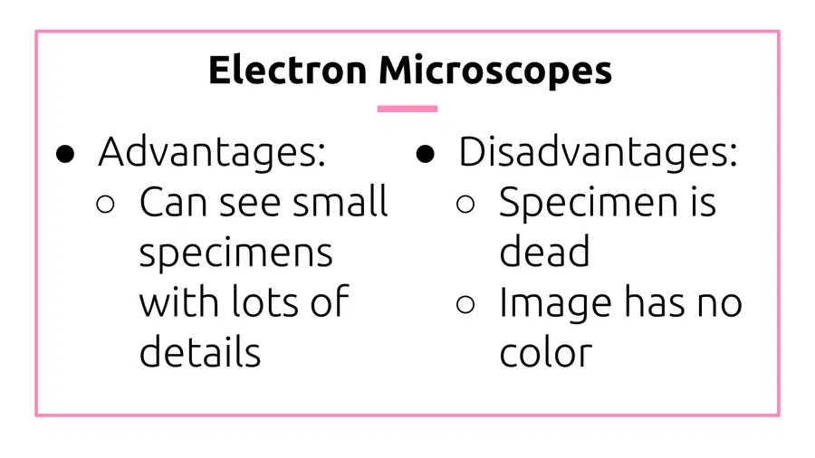
What are the advantages and disadvantages of using electron microscopes?

1

Can see small specimens with lots of details

2

Specimen is dead

3

Image has no color

4

All of the above

29

Multiple Choice

A student views cheek cells under low power. Before switching to high power, the student should:
1
adjust the ocular lens
2
center the image being viewed
3
adjust the course adjustment to move the stage away from the slide
4
remove the slide from the stage

30

Multiple Choice

Question image

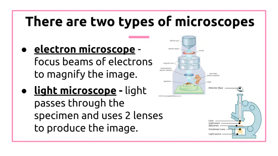
Electron microscopes differ from light microscopes in that they produce an image of a specimen by using a beam of electrons rather than

1

a beam of light.

2

a beam of compounds

3

a beam of cells

4

a beam of specimens

31

Multiple Choice

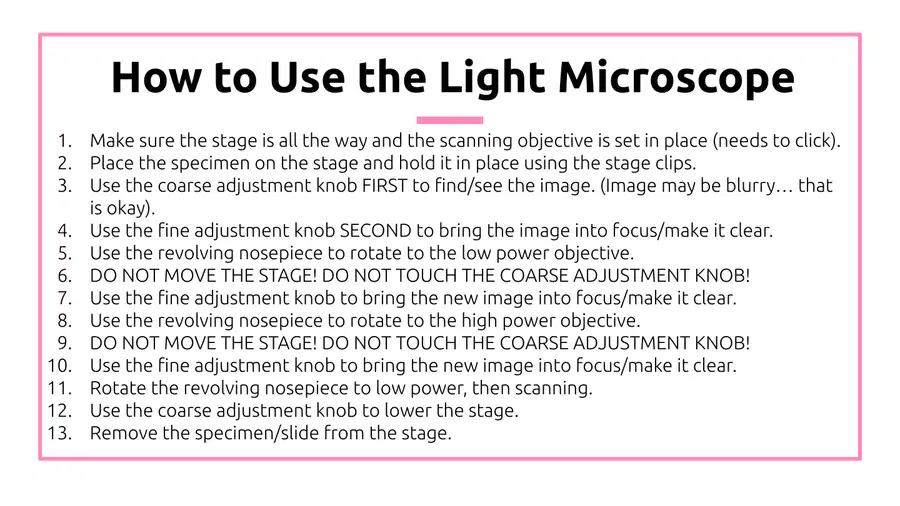
This bring the specimen into sharp focus
1
Coarse Adjustment Knob
2
Fine Adjustment Knob
3
Eyepiece
4
Diaphragm

32

Multiple Choice

What power do you start with when using a microscope?
1
High power
2
Medium power
3
Low power
4
Extra high power

33

Multiple Choice

Scientists must make informed choices about which tools to use for various tasks, including which kind of microscope is best suited to a particular investigation. Which of the following is NOT important to know in determining which kind of microscope should be used to view a particular specimen?

1

The type of material the specimen is made of

2

The age of the particular specimen

3

The size of the specimen being observed

4

The magnification required to view the specimen clearly

34

Dropdown

Question image
What is the name of the microscope part labeled as A? *Spelling counts & use all lowercase letters*

35

Labelling

Where do these labels go?

Drag labels to their correct position on the image

base

high power lens

nose piece

36

Labelling

Label these:

Drag labels to their correct position on the image

Stage

Eye piece

diaphragm

37

Fill in the Blanks

media image

38

Multiple Choice

Question image
Which part lets you turn up the power of magnification, such increasing from 40x to 100x?
1
coarse adjustment knob
2
diaphragm
3
field-of-view
4
revolving nosepiece

39

Multiple Select

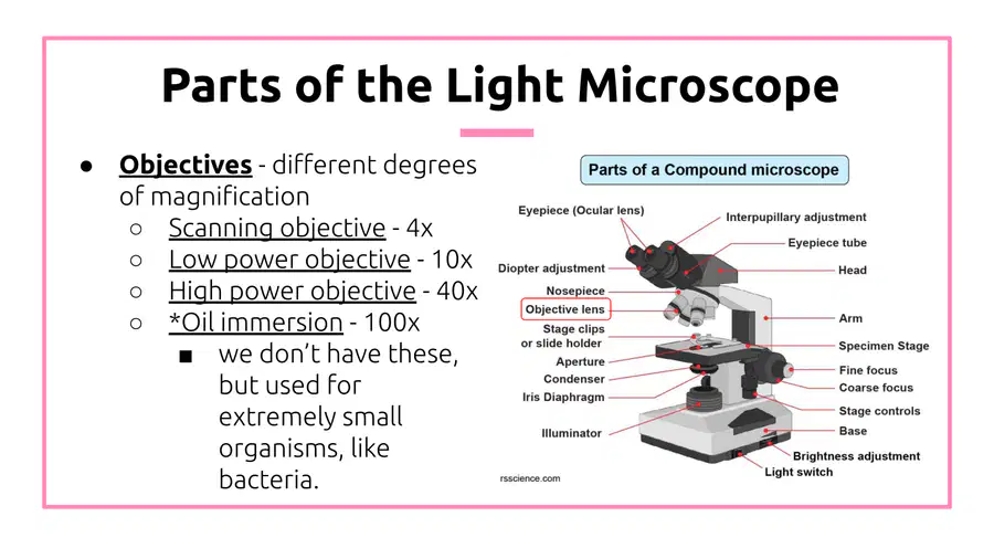
What are the different degrees of magnification in a light microscope? Check all that apply

1

Low power objective - 10x

2

High power objective - 40x

3

Oil immersion - 100x

4

SEM

40

Multiple Choice

Question image

Which objective is circled here

1

High

2

Medium

3

Low/Scanning

41

Fill in the Blanks

Type answer...

42

Fill in the Blanks

media image

Type answer...

43

Multiple Choice

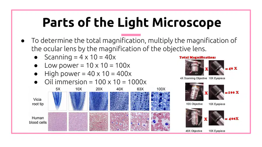
Find the total magnification: 
Eyepiece = 10X 
Objective lens = 55X
 
1
10x
2
55x
3
65x
4
550x

44

Multiple Choice

Question image

If the ocular lens has a magnification of 10x and the high power objective has a power of 40x, what is the total magnification of a microscope on high power?

1

50x

2

10x

3

40x

4

400x

45

Fill in the Blanks

media image

46

Labelling

What is the name of the microscope part labeled as L? *Spelling counts & use all lowercase letters*

Drag labels to their correct position on the image

Stage Clips

Course Adj.

47

Multiple Choice

Question image
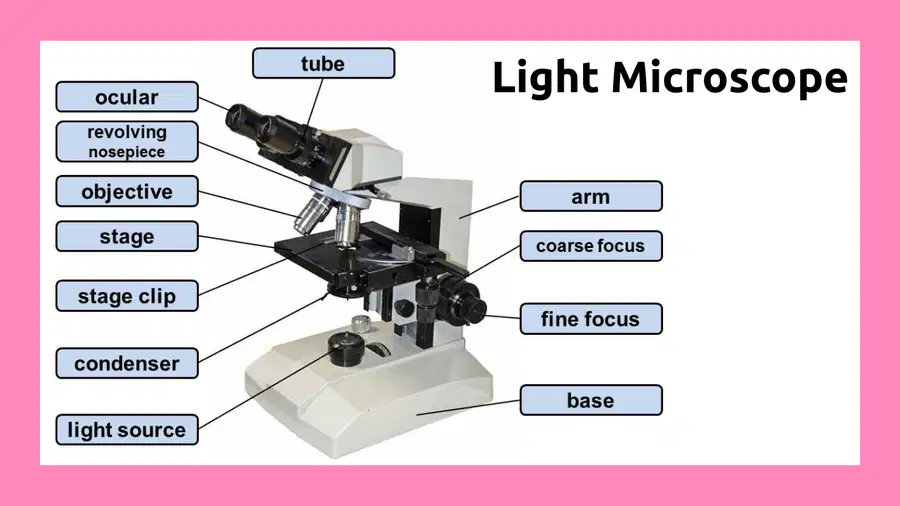
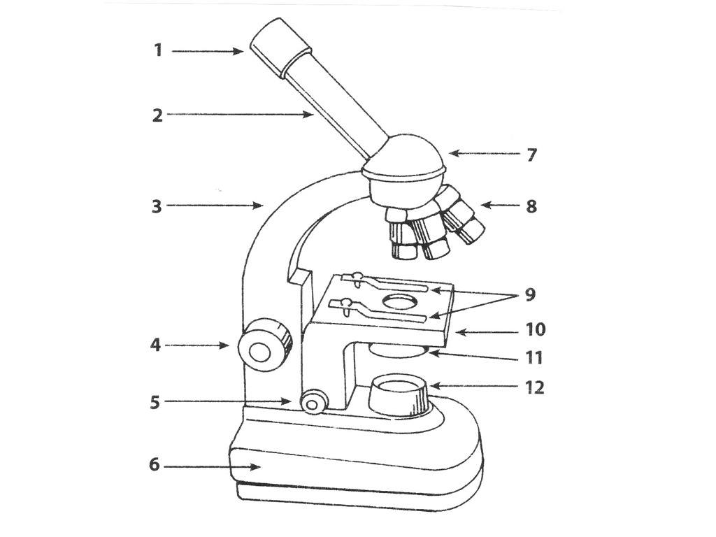
What part is 5?
1
coarse adjustment
2
fine adjustment
3
diaphragm
4
stage

48

Multiple Choice

Hold the slide in place on the stage
1
Stage
2
Stage Clips
3
Aperature
4
Light Source

49

Multiple Choice

Which of the following do you never use when on high power?

1

fine adjustment knob

2

diaphragm

3

coarse adjustment knob

4

stage clips

50

Multiple Choice

A student begins viewing a human cheek cell slide under a microscope and the image is very dim. What piece of the microscope, located under the stage, would the student adjust to alter the brightness?

1

diaphragm

2

ocular lens

3

substage

4

fine adjustment knob

media

Show answer

Auto Play

Slide 1 / 50

SLIDE