
 Worksheets
Benchmark 3 Review for Human Anatomy & Physiology
Total questions: 101
Worksheet time: 55mins
What type of muscle tissue is found in the heart?
skeletal
smooth
cardiac
voluntary
Muscles move bones by
pushing
pulling
both pushing and pulling
Muscles contract when
thin filaments slide past thin filaments
thick filaments slide past thick filaments
thin filaments slide past thick filaments
Which connective tissue surrounds fasicles?
epimysium
endomysium
perimysium
Which connective tissue surrounds the whole muscle?
epimysium
endomysium
perimysium
Which connective tissue surrounds individual muscle fibers?
epimysium
endomysium
perimysium
Actin myofilaments are
attached to the z disk
are found primarily in the h zone
are thicker than myosin myofilaments
all of these options
Which area of a sarcomere contains only actin?
Z disk
A band
H zone
I band
Which part of the sarcomere does not shorten during muscle contraction?
A band
H zone
I band
None of these
Which type of muscle cell is striated, voluntary and has multiple nuclei located at the periphery?
Cardiac
Smooth
Skeletal
Which type of muscle cell has one nucleus, is striated and has intercalated disks?
Skeletal
Cardiac
Smooth
Which of the following muscle cells are not striated, involuntary, and have one nucleus per cell?
Skeletal
Cardiac
Smooth

Which of the following puts the structures of the muscle in order from largest to smallest?
Whole muscle, muscle fiber, actin & myosin, myofibril, muscle fasicle
Whole muscle, muscle fasicle, muscle fiber, myofibril, actin & myosin
Whole muscle, muscle fiber, muscle fasicle, myofibril, actin & myosin
Whole muscle, muscle fasicle, muscle fiber, actin & myosin, myofibril

Identify the highlighted muscle
Deltoid
Biceps
Trapezius
Hamstring

Identify the highlighted muscle
Latissimus dorsi
Deltoid
External Oblique
Pectoralis Major

Identify the highlighted muscle
Biceps
Gluteus Maximus
Triceps
Deltoid

Identify the highlighted muscle
Extensor Carpi
Extensor Digitorum longus
Gastrocnemius
Tibialis anterior

Identify the highlighted muscle
Deltiod
Trapezius
Latissimus Dorsi
Sartorius

Identify the highlighted muscle
Hamstrings
Deltoid
Gluteus Maximus
Oblique

Identify the highlighted muscle
illopsoas
Biceps
Sartorius
Trapezius

Identify the highlighted muscle
Tibialis Anterior
Gastrocneminus
Deltoid
Biceps

What is the name of the blue structure
axon terminal
dendrite
schwann cell
axon

Which of the following cells protect the nervous system from infection
microglial
astrocyte
oligodendrocyte
ependymal cell

Which of the following glial cells make spinal fluid
astrocyte
oligodendrocyte
microglial cell
ependymal cell

What part of a neuron receives messages from other neurons?
dendrite
cell body
axon
axon terminal

what might be a job of a motor neuron?
connecting to a muscle and helping you move
connecting within the brain to help you make a decision
sending a message toward your brain from a pain receptor in your skin
all of these

Which best describes myelin?
it is a neurotransmitter that triggers muscle contraction
it is a protein that builds the axon
it is a fat that speeds the message along the axon
it is a fat that helps the neuron "hear" messages
which of the following is true?
neuroglial cells protect and support neurons
neurons help and support neuroglial cells
neurons transmit the electochemical message
neuroglial cells make myelin in the PNS
The PNS (peripheral nervous system)
contains the brain and spinal cord
contains all the nerves of the body
is divided into the somatic and autonomic systems
is divided into the sympathetic and parasympathetic divisions

The pink structure is the.....
frontal lobe
occipital lobe
parietal lobe
temporal lobe
The two major parts of the nervous system are the
brain and spinal cord
central and somatic nervous systems
sympathetic and parasympathetic systems.
peripheral and central nervous systems
The speed of neural conduction between neurons will result in difficulties in speaking, vision, and balance if the ________________ insulating the axons is damaged or removed.
Axon
Myelin Sheath
Nucleus
Brain
Dr. Vazquez is examining a cell from the nervous system of an animal. He notices that at one end of the cell body is a long, fibrous strand of tissue. He immediately recognizes this as an axon that is responsible for
carrying signals away from the cell body
receiving signals from other cells and carrying them toward the cell body.
determining the speed at which an action potential will travel
determining whether the cell inhibits or excites neighboring neurons
Which of the following is regulated by the autonomic nervous system
Breathing
Kicking a soccer ball
Balancing on one foot
Raising your hand
Neurotransmitters are ______________ that travel across the ______________ to another cell.
electrical signals; receptors
electrical signals; synapse
chemicals; receptors
chemicals; synapse

What is number 3 pointing at on the neuron?
Dendrite
Axon
Myelin Sheath
Soma

What part of the neuron is number 4 pointing at?
Dendrite
Axon Terminal
Axon
Myelin Sheath

Which part of the Neuron is number 1 pointing at?
Dendrite
Cell body
Axon
Nucleus
What is a nerve cell?
axon
neuron
dendrite
myelin
Which 2 are parts of the central nervous system?
brain
nose
spinal cord
left big toe
The brain is covered in a membrane called:
choroid plexus.
meninges.
ventricles.
cerebral aqueduct.
Cranial nerves and spinal nerves are part of the peripheral nervous system.
True
False
What is the length between Z-lines of an actin molecule called?
fascicle
sarcomere
myosin binding site
sarcoplasmic length
____________ attaches to actin to form the cross bridge.
Troponin
Tropomyosin
Myosin
Calcium

___ takes place when blood travels from the heart, through the lungs, and back to the heart.
pulmonary circulation
systemic circulation
___ takes place when oxygen-rich blood travels to all body tissues except the lungs.
pulmonary circulation
systemic circulation
What prevents blood from flowing back into the atria when the ventricles contract?
semilunar valves
sinoatrial node
atrioventricular node
tricuspid and bicuspid valves
When blood leaves this chamber of the heart, it heads out to the body (not the lungs)
left atrium
right atrium
left ventricle
right ventricle
One of the body's largest blood vessels that transports blood from the body region below the diaphragm to the heart
superior vena cava
inferior vena cava
pulmonary trunk
pulmonary veins
When this chamber contracts, it pushes blood through the tricuspid valve
right atria
right ventricle
left atria
left ventricle
This initiates the action potential that causes the heart to beat
Inferior vena cava
Atrioventricular node (AV node)
Superior vena cava
Sinoatrial node (SA node)
This is responsible for distributing electrical impulses through the heart
Atrioventricular node
Sinoatrial node
Inferior vena cava
Superior vena cava
This blood vessel splits in two and sends oxygen-poor blood away from the heart and to the lungs
aorta
pulmonary veins
superior vena cava
pulmonary trunk
This chamber of the heart receives oxygenated blood from the lungs
right atrium
right ventricle
left atrium
left ventricle
These are the smallest blood vessels with the thinnest walls in order to maximize diffusion of nutrients in and out of the interstitial fluid
arteries
veins
capillaries
aorta
Which best explains why capillary walls are so thin?
because they do not need to pump blood
because they are found only in the smallest organs and tissues
because they maximize diffusion of nutrients
because blood only travels through them slowly
These carry blood towards the heart
arteries
veins
These carry blood away from the heart
Arteries
Veins
Blood traveling through the pulmonary circuit goes through the right side of the heart and out to the _______
left side of the heart
body
lungs
tricuspid valve
Why is coronary circulation necessary?
because blood needs to be pushed throughout the entire body
because the heart itself needs nourishment
because the scalp requires an exceptional amount of nourishment
Blood in capillary beds flows from
arteries to veins
veins to arteries


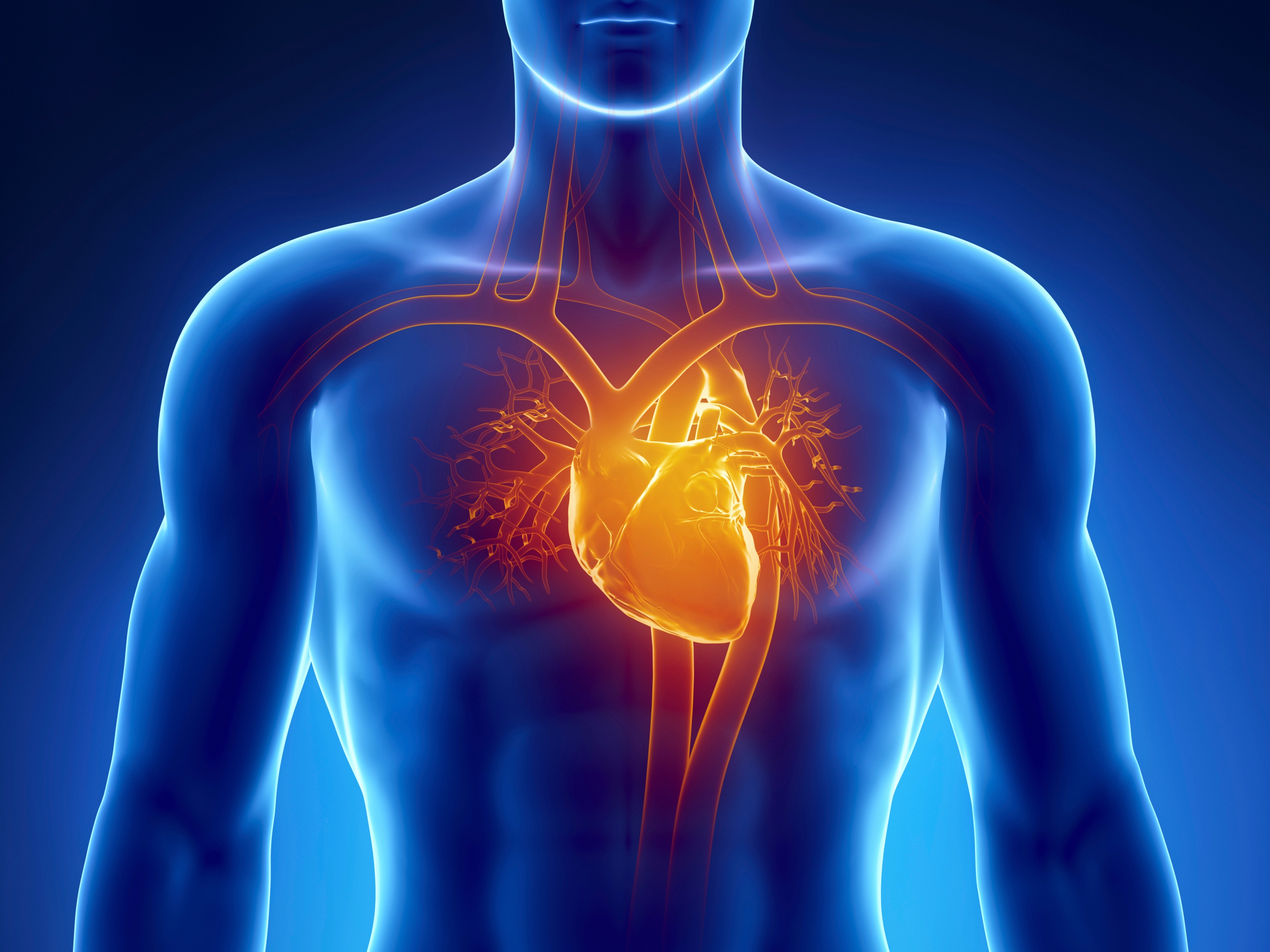

The double walled sac that surrounds the heart is called the:
Arteries
Valves
Pericardium
Chambers
What type of blood do the Vena Cava carry?
Oxygenated
Deoxygenated
What type of blood travels through the aorta?
Oxygenated
Deoxygenated
These blood cells protect from infections:
Red Blood Cells
White Blood Cells
Plasma
Platelets
These lower chambers of the heart pump blood out to the body through arteries:
Ventricles
Atrium
Vena cava
Aorta
Types of cells in the blood that causes blood clots to form
Platelets
Blood
Plasma
Hemoglobin
White blood cells
The oxygen-carrying protein in blood
Platelets
Blood
Plasma
Hemoglobin
White Blood Cells


Your heart is an involuntary muscle that is always working.
True
False
The AV valve on the right side is called the
tricuspid valve
bicuspid valve
pulmonary valve
aortic valve
The valve that regulates blood to the lungs is called
aortic
tricuspid
bicuspid
pulmonary
Which one of these valves is a semilunar valve?
tricuspid
bicuspid
aortic
atrium


