
 Worksheets
anatomy test #1
Total questions: 124
Worksheet time: 1hrs 3mins
A body part said to be furthest from point of attachment to trunk is said to be
proximal
distal
lateral
medial
Closest point to the attachment of the trunk is called
proximal
distal
cranial
caudal
On the inside; underneath another structure is known as
dorsal
ventral
anterior
deep
Your skin is ___________ to your bones
deep
lateral
superficial
dorsal
The location on our body that is directly down the middle of your body splitting your body into left and right is called
cranial
superior
inferior
midline
A body part said to be furthest from point of attachment to trunk is said to be
proximal
distal
lateral
medial
Closest point to the attachment of the trunk is called
proximal
distal
cranial
caudal
On the inside; underneath another structure is known as
dorsal
ventral
anterior
deep
Your skin is ___________ to your bones
deep
lateral
superficial
dorsal
The location on our body that is directly down the middle of your body splitting your body into left and right is called
cranial
superior
inferior
midline
Toward the midline of the body is called
medial
lateral
inferior
deep
Away from the midline of the body is called
medial
lateral
proximal
distal
Toward the head end is known as
caudal
distal
proximal
cranial
When we describe something as toward the feet not head it is called
superior
inferior
medial
lateral
Toward the head or above is what we describe as
inferior
superior
distal
proximal
In front of; toward the surface is known as
posterior
dorsal
anterior
inferior
In back of; toward the back surface is known as
cranial
caudal
anterior
posterior
At the back side of the human body is known as
caudal
cranial
dorsal
anterior
Which of these does not occur when in Anatomical Position
Face forward
Feet forward
Palms facing backward
Palms facing forward

Which side is facing you?
Anterior
Posterior
Lateral
Medial
The chin is _____ to the eye.
superior
deep
inferior
lateral

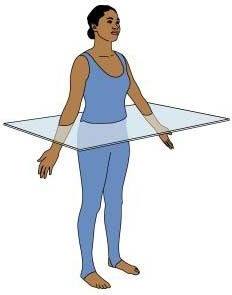

What does E indicate in the image?
the transverse plane of the feet
the coronal plane of the feet
the median plane of the feet
What does E indicate in the image?
the transverse plane of the feet
the coronal plane of the feet
the median plane of the feet

What is the anatomical name for the sole of the foot?
the dorsum
the plantar surface
the palmar surface

Which of the following describes the position of point 1 in comparison with point 2?
point 1 is medial
point 1 is lateral
point 1 is anterior

Compared to point 3, point 2 is ...
distal
lateral
proximal

Which surface of the hand is shown by point 3?
the plantar surface
the dorsal surface
the palmar surface

Something closer to the head (shown as direction A on the image) is referred to as ...
anterior
medial
superior

Compared to point 5, point 4 is ...
superior
medial
inferior

Compared to point 4, point 5 is ...
proximal
anterior
distal

A sagittal plane runs parallel to ...
the transverse plane
the median plane
the coronal plane

The toes are anterior to the ankle : true or false?
true
false
neither

The median plane passes through the body at right-angles to ...
the sagittal and coronal planes
the horizontal and coronal planes
the horizontal and sagittal planes

Which of the following terms refers to the inferior end of the vertebral column?
cranial
ventral
caudal

Two areas located on the same side of the body (left or right) are said to be ...
ipsilateral
contralateral
mediolateral

The elbow is proximal to the wrist : true or false?
true
false
neither

Two areas located on opposite sides of the body are said to be ...
circumlateral
contralateral
ipsilateral
What type of protein fibers are present in areolar CT?
Collagen
Reticular fibers
Elastic Fibers
All three
What is the function of areolar CT?
Binds skin and epithelia to deeper tissue.
Attach bone to bone or bone to muscle.
Withstands stress.
Provides supportive framework.
What cell type is present in adipose tissue?
Fibroblasts
Adipocytes.
Mesenchymal cells.
Macrophages.
Identify the adipose CT:


What cell type is present in reticular CT?
Fibroblasts
Adipocytes.
Mesenchymal cells.
Macrophages.
What is the function of reticular CT?
Binds skin and epithelia to deeper tissue.
Attach bone to bone or bone to muscle.
Withstands stress.
Provides supportive framework.
What is the function of dense regular CT?
Binds skin and epithelia to deeper tissue.
Attach bone to bone or bone to muscle.
Withstands stress.
Provides supportive framework.
What cell type is present in dense regular CT?
Fibroblasts
Adipocytes.
Mesenchymal cells.
Macrophages.
What type of protein fibers are present in dense irregular CT?
Collagen
Reticular fibers
Elastic Fibers
All three
What is the function of dense irregular CT?
Binds skin and epithelia to deeper tissue.
Attach bone to bone or bone to muscle.
Withstands stress.
Provides supportive framework.
What type of protein fibers are present in elastic CT?
Collagen
Reticular fibers
Elastic Fibers
All three
What is the function of elastic CT?
Provides supportive framework.
Allows for stretching and recoiling.
Withstands stress.
Transport
What is the cell type of hyaline cartilage?
Fibroblast
Chondrocytes
Osteocytes
Lacunae.
Which is hyaline cartilage?


What type of protein fibers are visible in fibrocartilage CT?
Collagen
Reticular fibers
Elastic Fibers
All three
Which is fibrocartilage CT?
Which is elastic cartilage CT?
What is the cell type of elastic cartilage CT?
Fibroblast
Chondrocytes
Osteocytes
Lacunae.
What is the cell type of bone CT?
Fibroblast
Chondrocytes
Osteocytes
Lacunae.
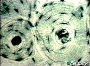
Which is bone CT?
What is the function of blood CT?
Provides supportive framework.
Allows for stretching and recoiling.
Withstands stress.
Transport
What are formed elements of blood CT?
Dissolved protein fibers.
Liquid nature of the ground substance.
A method of transporting nutrients.
Erythrocytes, leukocytes, and platelets.
Single layer of flattened cells with disc-shaped central nuclei; the simplest of the epithelium.
simple cuboidal
simple squamous
simple columnar
stratified squamous

What tissue type is this
dense connective
smooth muscle
cardiac muscle
areolar tissue
Location: Under skin; around kidney and eyeballs; in bones and within abdomen; in breasts.
bone
areolar
simple columnar
adipose

blood
adipose
simple squamous
stratified cuboidal
Wraps and cushions organs; its macrophages phagocytize bacteria; plays important role in inflammation; holds and conveys tissue fluid.
adipose
pseudostratified squamous
areolar
dense connective
The nucleoplasm and cytosol make up the ________ fluid.
extracellular fluid
intracellular fluid
intercellular fluid
nuclear
The movement of substances through the cell membrane against their concentration gradient is a type of ________ process
active
passive
diffusion
filtration

blood
reticular
Areolar
bone
Epithelial tissues have one free surface or edge known as the ________ surface.
basal
apical
free
basement

cardiac muscle
dense connective
skeletal muscle
smooth muscle
Osmosis transports water across membranes using ________.
nothing
aquaporins
solute pumping
ATP
The type of epithelial tissue found lining organs of the digestive system such as the small intestines is ________.
simple columnar
pseudostratified columnar
simple squamous
simple cuboidal
The component of a control system that provides the means for the control center's response (output) is called the ________.
stimulus
effector
receptor
control center
The hypogastric (pubic) region is ________ to the umbilical region.
superior
inferior
proximal
posterior
The diaphragm creates a ________ section between the thoracic and abdominal body cavities.
coronal (frontal)
sagittal
transverse
median
The right and left iliac (inguinal) regions are lateral to the ________ region.
lumbar
umbilical
iliac
hypogastric
In order to break a disaccharide down into simple sugar units ________.
water molecules must be added to each bond
water molecules must be removed from each bond
carbon atoms must be added to each bond
water molecules and carbon atoms must be removed from each bond
Which of the following is the correct order of elements in a control system?
receptor, stimulus, afferent pathway, control center, efferent pathway, effector, response
receptor, stimulus, efferent pathway, control center, afferent pathway, effector, response
effector, stimulus, efferent pathway, control center, afferent pathway, receptor, response
stimulus, receptor, afferent pathway, control center, efferent pathway, effector, response

simple cuboidal
simple columnar
simple squamous
stratified squamous
Jerome plays football and recently broke a bone in the tarsal region. This region is in the ________.
foot
wrist
ankle
shin


The cheeks are _______ to the tongue.
Medial
Lateral
Dorsal
Inferior
A transverse plane divides the body into _______ segments.
equal right and left
superior and inferior
anterior and posterior
both right and left and anterior and posterior segments
______ is the study of the structure and shape of the body.
Biology
Chemistry
Anatomy
Physiology
The anatomical position is characterized by all of the following EXCEPT _______.
arms at sides
feet parallel
thumbs pointed laterally
palms turned toward one's back
If you wish to make a drawing of a section through the human body that showed the heart and both of the lungs, your section could be in which of the following planes? (1) Frontal (coronal); (2) Transverse; (3) Sagittal
1 only
2 only
3 only
1 and 2
2 and 3
Which of these is found in the dorsal body cavity?
Kidneys
Brain
Lungs
Heart
Which of the following is NOT true of a sagittal plane?
Both the sagittal and coronal planes represent a cut made along a lengthwise plane.
A sagittal section is a cut made along the lengthwise plane of the body, dividing the body into right and left parts.
A sagittal section divides the body into anterior and posterior parts.
A midsagittal plane divides the body into equal right and left parts.
Which term(s) is/are defined as being close to the origin of the body part or the point of attachment of a limb to the body trunk?
Superficial
Superior, cranial
Proximal
Distal
Lateral
In proper anatomical positioning the palms face _______.
posteror
anterior
medial
superior
inferior
The heart is contained in which cavity?
Pelvic
spinal
thoracic
cranial

What is the name of this plane?
Transverse
Sagittal
Midsagittal
Coronal

Name the green cavity
Thoracic
Cranial
Abdominal
Pelvic

What is A?
Dorsal/posterior
spinal
cranial
ventral/anterior
What is B?
Dorsal/posterior
spinal
cranial
ventral/anterior
What is C?
Dorsal/posterior
spinal
cranial
ventral/anterior
What is L?
Dorsal/posterior
thoracic
abdominopelvic
ventral/anterior
What is K?
abdominal
thoracic
abdominopelvic
ventral/anterior
What is J?
abdominal
thoracic
abdominopelvic
pelvic
What is i?
abdominal
thoracic
abdominopelvic
pelvic
What is G?
abdominal
thoracic
abdominopelvic
diaphragm

