
 Worksheets
Module 8
Total questions: 52
Worksheet time: 27mins
(responsiveness or irritability) ability to receive and respond to stimuli
Excitability
Contractility
Extensibility
Elasticity
ability to shorten when stimulated
Excitability
Contractility
Extensibility
Elasticity
ability to be stretched
Excitability
Contractility
Extensibility
Elasticity
ability to recoil to resting length
Excitability
Contractility
Extensibility
Elasticity
dense regular connective tissue surrounding entire muscle
Epimysium
Perimysium
Endomysium
fibrous connective tissue surrounding fascicles groups of muscle fibers (cell)
Epimysium
Perimysium
Endomysium
fine areolar connective tissue surrounding each muscle fiber (cell)
Epimysium
Perimysium
Endomysium
run the entire length of an A band
Thick (myosin) filaments
Thin (actin) filaments
Z disc
H zone
M line
run the length of the I band and partway into the Aband
Thick (myosin) filaments
Thin (actin) filaments
Z disc
H zone
M line
coin-shaped sheet of proteins that anchors the thin filaments andconnects myofibrils to one another
Thick (myosin) filaments
Thin (actin) filaments
Z disc
H zone
M line
lighter midregion where filaments do not overlap
Thick (myosin) filaments
Thin (actin) filaments
Z disc
H zone
M line
line of protein myomesin that holds adjacent thick filaments together
Thick (myosin) filaments
Thin (actin) filaments
Z disc
H zone
M line
basic functional unit of muscle fiber
(a)
elastic chains of amino acids; make muscles extensible and elastic
A-band
I-bands
Titin filaments
Myosin tails
Attached to bone or (some facial muscles) to skin
Skeletal
Cardiac
Smooth
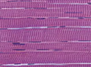
Single, very long, cylindrical, multinucleuated cells
Skeletal
Cardiac
Smooth
Walls of heart
Skeletal
Cardiac
Smooth
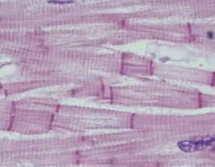
Branching chains of cells ; uni- or bi-nucleate; striations
Skeletal
Cardiac
Smooth
Single wall muscle in walls of hollow-visceral organs (other than the heart)
Skeletal
Cardiac
Smooth

Single, fusiform, uninucleate ; no striations
Skeletal
Cardiac
Smooth
muscle does not have time to completely relax between stimuli
Increase frequency of stimulus
Ca2+ release stimulates further contraction
Further increase in stimulus frequency
temporal (wave) summation
Increase frequency of stimulus
Ca2+ release stimulates further contraction
Further increase in stimulus frequency
unfused (incomplete) tetanus
Increase frequency of stimulus
Ca2+ release stimulates further contraction
Further increase in stimulus frequency
the muscle shortens and performs work
Concentric contractions
Eccentric (stretch) contractions
Isotonic contraction
Isometric contractions
the muscle contracts as it lengthens
Concentric contractions
Eccentric (stretch) contractions
Isotonic contraction
Isometric contractions
Muscle changes in length and moves the load
Concentric contractions
Eccentric (stretch) contractions
Isotonic contraction
Isometric contractions
The load is greater than the tension the muscle is able to develop
Concentric contractions
Eccentric (stretch) contractions
Isotonic contraction
Isometric contractions
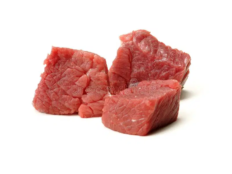
contract slowly, have slow acting myosin ATPases, and are fatigue resistant
Slow oxidative fibers
Fast oxidative fibers
Fast glycolytic fibers

contract quickly, have fast myosin ATPases, and have moderate resistance to fatigue
Slow oxidative fibers
Fast oxidative fibers
Fast glycolytic fibers

contract quickly, have fast myosin ATPases, and are easily fatigued
Slow oxidative fibers
Fast oxidative fibers
Fast glycolytic fibers
control fine movements (fingers, eyes)
Small motor units
Large motor units
large weight-bearing muscles (thighs, hips)
Small motor units
Large motor units
First step in skeletal muscle contraction
A thought in the brain results in a impulse traveling down a nerve to a muscle
At the neural motor end plate, the impulse causes calcium to enter the nerve
Calcium results in exocytosis of the neurotransmitter Acetylcholine(Ach) from the end plate into the neuromuscular synapse
Acetylcholine binds to the Sodium channels on the sarcolemma of the muscle
Second step in skeletal muscle contraction
A thought in the brain results in a impulse traveling down a nerve to a muscle
At the neural motor end plate, the impulse causes calcium to enter the nerve
Calcium results in exocytosis of the neurotransmitter Acetylcholine(Ach) from the end plate into the neuromuscular synapse
Acetylcholine binds to the Sodium channels on the sarcolemma of the muscle
Third step in skeletal muscle contraction
A thought in the brain results in a impulse traveling down a nerve to a muscle
At the neural motor end plate, the impulse causes calcium to enter the nerve
Calcium results in exocytosis of the neurotransmitter Acetylcholine(Ach) from the end plate into the neuromuscular synapse
Acetylcholine binds to the Sodium channels on the sarcolemma of the muscle
Fourth step in skeletal muscle contraction
A thought in the brain results in a impulse traveling down a nerve to a muscle
At the neural motor end plate, the impulse causes calcium to enter the nerve
Calcium results in exocytosis of the neurotransmitter Acetylcholine(Ach) from the end plate into the neuromuscular synapse
Acetylcholine binds to the Sodium channels on the sarcolemma of the muscle
Fifth step in skeletal muscle contraction
Acetylcholine binding to the sodium channel receptor opens the channel allowing Sodium(+) to diffuse into the muscle thereby reversing the membrane electrical potential from positive on the outside and negative on the inside of the sarcolemma to positive on the inside and negative on the outside
At the neural motor end plate, the impulse causes calcium to enter the nerve
Calcium results in exocytosis of the neurotransmitter Acetylcholine(Ach) from the end plate into the neuromuscular synapse
Acetylcholine binds to the Sodium channels on the sarcolemma of the muscle
Sixth step in skeletal muscle contraction
Acetylcholine binding to the sodium channel receptor opens the channel allowing Sodium(+) to diffuse into the muscle thereby reversing the membrane electrical potential from positive on the outside and negative on the inside of the sarcolemma to positive on the inside and negative on the outside
This membrane potential reversal results in a action potential (impulse) that travels along the sarcolemma until it reaches a T-tubule where it proceeds down
Calcium results in exocytosis of the neurotransmitter Acetylcholine(Ach) from the end plate into the neuromuscular synapse
Acetylcholine binds to the Sodium channels on the sarcolemma of the muscle
Seventh step in skeletal muscle contraction
Acetylcholine binding to the sodium channel receptor opens the channel allowing Sodium(+) to diffuse into the muscle thereby reversing the membrane electrical potential from positive on the outside and negative on the inside of the sarcolemma to positive on the inside and negative on the outside
This membrane potential reversal results in a action potential (impulse) that travels along the sarcolemma until it reaches a T-tubule where it proceeds down
The impulse stimulates electrical sensitive gaited voltage sensors to open the doors of the terminal cisternae of the sarcoplasmic reticulum and thereby releasing Calcium onto the sarcomere of the muscle
Acetylcholine binds to the Sodium channels on the sarcolemma of the muscle
Eighth step in skeletal muscle contraction
Acetylcholine binding to the sodium channel receptor opens the channel allowing Sodium(+) to diffuse into the muscle thereby reversing the membrane electrical potential from positive on the outside and negative on the inside of the sarcolemma to positive on the inside and negative on the outside
This membrane potential reversal results in a action potential (impulse) that travels along the sarcolemma until it reaches a T-tubule where it proceeds down
The impulse stimulates electrical sensitive gaited voltage sensors to open the doors of the terminal cisternae of the sarcoplasmic reticulum and thereby releasing Calcium onto the sarcomere of the muscle
Calcium then binds with the troponin complexes whichresults in tropomyosin being pulled off the myosin bindingsites of G-actin
Ninth step in skeletal muscle contraction
The Myosin heads containing ATP then bind to G-actin
This membrane potential reversal results in a action potential (impulse) that travels along the sarcolemma until it reaches a T-tubule where it proceeds down
The impulse stimulates electrical sensitive gaited voltage sensors to open the doors of the terminal cisternae of the sarcoplasmic reticulum and thereby releasing Calcium onto the sarcomere of the muscle
Calcium then binds with the troponin complexes whichresults in tropomyosin being pulled off the myosin bindingsites of G-actin
Tenth step in skeletal muscle contraction
The Myosin heads containing ATP then bind to G-actin
ATP is then broken into ADP thereby causing the myosin head-Gactin to be putin a cocked position. When ADP and the P are released from the myosin head, theenergy causes the myosin head to pull the actin filament to the center of thesarcomere and results in a contraction
The impulse stimulates electrical sensitive gaited voltage sensors to open the doors of the terminal cisternae of the sarcoplasmic reticulum and thereby releasing Calcium onto the sarcomere of the muscle
Calcium then binds with the troponin complexes whichresults in tropomyosin being pulled off the myosin bindingsites of G-actin
Eleventh step in skeletal muscle contraction
The Myosin heads containing ATP then bind to G-actin
ATP is then broken into ADP thereby causing the myosin head-Gactin to be putin a cocked position. When ADP and the P are released from the myosin head, theenergy causes the myosin head to pull the actin filament to the center of thesarcomere and results in a contraction
A second ATP attaches to the myosin head and separates it from G-actin and calcium is released from troponin and tropomyosin slides back over G-actin releasing the myosin head from the G-actin and covering the binding site. This results in a relaxation
Calcium then binds with the troponin complexes whichresults in tropomyosin being pulled off the myosin bindingsites of G-actin
twelfth step in skeletal muscle contraction
The Myosin heads containing ATP then bind to G-actin
ATP is then broken into ADP thereby causing the myosin head-Gactin to be putin a cocked position. When ADP and the P are released from the myosin head, theenergy causes the myosin head to pull the actin filament to the center of thesarcomere and results in a contraction
A second ATP attaches to the myosin head and separates it from G-actin and calcium is released from troponin and tropomyosin slides back over G-actin releasing the myosin head from the G-actin and covering the binding site. This results in a relaxation
This whole process of skeletal muscle contraction is called the sliding scale model
What is a thick filament?
Myosin
Actin
What is a thin filament?
Myosin
Actin

Myosin
Acin
I band
H band

Myosin
Acin
I band
H band
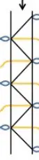
A band
Z-disk
I band
H zone
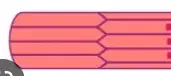
A band
Z-disk
I band
H zone
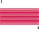
A band
Z-disk
I band
H zone
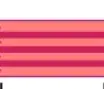
A band
Z-disk
I band
H zone

