
 Worksheets
A&P 1 2113- Ch 11 and 12
Total questions: 144
Worksheet time: 1hrs 28mins
When Na+ is diffusing IN
resting potential
depolarization
action potential
repolarization
refractory period
When K+ is diffusing OUT
resting potential
depolarization
action potential
repolarization
refractory period
When the Na/K pump is replacing the ions
resting potential
depolarization
action potential
repolarization
refractory period
The peak change in membrane potential
resting potential
depolarization
action potential
repolarization
refractory period
specifically -70 mV
resting potential
depolarization
action potential
repolarization
refractory period

The picture shows....
resting potential
depolarization
action potential
repolarization
refractory period

The picture shows....
resting potential
depolarization
action potential
repolarization
refractory period

The picture shows....
resting potential
depolarization
action potential
repolarization
refractory period

The picture shows....
resting potential
depolarization
action potential
repolarization
refractory period
Stronger stimuli result in
stronger action potentials
more frequent action potentials
The difference between electrical potential between ANY two points
voltage
amperage
charge
membrane potential
The difference between electrical potential between the inside and outside of a biological cell
voltage
amperage
charge
membrane potential
What does the myelin sheath do the the action potential carried on an axon?
increases its frequency
increases its amplitude
increases the velocity
When Na+ is diffusing in
the membrane potential increases
the membrane potential decreases
When K+ is diffusing out...
the membrane potential increases
the membrane potential decreases
All action potentials have the SAME
magnitude
velocity
frequency
The time after one action potential before a second action potential can be conducted.
resting potential
depolarization
action potential
repolarization
refractory period
The charge on the outside of a neuron cell membrane at rest is
Positive
Negative
Neutral


Put the following structures in order from superficial to deep.
1. muscle fiber
2. perimysium
3. myofibril
4. fascicle
5. endomysium
6. epimysium
6, 2, 4, 5, 1, 3
1, 3, 5, 6, 4, 2
1, 5, 4, 3, 2, 6
6, 2, 5, 4, 1, 3
A muscle that assists the muscle that is primarily responsible for a given action is a(n)
antagonist.
synergist.
agonist.
originator.
The end of a muscle that remains stationary when the muscle contracts is called the
origin.
aponeurosis.
insertion.
tendon.
The powerful abductor muscle of the upper arm is the
supraspinatus.
subscapularis.
deltoid.
coracobrachialis.
The long strap-like muscle on the anterior thigh is the ________ muscle.
gracilis
sartorius
iliotibial tract
adductor longus
Which of the following controls adduction at the shoulder?
supraspinatus
subscapularis
infraspinatus
teres major
When a muscle is stimulated repeatedly at a high rate, the amount of tension gradually increases to a steady maximum tension. This state of maximum tension is called
complete tetanus.
incomplete tetanus.
wave summation.
a twitch.

What muscle is highlighted?
trapezius
masseter
temporalis
sternocleidomastoid

What is the muscle?
orbicularis oculi
orbicularis oris
frontalis
temporal

What is the muscle?
masseter
zygomaticus
temporalis
orbicularis oris

What is this muscle?
temporalis
orbicularis oculi
frontalis
zygomaticus

What muscle is this?
sternocleidomastoid
masseter
zygomaticus
temporalis

What is this muscle?
masseter
zygomaticus
orbicularis oris
orbicularis oculi

What is this muscle?
sternocleidomastoid
trapezius
latissimus dorsi
temporalis

Which muscle turns the head?
trapezius
sternocleidomastoid
frontalis
temporalis

Which muscle is responsible for chewing?
masseter
orbicularis oris
orbicularis oculi
zygomaticus
Which muscle elevates the shoulders in a shrug?
latissimus dorsi
masseter
sternocleidomastoid
trapezius
Which muscle is used when smiling?
zygomaticus
orbicularis oris
temporalis
masseter
Which muscle is responsible for opening and closing the eyes?
orbicularis oculi
orbicularis oris
frontalis
masseter
Which muscle is used when puckering the lips?
zygomaticus
orbicularis oculi
orbicularis oris
masseter
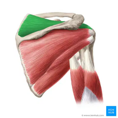
Teres Minor
Subscapularis
Supraspinatus
Infraspinatus
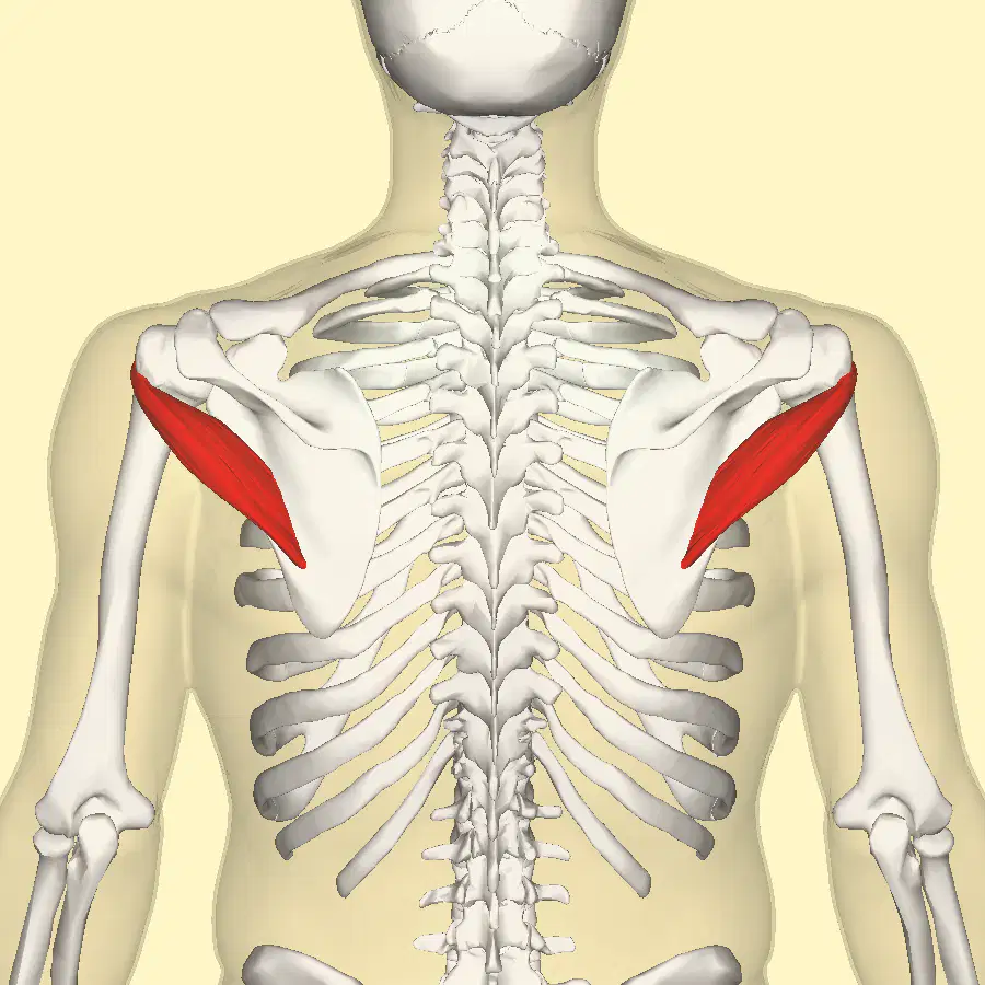
Teres Major
Teres Minor
Subscapularis
Infraspinatus
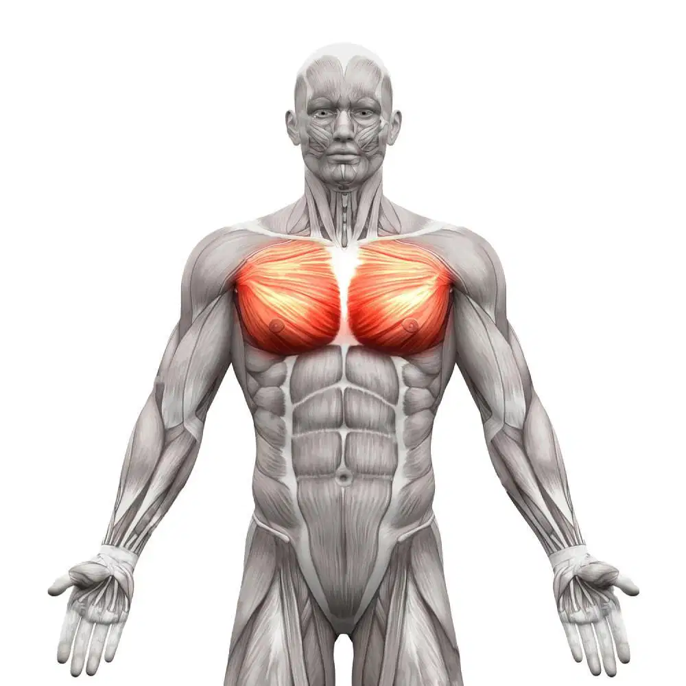
Latissimus dorsi
Trapezius
Pectoralis Major
Biceps
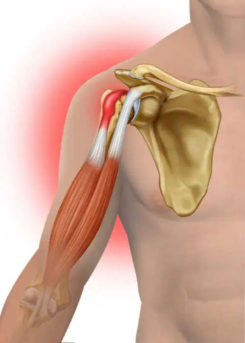
Triceps
Biceps
Sternocleidomastoid
Anterior deltoid

Pectoralis minor
Supraspinatus
Anterior deltoid
Biceps tendon
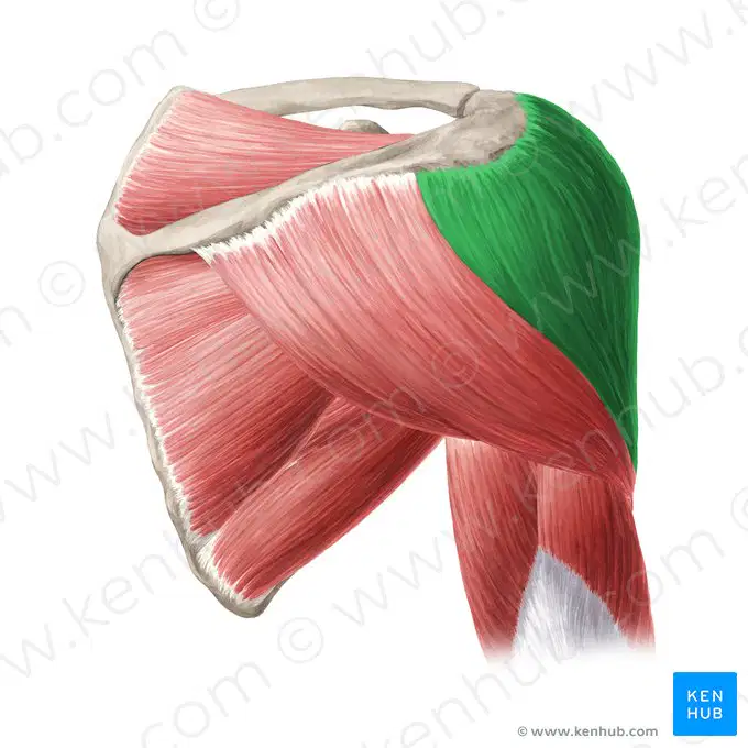
Which deltoid muscle is highlighted in green?
Biceps tendon
Anterior deltoid
Middle deltoid
Posterior deltoid
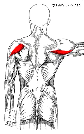
Anterior deltoid
Posterior deltoid
Middle deltoid
Sternocleidomastoid

Trapezius
Latissimus dorsi
Pectoralis Major
Triceps

trapezius
latissimus dorsi
levator scapulae
rhomboids
deltoid

What action does the Latissimus Dorsi perform?
Shoulder Extension, adduction, medial rotation, hyperextension
Shoulder adduction, medial rotation and horizontal adduction
Shoulder Lateral Rotation, horizontal abduction
Shoulder Flexion

Latissimus dorsi is inserted into
Lesser tubercle of humerus
Lateral lip of bicipital groove
Medial lip of bicipital groove
Medial floor of bicipital groove of humerus

The chest muscle that originates at the sternum, costal cartilage of the first 6 ribs, and medial third of clavicle
Deltoid
Bicep brachii.
Pectoralis major
Pectoralis minor


What is the name of this joint?
Glenoid fossa joint
Glenohumeral joint
Glenoclavicular joint
Coracohumeral joint
Lifting your arm straight up from the side is called what?
Shoulder extension
Shoulder adduction
Shoulder flexion
Shoulder abduction
Is the latissimus dorsi located on the anterior or posterior side of the human body?
anterior
posterior
Which of the following muscles originates on the humerus and inserts on the ulna
biceps brachii
triceps brachii
brachialis
brachioradialis

What muscle is shown here?
the deltoid
the external obliques
the rectus abdominis
the sternocleidomastoid
Is the serratus anterior located medial or lateral to the rectus abdominis?
medial
lateral


Muscle A?
rectus abdominus
external obliques
internal obliques
transverse abdominus

Muscle B?
rectus abdominus
external obliques
internal obliques
transverse abdominus

Identify the muscle in the picture
Trapezius
Erector Spinae
Rectus Femoris
Obliques

functions of this muscle is
Hip Extension, Adduction and Internal Rotation
Hip Extension, Abduction and External Rotation
hip flexion, abduction and external rotation
Hip flexion, adduction and internal roation

name the muscle
Glute max
Glute Min
Glute med
Piriformis

name the muscle
Rectus Femoris
Biceps femoris
Semimembranosus
Semitendinosus

name the muscle
Rectus Femoris
Biceps femoris
Semimembranosus
Semitendinosus

name the muscle
Rectus Femoris
Biceps femoris
Semimembranosus
Semitendinosus

name the muscle
achilles
Gastrocnemius
Anterior crus
Soleus

name the muscle
achilles
Gastrocnemius
Anterior crus
Soleus

name the muscle
sartorius
Rectus ABdominis
Iliacus
Psoas Major

name the muscle
sartorius
Rectus ABdominis
Iliacus
Psoas Major

name the muscle
sartorius
Vastus Intermedius
Rectus Femoris
Vastus Lateralis

name the muscle
sartorius
Vastus Intermedius
Rectus Femoris
Vastus Lateralis

name the muscle
Vastus Medialis
Vastus Intermedius
Rectus Femoris
Vastus Lateralis

name the muscle
Vastus Medialis
Vastus Intermedius
Rectus Femoris
Vastus Lateralis

name the muscle
Vastus Medialis
Vastus Intermedius
Rectus Femoris
Vastus Lateralis
Which of the following are in your true hip flexors?
Glute Max
Iliacus
Sartorius
Piriformis
What are the three functions of your sartorius
hip flexion, knee flexion and hip ER
hip extension, knee flexion and hip ER
hip flexion, knee extension and hip ER
hip flexion, knee flexion and hip IR
Which of the following muscles DOES NOT flexion the hip
Rectus Femoris
Biceps Femoris
Sartorius
Psoas Major
Which of following muscles is medial and deep on the hamstrings
Semimembranosus
Semitendinosus
Biceps Femoris
Vastus Medialis

This movement of the hip is called
extension
flexion
adduction
abduction
The most medial thigh muscles cause ___ of the hip
rotation
flexion
abduction
adduction

What "relationship" word describes the tensor fasciae latae and the adductor group?
synergists
antagonists
stabilizers
agonists
The muscle moving your hip forward as you walk
iliopsoas
gluteus maximus
gluteus medius
gracilis
Which relationship describes the gracilis and adductors?
synergists
antagonists
prime mover

Select the muscle(s) working in the image.
Iliopsoas
Gluteus medius
adductors
Tensor Fasciae Latae

This action is called
hip flexion and lateral rotation
hip extension and lateral rotation
hip flexion and medial rotation
hip extension and medial rotation
Which of the following muscles does NOT contribute to hip adduction?
Gracilis
Adductor longus
Gluteus maximus
Adductor brevis
extends leg at knee
check all that apply
vastus lateralis
sartorius
vastus medialis
adductor longus
flexes leg at knee, extends thigh at hip
check all that apply
rectus femoris
semitendinosus
semimembranosus
biceps femoris

O: Lateral tibia and interosseous membrane
I: First cuneiform and first metatarsal
A: Ankle inversion and dorsiflexion
Main muscle of the front compartment.
Flexor Hallucis Longus
Flexor Digitorum Longus
Tibialis Anterior
Extensor Hallucis Longus

__________ tendon sits on the posterior of the lower leg and ankle
Soleus
Achilles
Gastrocnemius
Tibialis Anterior


synaptic knobs
Myelin, an insulating material, is found in
cell bodies
dendrites
axon terminals
Schwann cells (for PNS)
Gaps between Schwann cells that increase the speed of the transmission of the signal down the neuron are called
axon terminals
dendrites
myelin
nodes of Ranvier
Sends impulses TOWARDS the CNS from receptors.
Motor Neuron
Sensory Neuron
Interneuron
Sends impulses AWAY from CNS to muscle and glands.
Motor Neuron
Sensory Neuron
Interneuron
What direction does an electrical signal move?
Dendrite, Cell body, Axon
Cell body, Axon, Dendrite
Cell body, Dendrite, Axon
Axon, Cell Body, Dendrite
What is the term for the propagation of an action potential from one gap in the myelin to the next?
Saltatory conduction
Voltage-gated conduction
Graded conduction
Mechanically-gated conduction
Where would we find ligand-gated channels?
On the dendrite
On the axon
Where we would find voltage regulated channels?
On the dendrite
On the axon
Neuroglial cells that form the fatty myelin sheath on neurons in the CNS are ___________ in the PNS are ________________?
ganglia and oligodendroglia
astrocytes and schwann cells
schwann cells and oligodendroglia
oligodendroglia and schwann cells
Small cells that engulf and destroy microbes and cellular debris in the CNS
microglia
horns
neuroglia
astrocytes

Produce cerebrospinal fluid in the brain & spinal cord
ependymal cells
oligodendrocytes
microglia
astrocytes
If you picked up a hot coal, the signal from the sensory neurons in your fingertips would travel to ____________ in your spinal cord.
Sensory Neurons
Motor Neurons
Interneurons
Type of neuron that perceive information about your environment.
Motor Neuron
Interneuron
Sensory Neuron
Type of neuron that sends nerve impulses to muscles and gland, by the command of other neurons.
Motor Neurons
Interneurons
Sensory Neurons
Neurons that only have on process?
Multipolar
Bipolar
Unipolar
Unpolar

multipolar neuron
A
B
C
bipolar neuron
A
B
C
unipolar neuron
A
B
C
The voluntary part of your nervous system is the
autonomic system
somatic system
sympathetic system
parasympathetic system
myelinated nerve fibers appear
white
gray
brown
clear
Neurons that carry information from the sensory receptors to the CNS:
Sensory, afferent
Sensory, efferent
Moror, afferent
Motor, efferent
bundles of nerve fibers running through the CNS
tracts
nerves
white matter
gray matter
bundles of nerve fibers running through the PNS
synapse
ganglia
tracts
nerves
part of the Schwann cell which is external to the myelin sheath
neurilemma
axon hillock
cell body
synapse
metabolic center of the neuron
cell body
axon
dendrites
synapse
conelike region of cell body where axon is attached
synapse
dendrite
axon hillock
neurilemma
The Autonomic Nervous System is part of the___________.
Somatic Nervous System
Peripheral Nervous System
Central Nervous System
Sensory Nervous System

