
 Worksheets
EOC Review HBS Unit1
Total questions: 116
Worksheet time: 58mins
lateral
posterior
medial
posterior
medial
ventral
The elbow is __________ to the hand.
Superior
Distal
Proximal
Superficial
The chest is __________ to the head.
Inferior
Deep
Distal
Superior
The foot is _____________ to the knee.
Inferior
Distal
Proximal
Lateral
_____________________: closer to the trunk of the body.
Superior
Inferior
Distal
Proximal
______________: Further away from the trunk of the body.
Proximal
Distal
Lateral
Medial
Front Side of the body
Posterior
Anterior
Lateral
Superior
Further from the midline:
Lateral
Medial
Superficial
Posterior
Toward the midline:
Medial
Lateral
Distal
Superior
The skin is __________ to the bone.
Superficial
Deep
Superior
Inferior
The sternum (chest bone) is __________ to the shoulder.
Distal
Lateral
Medial
Superior
The Pelvic Cavity is _____ to the Abdominal Cavity.
Superior
Proximal
Inferior
Lateral
The backside of the body is also referred to as what?
Posterior
Anterior
Dorsal
Both Posterior and Dorsal
Both Anterior and Posterior
Give the anatomical term for the thigh
popliteal
patellar
crural
femoral
What is the anatomical term for the mouth?
nasal
oral
buccal
orbital
none of the above
What is the anatomical term for the breastbone?
acromial
axillary
thoracic
sternal
none of the above
What is the anatomical term for the base of the skull?
occipital
cephalic
orbital
frontal
What is the anatomical term for the the lower back?
deltoid
scapular
vertebral
lumbar
sacral
What is the anatomical term for the back of the elbow?
olecranal
brachial
carpal
digital
What is the anatomical term for the armpit?
sternal
axillary
acromial
brachial
What is the anatomical term for the sole of the foot?
tarsal
digital
plantar
calcaneal
What is the anatomical term for the heel?
calcaneal
plantar
tarsal
crural
What is the common name for the otic?
nose
ears
eyes
mouth
What is the common name for the tarsal?
foot
ankle
toe
heel
What is the common name for the carpal?
ankle
thumb
shoulder
fingers
none of the above
Chin area
Buccal
Mental
Otic
Cephalic
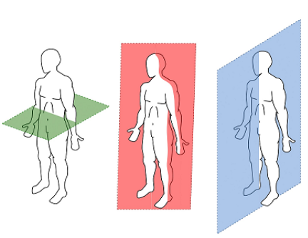
Divides the body into left and right portions.
Frontal
Transverse
Sagittal.
Ventral
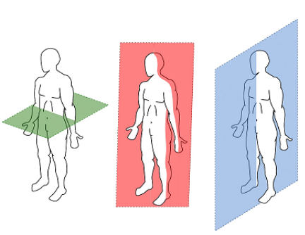
Divides the body into front and back portions.
Sagittal
Frontal
Transverse
Lateral
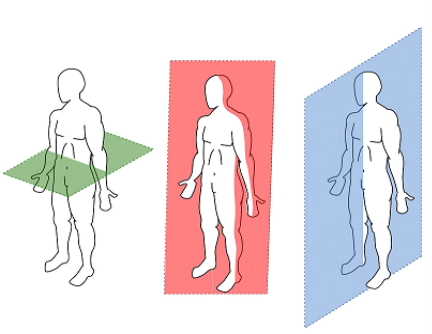
Divides the body into top and bottom portions.
Sagittal
Frontal
Transverse
Anterior

What is K?
abdominal
thoracic
abdominopelvic
ventral/anterior
What is i?
abdominal
thoracic
abdominopelvic
pelvic
What is G?
abdominal
thoracic
abdominopelvic
diaphragm

Identify the structures that make up the axial skeleton,
talus, calcaneus, navicular, cuboid, and cuneiform
limbs and girdles
skull, vertebrae, ribs and sternum
hands, arms and shoulder girdle
Your sister hurt her thoracic vertebrae in a car accident. Which skeleton is injured?
Axial skeleton
Appendicular skeleton
Axial and Appendicular
Neither Axial nor Appendicular
Your father broke his femur, which is the long bone in the upper thigh.
Axial skeleton
Appendicular skeleton
Axial and Appendicular
Neither Axial nor Appendicular
Which type of tissue is a communication system for the body?
Epithelial
Connective
Muscle
Nervous
NA - connective tissue
Voluntary
Voluntary
Ligaments
What is the function of elastic connective tissue?
Provides supportive framework.
Allows for stretching and recoiling.
Withstands stress.
Transport
A form of connective tissue in which the cells are specialized for the synthesis and storage of energy-rich reserves of fat, or lipid.
Hyaline cartilage
Adipose tissue
Connective tissue
Areolar tissue

Bone #3 is...
Parietal Bone
Zygomatic Bone
Sphenoid Bone
Maxilla

Bone #4 is...
Mandible
Occipital Bone
Maxilla
Temporal Bone

Bone #5 is...
Maxilla
Mandible
Frontal Bone
Occipital Bone

Bone #6 is...
Frontal Bone
Parietal Bone
Sphenoid Bone
Occipital Bone

Bone #7 is...
Hyoid Bone
Occipital Bone
Temporal Bone
Zygomatic Bone

Sphenoid
Temporal
Parietal
Zygomatic

Identify muscle #2
Occipitalis
Frontalis
Temporalis
Trapezius
Identify muscle #3
Occipitalis
Frontalis
Orbicularis oculi
Trapezius
Orbicularis oris
Identify muscle #8
Buccinator
Masseter
Orbicularis oculi
Zygomaticus
Orbicularis oris
Which bone structure makes new blood cells?
Compact
Spongy
Red Marrow
Yellow Marrow

Identify the structure circled in red.
osteon
Haversian system
central Haversian canal
perforating Volkmann's canal
osteocyte in lacuna

Identify the structures circled in red.
osteons
medullary cavity
central Haversian canal
perforating Volkmann's canal
osteocytes in lacunae

Identify the specific structures indicated by the arrows on the right side of the diagram.
osteons
perforating Volkmann's canals
concentric lamellae
trabeculae of spongy bone tissue
medullary cavity

Identify the structures at the red arrows.
lacunae
osteons
interstitial lamellae
central Haversian canals
canaliculi

What type of bone tissue is pictured here?
compact
spongy
smooth
adipose


What type of fracture is this?
Compound
Comminuted
Simple
Spiral

What type of fracture is this?
Compound
Hairline
Simple
Spiral

What type of fracture is this?
Comminuted
Open
Closed
Spiral

The ENDS of the long bone are anatomically referred to as the:
Epiphyseal plate
Medulary Cavity
Periosteum
Epiphysis

Identify 4
Articular Cartilage
Proximal Epiphysis
Distal Epiphysis
Epiphyseal Line
Identify 5, the type of bone tissue with trabeculae is called
Spongy Bone
Compact Bone
Articular Cartilage
Epiphyseal Line
Structure 7 is the outer covering of the bone made up of fibrous connect tissue called
Medullary Cavity
Compact Bone
Yellow Marrow
Periosteum
This is the second step of fracture repair
hematoma forms
bony callus forms
fibrocartilage callus forms
bone remodeling
This is the last step of fracture repair
hematoma forms
bony callus forms
fibrocartilage callus forms
bone remodeling
During fracture repair, the fibrocartilage callus is replaced with
compact bone
cartilage and collagen
spongy bone
compact bone and collagen
What is the first stage of bone growth and repair?
Bone Generation
Bone Remodeling
Bony Callus Formation
Hematoma Formation
During Hematoma Formation, What happens at the site of the break?
Blood Clot or a Hematoma
Fibroblasts Produce Collagen Fibres
Osteoblasts Form Spongy Bone
Air gets to it and everything is fine
During Bony Callus Formation, What is Fibrocartilaginous Callus Converted Into?
Compact Bone
Bone Marrow
Bony Callus of Spongy Bone
Red Blood Cells
Muscles are attached to bones by _____.
Ligaments
Tendons
Cartilage
Axons
Thick filaments are in a myofibril are
ATPase
Sarcoplasmic reticulum
Actin
Myosin
Thin filaments in a myofibril are
Sarcomeres
Sarcoplasm
Actin
Myosin
Which filament slides inward during muscle contraction
Actin
Myosin
Smooth
Skeletal
What layer of connective tissue covers the muscle fiber?
endomysium
epimysium
perimysium
What layer of tissue surrounds the entire muscle?
Endomysium
Epimysium
Perimysium
What two chemicals are necessary for muscle contraction to occur?
Niacin and Potassium
Calcium and ATP
Sodium and Potassium
Acetylcholine and Potassium
The joints that allow the most movement are called
Fixed
Fibrous
Cartilaginous
Synovial
An example of a Saddle joint is in the
Knee
Hip
Thumb
Vertebral column
When kicking a football the knee
Flexes
Extends
Abducts
Adducts


The ends of long bones are covered with a type of hyaline cartilage called...
connective tissue
ligaments
tendons
articular cartilage
The cartilage which is most abundant and varies the most within the body is...
articular cartilage
hyaline cartilage
elastic cartilage
fibrocartilage
The cartilage which is the most flexible and least abundant in the body is....
hyaline cartilage
elastic cartilage
fibrocartilage
articular cartilage
Elastic cartilage can be found in your....
Intervertebral discs
Ears
Ribs
Throat
Short bones can be found in your ___________ and __________
ribs, neck
wrists, ankles
feet, hands
fingers, toes
The Greater Trochanter is a _______ ________ on the _________
small bump, fibula
large bump, radius
small bump, ulna
huge bump, femur

