
 Worksheets
Assignment | Unit 4C.1 | The Heart | Bio 20
Total questions: 50
Worksheet time: 3hrs 0mins
The type of muscle found only in the heart is (a) .
Match the following muscle types with their control type.
Cardiac muscle
Involuntary control in the heart
Smooth muscle
Involuntary control in organs
Skeletal muscle
Voluntary control in the body
Myocardial muscle
Involuntary control in the heart
Which chamber of the heart receives deoxygenated blood from the superior and inferior vena cavae?
Left atrium
Right atrium
Left ventricle
Right ventricle
Match the following heart components with their functions.
Receive blood from veins
Atrium
Pump blood out of the heart
Ventricle
Filter the blood
Kidney
Oxygenate the blood
Lungs
Which blood vessel carries oxygenated blood away from the heart to the body?
Pulmonary vein
Superior vena cava
Inferior vena cava
Aorta
Match the following parts of the body with the blood vessels that return blood from them:
The lungs
Pulmonary veins
The upper body
Superior vena cava
The lower body
Inferior vena cava
The heart muscle itself
Coronary sinus
The inferior vena cava delivers blood into the (a) of the heart.
Match the following heart functions with their corresponding atrioventricular valve actions.
Blood from entering the aorta
Prevents blood from flowing backward into the atria
Blood from flowing backward into the atria
Prevents blood from entering the aorta
Blood from mixing between ventricles
Prevents blood from mixing between ventricles
Blood from exiting the heart too quickly
Prevents blood from exiting the heart too quickly
Which of the following is an example of a semilunar valve?
Mitral valve
Tricuspid valve
Aortic valve
AV valve
What acts as the natural pacemaker of the heart?
AV node
Purkinje fibres
SA node
Bundle of His
The atrioventricular node (AV node) delays the impulse to:
Allow the atria to contract fully
Speed up ventricular contraction
Stop atrial contraction
Stimulate the Purkinje fibres immediately
The structure that ensures rapid conduction of the electrical signal throughout the ventricles is (a)
What is the function of the septum in the heart?
Pump blood to the lungs
Separate oxygenated and deoxygenated blood
Control heart rate
Open and close valves
True or False: Cardiac muscle is an involuntary muscle found only in the heart.
True
False
True or False: Smooth muscle is responsible for voluntary movements like walking and running.
True
False
Skeletal muscle contracts (a) without conscious control.
True or False: The atria are the lower chambers of the heart.
True
False
True or False: The ventricles pump blood out of the heart to the lungs and the rest of the body.
True
False
The superior vena cava returns deoxygenated blood from the (a) body to the heart.
True or False: The inferior vena cava drains deoxygenated blood from the lower parts of the body into the right atrium.
True
False
True or False: The aorta carries deoxygenated blood from the heart to the lungs.
True
False
True or False: The atrioventricular valves prevent backflow of blood into the atria when the ventricles contract.
True
False
True or False: Semilunar valves are located between the atria and ventricles.
True
False
True or False: The sinoatrial (SA) node acts as the heart’s natural pacemaker.
True
False
True or False: The atrioventricular (AV) node directly initiates the heartbeat without input from the SA node.
True
False
Purkinje fibres help coordinate ventricular contraction by rapidly conducting the electrical impulse: (a)
True or False: The septum prevents the mixing of oxygenated and deoxygenated blood within the heart.
True
False
Identify and explain the three main roles of the circulatory system?
Label the four chambers of the heart.
Right ventricle
Left ventricle
Right atrium
Left atrium
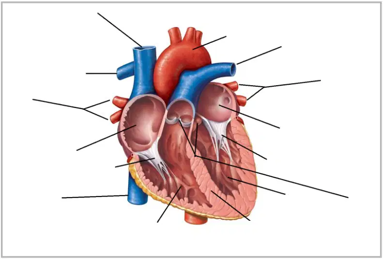
Label the parts of the heart's anatomy by using the label option provided and dragging them to the corresponding numbered parts on the diagram.
aorta
vena cava
pulmonary veins
right ventricle
pulmonary arteries
The Heart of a Swimmer vs. the Heart of a Runner
Regular exercise changes the look and workings of the human heart. And researchers are discovering that different sports affect the heart differently. Do world-class swimmers’ hearts function differently than the hearts of elite runners? A new study finds that the answer may be yes, and the differences, although slight, could be telling and consequential, even for those of us who swim or run at a much less lofty level.
Cardiologists and exercise scientists already know that regular exercise changes the look and workings of the human heart. The left ventricle, in particular, alters with exercise. This chamber of the heart receives oxygen-rich blood from the lungs and pumps it out to the rest of the body, using a rather strenuous twisting and unspooling motion, as if the ventricle were a sponge being wrung out before springing back into shape.
Exercise, especially aerobic exercise, requires that considerable oxygen be delivered to working muscles, placing high demands on the left ventricle. In response, this part of the heart in athletes typically becomes larger and stronger than in sedentary people and functions more efficiently, filling with blood a little earlier and more fully and untwisting with each heartbeat a bit more rapidly, allowing the heart to pump more blood more quickly.
Based on what you know about exercise and changes to skeletal muscle, what do
you think will happen to the wall of the left ventricle as a result of exercise – explain
why you think it will get thicker or it will get thinner.
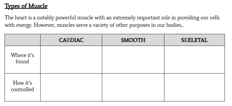
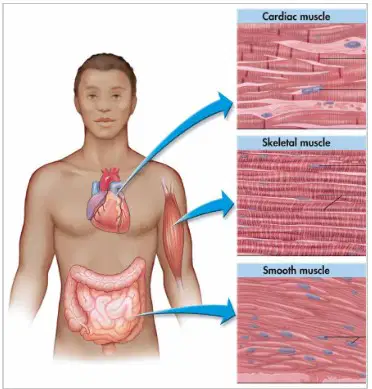
Where is the Cardiac Muscle Found?
Where is the Smooth Muscle Found?
Where is the Skeletal Muscle Found?
How is the Skeletal Muscle Controlled?
How is the Smooth Muscle Controlled?
How is the Cardiac Muscle Controlled?
What stimulates skeletal muscles to contract, and what substance is released in the muscle fibres as a result?
Explain the role of calcium ions (Ca2+) in muscle contraction. Which two protein filaments interact, and how does their movement result in muscle contraction?
Describe what happens to the actin and myosin filaments during the relaxation phase of muscle movement. What is the outcome for the muscle as these filaments disengage?
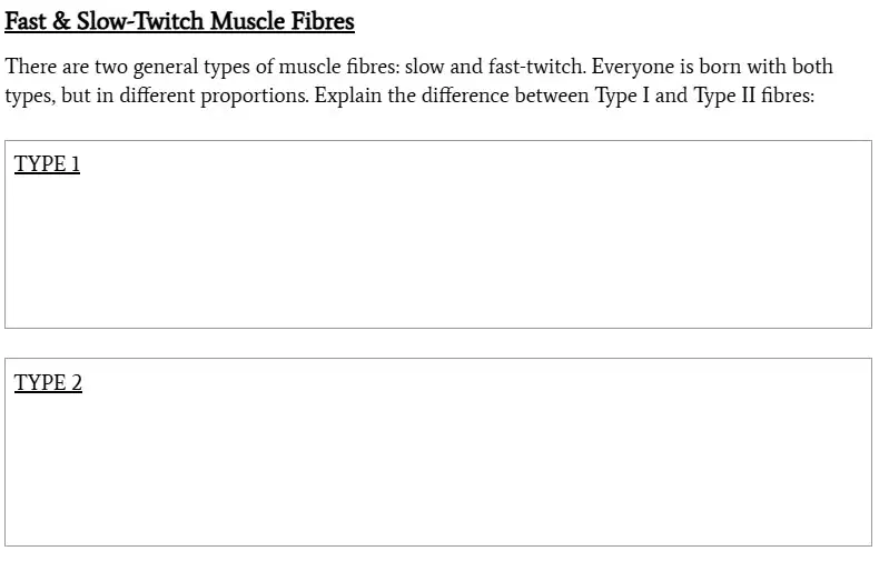
Fast & Slow-Twitch Muscle Fibres
There are two general types of muscle fibres: slow and fast-twitch. Everyone is born with both types, but in different proportions. Explain the difference between Type I and Type II fibres:
Explain the Three Main Pathways of the Circulatory System
PULMONARY PATHWAY
SYSTEMIC PATHWAY
CORONARY PATHWAY
What is the function of valves within the heart?
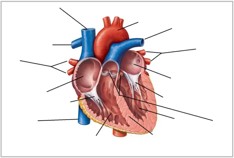
What is the function of the septum?
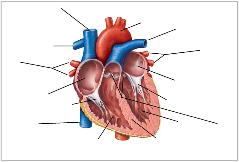
Why is the muscle surrounding the left ventricle so much thicker than the muscle surrounding the right ventricle?
Often, arteries are thought of as blood vessels that carry oxygenated blood, while veins carry deoxygenated blood. However, this is not always the case, as demonstrated by the pulmonary arteries & veins. What is the better way to define an artery vs. a vein?
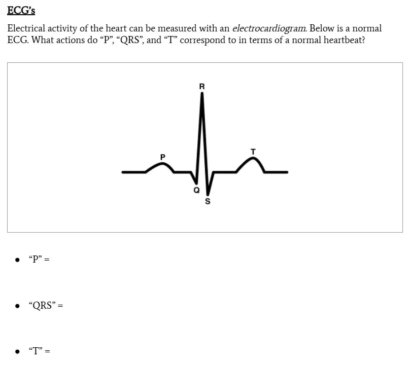
Electrical activity of the heart can be measured with an electrocardiogram. Below is a normal ECG.
What actions do “P” correspond to in terms of a normal heartbeat?
Electrical activity of the heart can be measured with an electrocardiogram. Below is a normal ECG.
What actions do “QRS” = correspond to in terms of a normal heartbeat?
Electrical activity of the heart can be measured with an electrocardiogram. Below is a normal ECG.
What actions do “T” = correspond to in terms of a normal heartbeat?
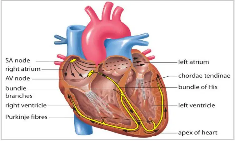
Delineate the steps involved in the process that signals the heart to beat. Be sure to explain how the electrical impulses move through the heart and lead to the contraction of the atria and ventricles.

