
 Worksheets
arterial: non-imaging testing
Total questions: 148
Worksheet time: 2hrs 30mins
What are the key aspects to know for all types of testing in arterial testing?
Capabilities and limitations, physical principles, technique, interpretation.
Only physical principles and technique.
Interpretation and technique only.
Capabilities and limitations only.

compression of the popliteal artery by the medial head of the gastrocnemius muscle or fibrous bands. Usually young male pt complaining of claudication in calf w/ exercise.
Eval: PPG on tor. plantar flexion and dorsiflexion
findings: normal at rest. Only compressed when muscle is activated. Waveforms will diminish or flatten upon flexions.
popliteal entrapment syndrome
thoracic outlet syndrome

compression of neuromuscular bundle by shoulder structures (ribs, ligaments, muscles). pt complain pain, tingling, weakness in arm when in certain positions. most common cause is neurogenic. must also test when replicating compression.
technique: PPG sensors on index finger
rest w/ hand in lap, arm raised at 90degr. to body! 180 degrees, exagerated miltary stand (shoulders back), adson maneuver (exaggerated military stance w/ head turned sharply toward arm being tested & then switched) , caustive position
popliteal entrapment syndrome
thoracic outlet syndrome
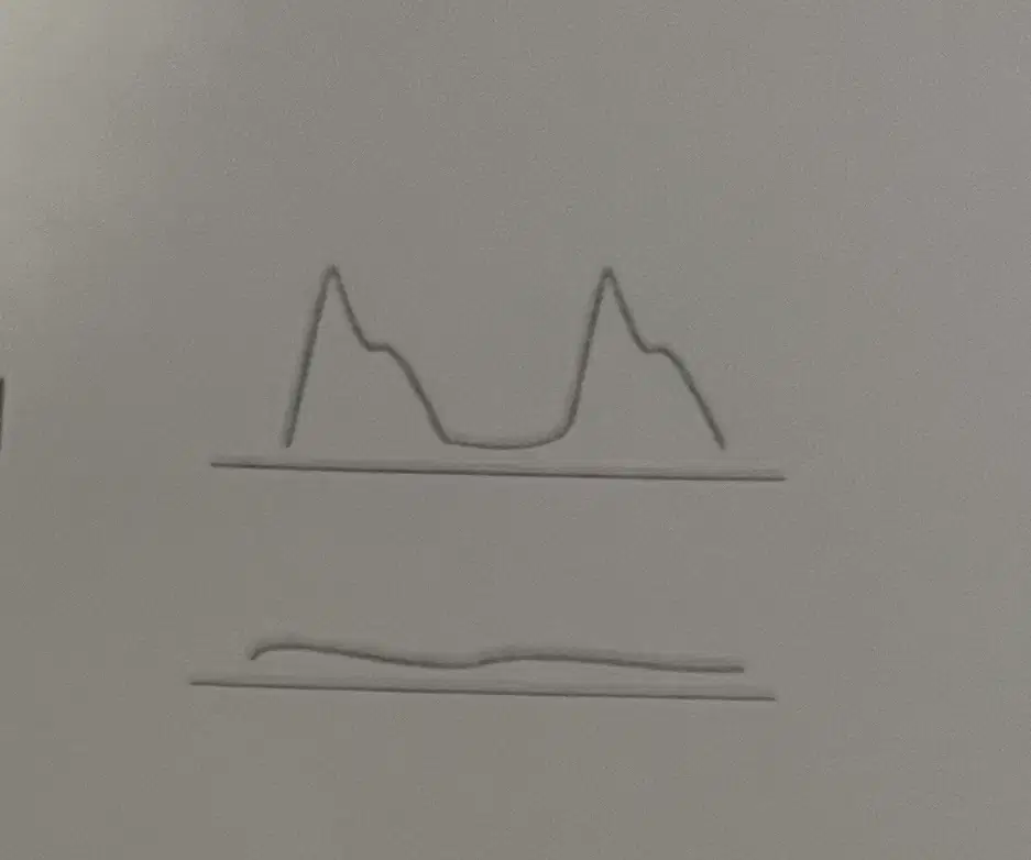
In regards of compression disorders if there is arterial compression = _____
A. normal in position
B. normal at rest, attenuated/flatted in position
C. attenuated/flattened at rest
D. A and C
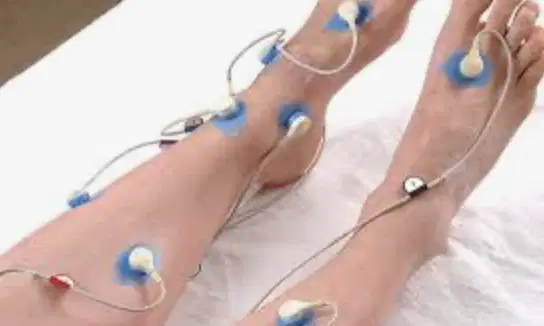
TRASCUTANEOUS OXIMETRY (TcPO2)
T/F
evaluates wound healing potential and also determines amputation level (special sensors measure oxygen tension)
normal >30mmHg
poor <40mmHg (impaired wound healing)
critical <50mmHg (Non-healing)
TRUE
FALSE
what are the physical principles of digital pressure and PPG?
use pneumatic cuffs (BP cuffs w/ special sensor) detects changes in volume
when volume increases in systole sensor shows peak on PVR
when volume decrease in diastole less pressure on cuff so there is downslope on PVR
small sensor on digit sends light and the capillary pulsations reflects the light.
what are some techniques used with digital pressure and PPG
obtain bilat brach pressures
use PPG to obtain digital pressures (when pulsation returns record pressure)
for raynauds - cold stress: 1st perform at rest, 2nd soak in ice water for 3min or to tolerance obtain pressures & waveforms
use same cuffs as segmental pressure study. cuff inflated to 40-65mmHg (not to occlude vessel)
brach cuff and forearm cuff, obtain pressures from the vessels in that area (brach & radial)
a normal interpretation of digital pressure & PPG w/in the finger(FBI)
>0.8
>0.6
>0.3
<0.8
<0.9
what is a normal interpretation of digital pressure & PPG of the toes (TBI)
>0.8
>0.6
>0.3
<0.8
<0.9
if there is obstructive disease (secondary raynauds) within a digital pressure or PPG exam there will be _______ and ______
dropped pressures
dampended waves
elevated pressures
peaked waves
what are some capabilities and limitations of penile pressure and testing?
evaluate for vasculogenic impotence (erectile dysfunction caused by vascular inflow or outflow)
leriche syndrome: terminal Ao. obstruction impotence, hip, and thigh claudication
performed with segmental pressure study and not vessel specific = understimate disease.
useful in PT with cold sensitivity and medial calcinosis
_____ are capabilities of digital pressure and ppg.
detects presence of arterial disease
useful in patients with cold sensitivity & medial calcinosis
dx fixed arterial obstruction vs vasospasm
assesses effect of treatment
vessel specific
____ are capabilities of plethsmography
dx true arterial claudication vs nonvascular related
evaluates functional aspects of disease
performed with Segmental pressure
localizes level of obstruction
assess results of medical and or exercise therapy

in regards of plethysmography what is its physical principles?
(image shows when the grey area(vesse) dilates with higher volume of blood flow it increases peak amplitude) vise versa
uses pneumatic cuffs( BP cuffs w/ special spensors to detect changes in volume)
1
air is introduced to cuff before exam 40-65mmhg
2
volume increase under cuff bladder at systole sensor shows peak in PVR/c, decrease in diastole less pressure in cuff so PVR/c downslopes
3
paired w/ Segmental pressure or accuracy
4
if paper is used to chart recording speed is set at 25mm/sec
5
what is the proper technique to perform a plethysmography exam?
use same cuff as segmental pressure study
cuff inflated to 40-65mmHg (not to occlude vessel)
obtain bilateral brachial pressures use ppg to obtain digital pressures
none of these
match the proper interpretation for plethysmography disease limits

normal, sharp peak dicrotic notch

mild sharp peak, absent dicrotic notch

flattened peak, slow upstroke/downslope

flattened, dampened pulse
in regards of segmental pressure testing of the upper extremities, what are some capabilities and limitations?
presence/absence of disease & approximate level and severity
not exact location or stenosis vs occlusion
medial calcinosis: incompressible vessels. often seen in diabetics & end stage renal disease. pressures will be inaccurately high
pt w/ dialysis fistula stents, DVT, or hx of lymphedema cannot have this exam
any surgical modifications
in regard of segmental pressure testing of the upper extremities, upper and lower extremities use the same cuff technique
TRUE
FALSE
in regards to segmental pressure testing within the lower extremities, what are some capabilities and limitations?
presence/absence of disease
approximate level and severity
not exact location or stenosis vs occlusion
medial calcinosis: incompressible vessels. often seen in diabetics and End stage renal disease.
pt w/ dialysis fistula, stents, DVT, or hx of lymphedema
in regard to segmental pressure testing of the lower extremities include all the techniques to complete this exam :
pt in basal state (resting for at least 20 mins)
- supine = no hydrostatic pressure
CUFFS: bilat brach cuffs
4 cuff method: 2 thighs, calf, ankle
3 cuff method: 1 large thigh, calf , ankle
width of cuff: 1.2x greater than diameter of limb
too wide:false low pressure
too narrow: false high pressure
Use Cw probe to locate arterial pressure distally (ex. pta or dpa)
inflate cuff to suprasystolic pressure(30 ^ than last audible signal) , slowly deflate record pressure when pulsation returns
in regard to segmental pressure testing of the lower extremities include all the interpretations for this exam:
calculate ankle/brachial index ABI
ankle pressure divided by highest brachial pressure
normal ankle pressures should be about the same as the brachial
in presence of arterial disease - pressure drops
ankle< brachial = disease ABI would be <1.0
ankle> brachial would give an ABI >1.0 no disease proximally
Abi > 1.3 indicates the vessels are likely incompressible: NOT accurate
In regards to segmental pressure of the upper extremities, place the interpretation of this exam in order:
pressures DROP abnormally distal to disease
1
compare both brachial to each other. normally they should be within 20mmHg of each other. if greater than 20 diff. prox disease on arm w/ lower pressure
2
look for drops of 20 or more between consecutive vertical levels. 20 or more drop means disease between those levels.
3
always compare up. radial to brachial, ulnar to brachial.
4
remember: arterial obstructive disease will decrease the pressures. disease is proximal to the lower pressure.
5
in regards to the allen test, what is the purpose of an Allen test?
evaluate patency of the palmar arch, wrist and digital arteries
evaluate the patency of the brachial artery
evaluate the patency of the basilic artery
evaluate the patency of the ulnar artery
Allens test: useful to see if its possible to confirm usability of the _____ in cases of (harvesting for coronary bypass, hemodialysis access graft or angiography)
radial artery
ulnar artery
brachial artery
cephalic artery
in regards to the allen test what is the proper technique for this test?
apply pressure to radial artery to occlude it, make fist. hand will turn pale as blood is drained from hand open hand maintaining pressure on artery.
apply pressure to cephalic artery to occlude it, make fist. hand will turn pale as blood is drained from hand open hand maintaining pressure on artery.
apply pressure to ulnar artery to occlude it, make fist. hand will turn pale as blood is drained from hand open hand maintaining pressure on artery.
apply pressure to brachial artery to occlude it, make fist. hand will turn pale as blood is drained from hand open hand maintaining pressure on artery.
in regards to the allen test, what is the proper interpretation?
standard: color returns to normal
modified: PPG waveforms are maintained throughout compression
repeat w/ ulnar artery compression
calculate ankle/brachial index ABI , ankle pressure divided by highest brachial pressure.
compare both brachial to each other, normally they should be within 20mmHg of each other.
none of these
True or False
in regards to segmental pressure criteria
pressures drop abnormally distal to disease
True
False
What does qualitative analysis in arterial testing focus on?
How it looks, NOT measurable, waveform contour.
Quantitative measurements, numerical data, blood pressure levels.
Statistical data, measurable outcomes, heart rate.
Chemical composition, blood sugar levels, cholesterol.
in regard to segmental pressure criteria:
normal thigh pressures on the 3 cuff study type should:
A. thigh should have the same or higher than brachial
B. high thigh should be 30 or more higher than highest brachial
none of these
both A & B are normal thigh pressures
in regard to segmental pressure criteria:
normal thigh pressures on the 4 cuff study type should:
A. thigh should have the same or higher than brachial
B. high thigh should be 30 or more higher than highest brachial
none of these
both A & B are normal thigh pressures
in regard to segmental pressure criteria:
look for drops of ___ or more between consecutive vertical levels.
<(less than)__ difference between levels is NORMAL. ____ or more drop means disease between those levels.
30
40
35
45
reactive hyperemia is for pts that cannot do ______
treadmill
stair master
segmental pressures
all of the above
____ purpose is to produce ischemia and distal vasodilation. cutting off the flow forces of the body to vasodilate. then when the cuff is released we see how the hyperemia (increase in flow) reacts = how quick it gets down
reactive hyperemia
exercise testing
segmental pressure testing
none of these is correct
in regards to reactive hyperemia, what is the proper technique?
inflate thigh cuff to suprasystolic pressures 3-5min
- release cuff and record ankle pressures
inflate brachial cuff to suprasystolic pressures 3-5min
constant load treadmill(unchanging) <12% grade and 1.5 mph for a max of 5min
pt in a basal state (resting for atleast 20mins)
supine & no hydrostatic pressure
in regards to reactive hyperemia, what is considered normal?
transient drop in pressure that returns to resting w/in 1min(<34%)
no change or increase in ankle pressures
transient drop in pressure that returns to resting immediately
no change or decrease in ankle pressure
in regards to reactive hyperemia, what is considered abnormal:
big drop in pressures
<50% drop= single level
>50% drop= multi level
increase in ankle presure
arterial exercise testing abnormal results represent:
no change or increase in ankle pressures
decrease in ankle presures
repeat ankle pressures every 2 min until return to resting
if it return <(less than)6 min: single level
if it return >6min: multi level
what types of recording does CW doppler record?
audio: doppler shifts w/in audible range
analog: zero crossing frequency meter on strip chart recorder estimates freq shifts, not very sensitive
spectral analysis: displays true or individual freq shifts according to time. more sensistive
all of the above
What does quantitative analysis in arterial testing focus on?
How it measures, velocities, ratios, indices, % diameter reduction.
Blood pressure levels.
Heart rate variability.
Cholesterol levels.
Which of the following is included in non-imaging testing?
CW
Pressure testing
Plethysmographic studies
All of the above
What are capabilities of CW doppler analysis?
Exact location of disease
confirms arterial occlusive disease
provides follow up info about progression of disease
Depth specificity
gives an approx location of significant obstruction
what are some techniques for CW doppler?
supine
8-10MHz CW probe
45-60degree angle
9-14MHz
25-60degree angle
when interpreting a normal waveform which of the above are normal waveforms?
triphasic
biphasic
monophasic
continuous
CW doppler analysis requires how many crystals?
1
2
3
4
CW doppler analysis is a (a) technique.
Fill in the blank: CW doppler analysis has no depth specificity, also known as _______.
range resolution
depth ambiguity
frequency shift
velocity estimation
What is the probe angle for CW doppler analysis?
45-60 degrees to skin surface
30-45 degrees to skin surface
60-75 degrees to skin surface
75-90 degrees to skin surface
What is the normal qualitative interpretation of vascular signals?
A) Monophasic
B) Triphasic or Biphasic
C) Continuous
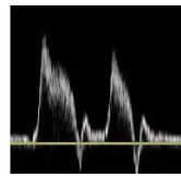
What does a deterioration in signal contour from one level to the next indicate?
Disease between those levels
Improvement in signal quality
No change in condition
Signal interference from external sources
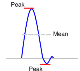
What does the pulsatility index measure?
Resistance
Velocity
Pressure
Flow
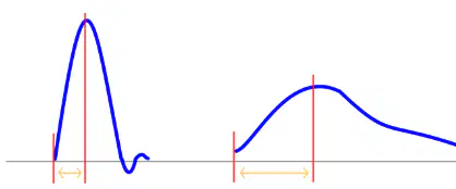
What does an increased acceleration time greater than 133 msec indicate?
A) Distal disease
B) Proximal (inflow) disease
C) No disease
time from beginning of systole to peak AKA upstroke
acceleration time
pulsatile index
peak systolic
none of these
how do you obtain pulsatility index?
peak to peak / mean
peak to end / mean
peak to end / peak
none of these
What is one of the capabilities of segmental pressure testing?
Exact location of stenosis
Presence/absence of disease
Cure for renal disease
Treatment for lymphedema
normal pulsatile index decreases as you go down the limb
TRUE/FALSE:
TRUE
FALSE
_____ causes inaccurately high pressures in segmental pressure testing?
hint: its associated to diabetic pts and pts w/ end stage renal disease due to incompressible vessels
Diabetes
Medial calcinosis
Hypertension
Lymphedema
Patients with which condition cannot have segmental pressure testing?
Diabetes
DVT
Hypertension
Obesity
pt w/ dialysis, fistula, stents, DVT or hx of lymphedema will have
innacurate results
cannot have exam
extremely low pressures
elevated pressures
Fill in the blank: The width of the cuff should be ______ greater than the diameter of the limb.
1.2x
1.5x
1.8x
2.0x
In segmental pressure testing, pressures must be taken distal to proximal. Arrange the following in the correct order:
ankle > calf > thigh
thigh > calf > ankle
calf > ankle > thigh
thigh > ankle > calf
What is the formula to calculate the Ankle/Brachial Index (ABI)?
Ankle pressure divided by HIGHEST brachial pressure.
Ankle pressure divided by LOWEST brachial pressure.
Brachial pressure divided by Ankle pressure.
Ankle pressure multiplied by Brachial pressure.

According to the ABI criteria, what ABI value indicates normal condition?
1.0
0.9-1.0
0.8-0.9
<0.5

According to the ABI criteria, what ABI value indicates claudication condition?
1.0
0.9-1.0
0.8-0.9
0.5-0.8
What does an ABI value of >1.3 indicate?
Normal
Vessels are likely incompressible
Mild disease
Severe disease
What is the purpose of exercise testing in vascular registry review?
Produces physiologic stress on circulation and reproduces ischemia.
Improves muscle strength and flexibility.
Increases heart rate variability.
Enhances respiratory function.
What is a contraindication for exercise testing?
Difficulty walking or breathing, severe HTN, cardiac issues.
Mild headache, slight fatigue, normal blood pressure.
Good physical fitness, regular exercise routine, healthy diet.
Normal heart rate, no history of heart disease, regular check-ups.
What is the technique used in exercise testing?
Constant load treadmill with <12% grade and 1.5 mph for a max of 5 min.
Variable load treadmill with >12% grade and 2.0 mph for a max of 10 min.
Constant load cycling with <10% resistance for a max of 5 min.
Variable load rowing with >15% resistance for a max of 8 min.
In exercise testing, what is considered normal?
No change or increase in ankle pressures.
Decrease in ankle pressures.
Significant increase in heart rate.
Drop in blood oxygen levels.
In reactive hyperemia, what indicates a multi-level disease?
A drop in pressure >50%
A rise in pressure >50%
No change in pressure
A drop in pressure <20%
In reactive hyperemia, what indicates a single-level disease?
A drop in pressure <50%
A rise in pressure >50%
No change in pressure
A drop in pressure <20%
In segmental pressure testing, what should be the normal difference in mmHg between both brachial pressures?
Within 20mmHg of each other.
Within 10mmHg of each other.
Within 30mmHg of each other.
Within 5mmHg of each other.
What happens to pressures distal to the disease in arterial obstructive disease?
Pressures DROP abnormally distal to disease.
Pressures INCREASE abnormally distal to disease.
Pressures remain UNCHANGED distal to disease.
Pressures FLUCTUATE abnormally distal to disease.
What is the standard interpretation of the Allen test?
Color returns to normal.
Color does not return to normal.
Pulse is absent.
Skin becomes pale.
____ testing: produces physiological stress on circulation & repodruces ischemia( reduced flow)
- ___ test Dx true vs Pseudo claudication
- contraindications: difficulty walk/breathing, HTN ,cardiac
technique: constant load treadmill ( unchanging) <12% grade and 1.5mph for a max of 5min.
- leave ankle cuffs on. after exercise, repeat ankle pressures.
exercise testing
segmental pressure testing
reactive hyperemia
none of these
a normal ______ exam will show no change or increase in ankle pressure
exercise testing
segmental pressure testing
reactive hyperemia
none of these
What is plethysmography usually performed with?
Segmental pressure study
Ultrasound imaging
MRI scan
CT scan
a abnormal ______ exam will show
- decrease in ankle pressure
- if abnormal repeat ankle pressures every 2 mins until return to resting
- if return <6mins = single level disease
- if return >6mins = multi level disease
exercise testing
segmental pressure testing
reactive hyperemia
none of these
Plethysmography is vessel specific and cannot underestimate disease.
True
False
What does plethysmography use to detect changes in volume?
Pneumatic cuffs (BP cuffs with special sensor)
Ultrasound waves
X-ray imaging
Magnetic resonance imaging (MRI)
Which of the following is NOT a term used in plethysmography interpretation?
Triphasic
Biphasic
Monophasic
High resistance
if cuff too wide: _____
falsely low pressure
falsely high pressure
undetermined pressure
if cuff is too narrow: _____
falsely high pressure
undetermined pressure
falsely low pressure
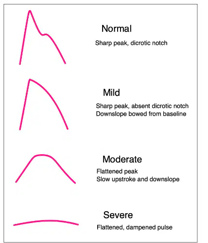
What is the interpretation of a waveform with a sharp peak and dicrotic notch?
Normal
Abnormal
Irregular
Erratic
when performing a segmental pressure exam patient must be
standing
supine
prone
basal state
What does PVR detect in the limb?
The overall volume of blood in the limb at that level.
The temperature of the limb.
The nerve activity in the limb.
The muscle strength in the limb.
What is indicated by a high amplitude 'fair' PVR contour in the presence of abnormal pressures?
Severe disease but presence of collateral development.
Mild disease with no collateral development.
Normal condition with no disease.
Moderate disease with partial collateral development.
What is the capability of Digital Pressure and PPG?
Same as plethysmography; useful in patients with cold sensitivity and medial calcinosis.
Measures blood glucose levels.
Monitors heart rate variability.
Assesses lung capacity.
What is the normal Finger FBI value?
Normal >0.8
Normal <0.8
Normal =0.8
Normal >=0.8
What does a 'Peaked pulse' after cold stress indicate?
Functional disease
Structural disease
Infectious disease
Genetic disorder
What is evaluated for penile vasculogenic impotence?
Vascular inflow or outflow
Hormonal levels
Neurological function
Psychological factors
What is the normal Penile/Brachial Index (PBI)?
Normal ≥ 0.65
Normal ≥ 0.75
Normal ≥ 0.55
Normal ≥ 0.45
What does a normal ABI with abnormal PBI indicate?
Disease isolated to internal iliac vessels
Normal vascular function
Disease isolated to external iliac vessels
No vascular disease
What is the significance of a dorsal venous velocity greater than 4cm/s?
Venous leak
Normal finding
Arterial insufficiency
Deep vein thrombosis
What is the normal range for Transcutaneous Oximetry TcPO2?
Normal >50mmHg
Normal <30mmHg
Normal 30-40mmHg
Normal 40-50mmHg
What is the common complaint of a young male patient with Popliteal entrapment syndrome?
Tingling in the arm
Claudication in calf with exercise
Headache
Chest pain
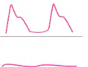
In Popliteal entrapment syndrome, what happens to the waveforms upon flexions?
Waveforms will increase
Waveforms will diminish or flatten
Waveforms will remain unchanged
Waveforms will become erratic
What is the most common cause of Thoracic outlet syndrome?
Muscular compression
Neurogenic (nerves)
Vascular compression
Bone fracture
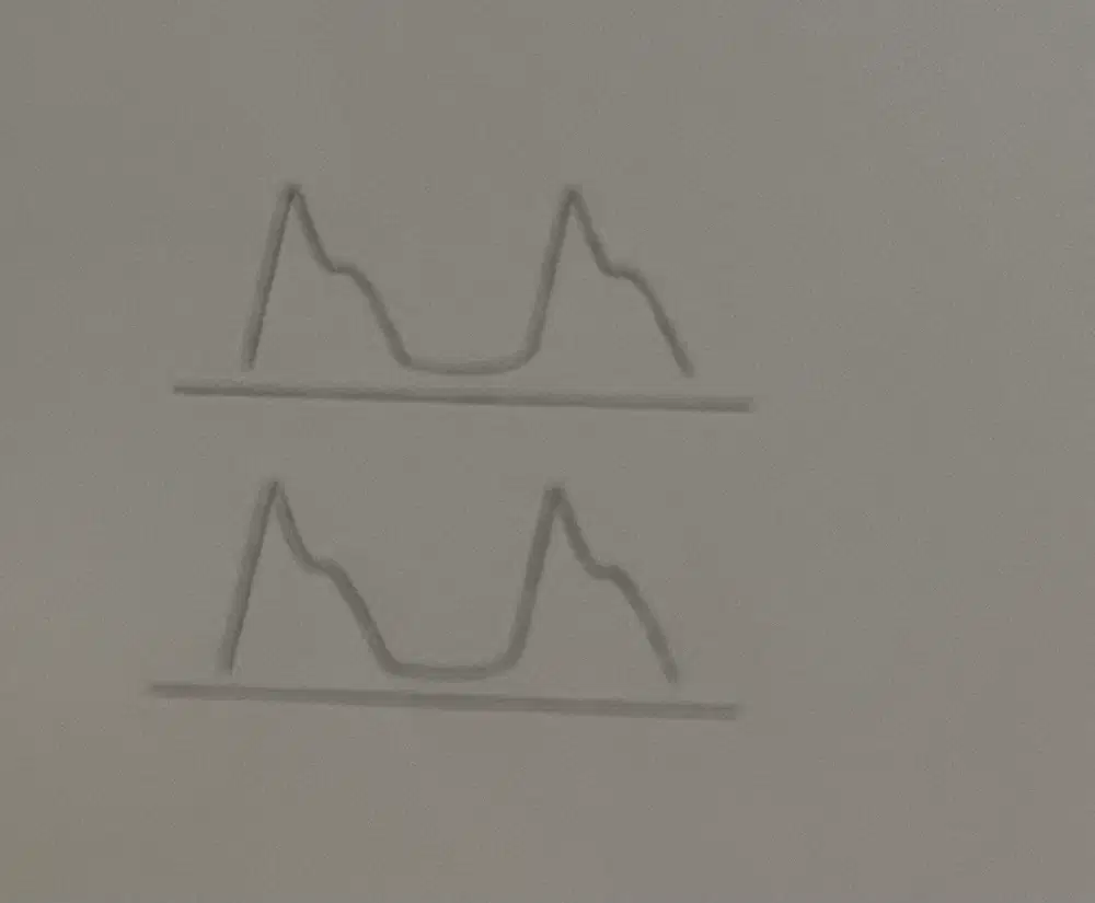
in compression disorders if only neurogenic (most likely finding) = ______
all normal w/ and w/o causative positions.
normal at rest
attenuated/flattened in position
attenuated/flattened at rest
What are Limitations of Continuous Wave doppler analysis?
CHF
(inflow&outflow)
temperature
Patients w/ bandages & casts cannot participate
cannot dx stenosis vs occlusion
tech dependent
Match the following
- X which is the line left to right = time
- Y which is the line up & down = frequency/amplitude

analog triphasic waveform

spectral analysis w/ triphasic waveform

analog w/ absence of flow reversal
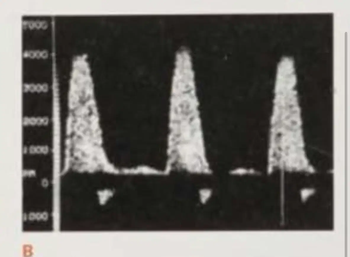
spectral analysis w/ flow reversal
for the audible signal method on continuous wave testing paired with segmental pressures,
how does the examiner know the signals that are being detected?
right headphone provides retrograde flow
left headphone provides retrograde flow
right headphone provides antegrade flow
left headphone provides antegrade flow
Quality interpretations for continuous wave testing includes _____ & _____ as NORMAL.
triphasic
multiphasic
sharp upstroke, sharp peak, rapid down stroke, short peak below baseline & resumption of forward flow above baseline
monophasic , biphasic
non-pulsatile
match the continuous wave doppler signals on analog that are abnormal or normal

abnormal, monophasic

abnormal/ biphasic
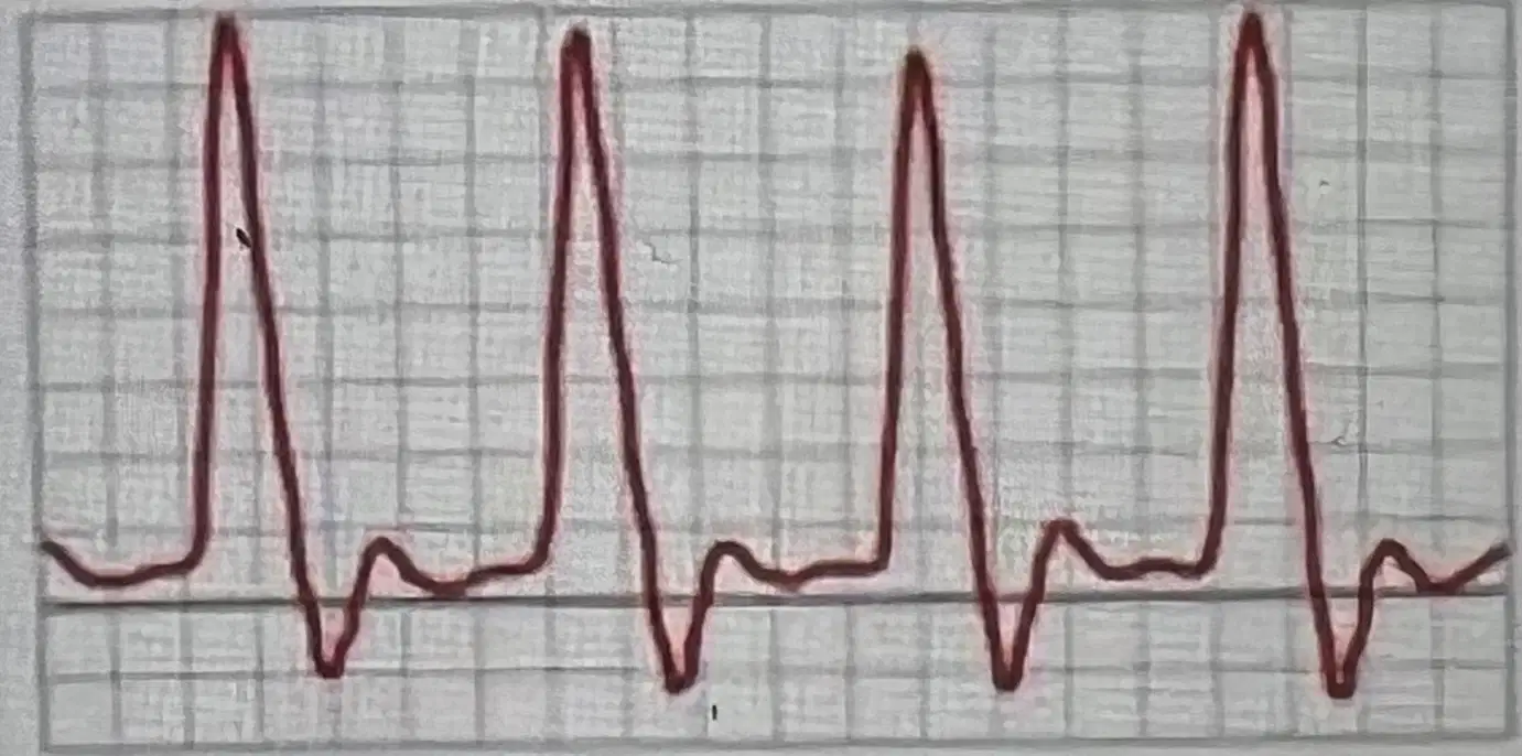
normal triphasic
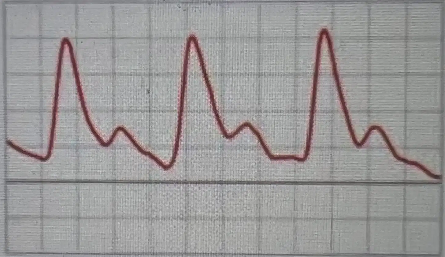
this image was obtained
post-exercise
post stenosis
prestenosis
all of these

this waveform was obtained in the common femoral artery what is the cause of this
normal waveform
iliac artery occlusion
post exercise
pre exercise

this flow is called ____ caused by 60-cycle noise & can be adjusted by increasing the filter or reducing gain.
thrill flow
trickle flow
monophasic flow
venous flow
T/F
patient with a warm relaxed extremity could also have normal monophasic low resistance continuous flow
true because it creates vasodilation
false because that only occurs when there's proximal obstruction or disease
the value of pulsatile index (PI) decreases in the presence of
proximal disease
distal disease
total occlusion
all of these

is the pulsatile index in this image increased or decreased?
decreased abnormally
decreased normally
increased normally
increased abnormally
continous wave is used for segmental pressure
true
false
In resting segmental pressures the patient should be resting ____ in supine position
20mins
30mins
45mins
1hr

where is the disease according to the segmental pressure
left iliofemoral disease
pelvic mass
popliteal disease
right iliofemoral disease

what is occuring in the segmental pressures on this patient.
within normal limits
pelvic pass
iliofemoral disease on the left exremity
none of these

what is occurring within these segmental pressures?
right femoral-popliteal occlusive disease
left femoral popliteal occlusive disease
pelvic mass
right iliofemoral occlusive disease
which pressures are most reliable and less likely to be misleading?
toes
ankles
calves
all of these are reliable
T/F
Resting segmental pressures are compared to pressures taken after exercise or after reactice hyperemia testing.
true
false
if the pressures taken after exercise testing have a drop and remain dropped for >6minutes it indicates
EXAMPLE: resting = 50mmhg
post exercise & 7minutes have passed = 45mmhg
single level disease
multilevel disease
entire leg disease
pelvic mass
if the pressures taken after exercise testing have a drop and remain dropped for >6minutes it indicates
EXAMPLE: resting = 50mmhg
post exercise & drop in pressures & 3 minutes have passed and then it = 50mmhg
single level disease
multilevel disease
entire leg disease
pelvic mass
in reactive hyperemia there are bilateral cuffs places at highest thighs & ankles
initial resting SPT are compared to post reactive hyperemia pressures (suprasystolic pressure >30mmhg)
Example: resting SPT : 120mmhg
post reactive hyperemia SPT: 60mmhg
what occured here?
single level disease
multilevel disease
entire leg disease
pelvic mass
in reactive hyperemia there are bilateral cuffs places at highest thighs & ankles
initial resting SPT are compared to post reactive hyperemia pressures (suprasystolic pressure >30mmhg)
Example: resting SPT : 120mmhg
post reactive hyperemia SPT: 90mmhg
what occured here?
transient drop of 17-34%
single level disease
multilevel disease
entire leg disease
in reactive hyperemia there are bilateral cuffs places at highest thighs & ankles
initial resting SPT are compared to post reactive hyperemia pressures (suprasystolic pressure >30mmhg)
what is considered normal?
transient drop in 17%
transient drop of 34%
big drop of >50
drop of <50
in reactive hyperemia there are bilateral cuffs places at highest thighs & ankles
initial resting SPT are compared to post reactive hyperemia pressures (suprasystolic pressure >30mmhg)
what is considered single level disease?
<50 drop in pressures
>50 drop in pressures
>6minutes
transient 17%-34% drop
in reactive hyperemia there are bilateral cuffs places at highest thighs & ankles
initial resting SPT are compared to post reactive hyperemia pressures (suprasystolic pressure >30mmhg)
what is considered multi level disease in reactive hyperemia?
<50 drop in pressures
>50 drop in pressures
>6minutes
transient 17%-34% drop
in Upper extremity SPT a difference of _____ between the radial & ulnar artery suggest obstruction in the artery w/ the lower pressure
≥ 20
≥ 30
≥ 45
≤ 20
arterial obstruction/disase occurs most frequently in the
upper ext
lower ext
abdomen
brain
there is an incomplete palmar arch about ____ of the time
50%
20%
30%
never
During the allen test limitations include
excessive ____ and _____ cause compression of radial or ulnary artery creating a false positive& pallor.
plantar flexions
dorsiflexions
exagerated hand/finger extensions
fist compression
wrist resting
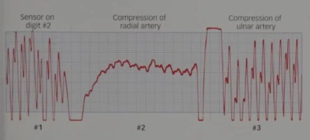
match the following for the allen test ( modified and standard testing)
- PPG on 2-3 fingers
- get Wave before/after comp.
- obtain tracing before comp. & during while the ppg is on
modified allen test
- compress radial a.
-make a fist = pallor
-keep compressed
-relax hand
standard allen test
- hand in fist = pale
- radial comp. & hand relaxed = color returns
= ulnar artery took over
Normal standard allens test
- radial a.= comp
- hand relaxed = remains pale
- radial a dependent
abnormal standar allens test
- waveform remains w/ radial a comp= ulnar a patent
- waveform distorts w/ radial a. ulnar a not patent
modified Allens interpretation
____ helps dx wound/ulcer healing
helps dx healing potential by amputation level
helps w/ calcified arteries in diabetics
these are capabilities of ;
laser doppler
resting SPT
reactive hyperemia SPT
exercise SPT testing
___ cannot be performed with ulcerated skin
patient cooperation of laying down quietly & w/o movement
- technician technique
laser doppler
resting SPT
reactive hyperemia SPT
exercise SPT testing
penile pressures are obtained by ONE of these
(meaning its an option but you can only use one to obtain pressures)
PPG
volume plethsmography
continuous wave doppler
duplex
a penile brachial pressure(PBI) of ____ is considered ABNORMAL in young adult with normal sexual response = vasculogenic impotence
>0.75
<0.65
>0.55
none of these
how is the nonimaging technique performed step by step in penile testing.
supine
1
cuffs on bilat arms & ankles & penile shaft cuff
2
calculate ABI to make sure it is normal for the study
3
ppg,plethsmography, or CW @ dorsal, ventral & medial aspects
4
penile divided by highest brachial
at or greater >0.75
5
Match the following plethysmographic waveforms
sharp, systolic peak w/ prominent dicrotic notch
normal
sharp peak absent dicrotic notch, downslow bowed away from baseline
mildy abnormal
flattened systolic peak, upslope/downslope time decreased, dicrotic absent
moderately abnormal
low amplitude equal upslope/downslope time or absent
severely abnormal
Match the following imaging technique to perform penile testing
supine w/ 7-10MHz probe
1
bilat PSV of cavernous & dorsal arteries obtained
2
vasodilator injected = erection
3
repeat PSV at prox cavernous a. & measure Dorsal vein velocity
4
POST injection normal PSV should incr: >30cm/s & venous flow should not go over <3cm/s
5
what may occur post-penile testing when guided injection is performed?
priapism
impotence
thrombus
irreversible damage

this image represents ____ penile testing results
abnormal
normal
mildly abnormal
highly abnormal
____ is the enlargement of veins of spermatic cord , occurs in young men, mostly on left side
patient standing or patient performing valsalva with reversal of flow indicated _______
varicocele
spider-veins
spermatocitis
hydrocele
____ are some limitation of plethsmography
not vesssel specific due to large area covered
difficult to dx major arteries & collaterals
obesity
cannot identify function aspect of disease or true vs false claudication
cannot localize or detect presence/absence of disease/obstruction
another name for plethsmography is
pneumo(air) plethsmography
true plethsmography
laser doppler
trickling doppler
photoplethsmography does not track the volume of blood flow, but instead tracks ____ and the infrared light reflected tracks blood flow.
- any increase in blood flow increases attenuation so it decreases reflection.
pulsations
obsutrction
disease
stenosis

moderate or severe abnormal volume plethsmography waveforms indicate _______ proximal to the level of the tracing.
hemodynamically significant disease
moderate disease
no disease
T/F
you can obtain a waveform of fair quality but an abnormal doppler segmental pressure at the same level due to collaterals
true
false

this plethsmography paired with SPT indicates what?
left sided inflow disease (iliofemoral)
bilateral inflow disease
right sided femoral disease
left sided outflow disease (femopopliteal)

this plethsmography(PVR) and SPT indicates disease beginning at
tibial
dpa
popliteal
ileofemoral
bilateral iliofemoral
_____ are limitations of digital pressure and ppg.
scaling/dry skin
ulcerations/gangrene affect placement of cuff or bandages
vasoconstriction affects results due to pt being cold, smoking,nervous
tight cuffs
not vessel specific
____ are limitations of TcpO2/PtcO2
electrolude not intact w/ skin
patient unable to lay still for 20minutes
tech dependent
low pressures in leg
the electrolude uses _____ degree celsuis to heat up skin in TcPo2/pCo2
44-45 degree celsius
50-55degree celsius
45-60 degree celsius
none of these

