
 Worksheets
MRSC5038 - WEEK 4 Fluoroscopy
Total questions: 100
Worksheet time: 1hrs 8mins
Which of the following procedures is NOT typically performed using fixed fluoroscopy X-ray equipment?
Barium Enemas
Barium Swallows
Lumbar Punctures
Eye Examinations
What do the terms "undercouch" and "overcouch" refer to in the context of X-ray equipment?
The type of patient bed used
The location of the X-ray tube
The size of the X-ray machine
The duration of the procedure
Which medical fields commonly use fluoroscopy X-ray equipment with iodine contrast agents?
Dermatology, Ophthalmology, Neurology
Cardiology, Vascular, Neuro-vascular
Gastroenterology, Endocrinology, Rheumatology
Pediatrics, Geriatrics, Psychiatry
At what frame rate are images typically displayed in fluoroscopy?
30 frames per second
60 frames per second
15 frames per second
24 frames per second
Why are images displayed at 30 frames per second in fluoroscopy?
The human eye perceives this as continuous motion
It is the maximum capacity of the monitor
It reduces radiation exposure
It enhances image resolution
What is the range of "beam-on" time for fluoroscopy?
A few seconds to a few hours
A few minutes to a few hours
A few seconds to a few minutes
A few minutes to a few days
How many images would a procedure with a "beam-on time" of 10 minutes at 30 f/s produce?
18,000 images
1,800 images
180,000 images
1,080 images
Why must fluoroscopic systems produce each image with a lower dose than radiography?
To reduce the cost of imaging
To keep patient and operator radiation dose ALARA
To increase the speed of imaging
To improve image quality
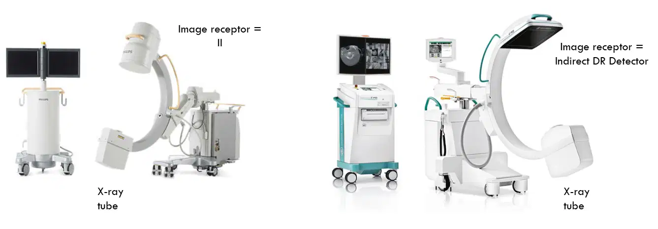
What are the two types of current image receptors?
Image intensifier (II) and Direct DR Detector
Image intensifier (II) and Indirect DR Detector
Direct DR Detector and Indirect DR Detector
Image intensifier (II) and Film-based Detector
What is the primary function of an Image Intensifier (II)?
To reduce the size of X-ray images
To amplify low signal levels obtained at low X-ray doses
To change the color of X-ray images
To increase the speed of X-ray processing
What shape is the image receptor of an Image Intensifier (II)?
Square
Triangular
Circular
Rectangular
What is the typical input diameter of a mobile Image Intensifier (II)?
15 cm
25 cm
35 cm
45 cm
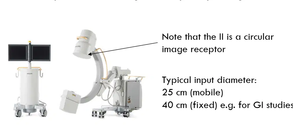
For what type of studies is a fixed Image Intensifier (II) with a 40 cm input diameter typically used?
Cardiac studies
GI studies
Neurological studies
Orthopedic studies
What is the function of the input phosphor in an image intensifier?
Converts electrons into visible light
Converts X-rays to light photons
Focuses and accelerates electrons
Converts light photons to electrons
Which component of the image intensifier converts light photons to electrons?
Input phosphor
Photocathode
Electronic lenses
Output phosphor
What role do electronic lenses play in an image intensifier?
Convert X-rays to light photons
Convert electrons into visible light
Focus and accelerate electrons towards the output layer
Convert light photons to electrons
Electronic lenses: focus and accelerate electrons towards output layer. These are the processes that achieve (a)
How are electrons converted into visible light in an image intensifier?
By the input phosphor
By the photocathode
By the electronic lenses
By the output phosphor
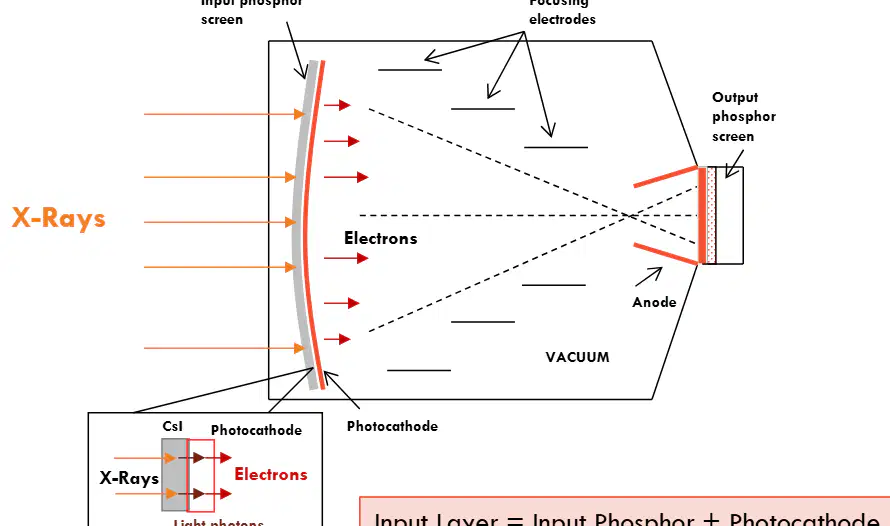
What components make up the input layer of an image intensifier?
Input phosphor and photocathode
Output phosphor and anode
Focusing electrodes and vacuum
X-rays and electrons
What is the primary function of the input phosphor in the input layer?
Absorbs light photons and converts them into electrons
Absorbs X-ray photons and converts them into light
Absorbs electrons and converts them into X-ray photons
Absorbs light and converts it into sound
Which material is used in the input layer for its column-like structure and good spatial resolution?
Sodium chloride (NaCl)
Calcium carbonate (CaCO3)
Caesium iodide (CsI)
Potassium iodide (KI)
What is the role of the photocathode in the input layer?
Converts electrons into X-ray photons
Absorbs light photons and converts them into electrons
Absorbs X-ray photons and converts them into light
Converts sound waves into light photons
What is the purpose of applying 25-35 kV in an image intensifier?
To decelerate electrons
To focus X-rays
To accelerate electrons
To cool the system
What are the two ways in which electron focusing causes intensification?
Electronic gain and magnification gain
Electronic gain and minification gain
Minification gain and light gain
Light gain and magnification gain
What is intensification more commonly known as?
Light gain
Power gain
Brightness gain
Energy gain
How is brightness gain calculated?
Electronic gain + Minification gain
Electronic gain - Minification gain
Electronic gain x Minification gain
Electronic gain / Minification gain
What is the range of voltage through which electrons from the photocathode are accelerated?
10 to 20 kV
25 to 35 kV
40 to 50 kV
5 to 15 kV
What is the effect of the applied voltage on the electrons from the photocathode?
It decreases their kinetic energy.
It causes electronic gain.
It stops their movement.
It reduces their speed.
By what factor does the electron energy increase due to electronic gain?
10
25
50
100
What is the role of focusing electrodes in minification gain?
To disperse the electron beam
To focus the electron beam from the large input layer onto the smaller output layer
To increase the size of the output layer
To invert the electron beam
How is the spatial pattern of electrons maintained at the output layer?
Enlarged and upright
Minified and inverted
Minified and upright
Enlarged and inverted
What is minification gain?
The process of enlarging an image
The image is much smaller at the output phosphor than at the input phosphor
The process of reducing energy concentration
The image is much larger at the output phosphor than at the input phosphor
What effect does minification gain have on energy?
It disperses energy over a large area
It reduces energy concentration
It intensifies energy by concentrating it on a small area
It has no effect on energy
What is the formula for calculating minification gain?
(dO/dI)2
(dI/dO)2
d_I * d_O
d_O - d_I
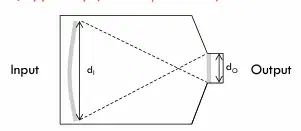
In the context of minification gain, what does 'd' represent?
Density
Diameter
Distance
Depth
Image Intensifier (II) Summary of Process
Input phosphor (CsI): converts the X-rays to light photons
Photocathode: converts light photons to electrons
Electronic lenses: focus and accelerate electrons towards output layer (minification and electronic gain)
Output phosphor: converts electrons into visible light
What is the primary use of INDIRECT DR Detectors in medical imaging?
As an alternative to MRI
As an alternative to the II in fluoroscopy
As an alternative to CT scans
As an alternative to ultrasound
In INDIRECT DR detectors for fluoroscopy:
Del size is usually (a) than radiography
Why is the del size in fluoroscopy usually larger than in radiography?
To achieve a reasonable data transfer rate for real-time imaging
To reduce the cost of equipment
To increase the resolution of images
To decrease the power consumption
What are the advantages of indirect DR detectors over the II?
Lower dose
Higher dynamic range
Less bulky
Lower dynamic range
What does film saturation result in?
Blackened image
Very bright image
Blurred image
Colorful image
What does II saturation result in?
Blackened image
Very bright image
Blurred image
Colorful image
What happens to IIs at high radiation levels?
They display all high radiation levels at maximum density.
They saturate and display all low radiation levels at the same minimum density.
They become transparent.
They reflect radiation.
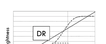
What effect does a higher X-ray dose have when passing through low-attenuation anatomy?
It causes the system to become more detailed.
It causes the system to saturate.
It reduces the image brightness.
It enhances color contrast.
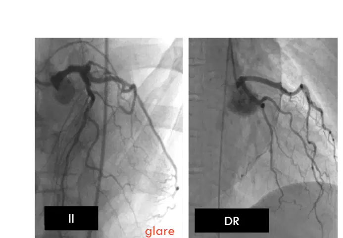
What is the effect of bright areas of saturation in a display on visualizing surrounding structures?
It enhances the visibility of structures
It makes it difficult to visualize surrounding structures
It has no effect on visualization
It improves the contrast of the image
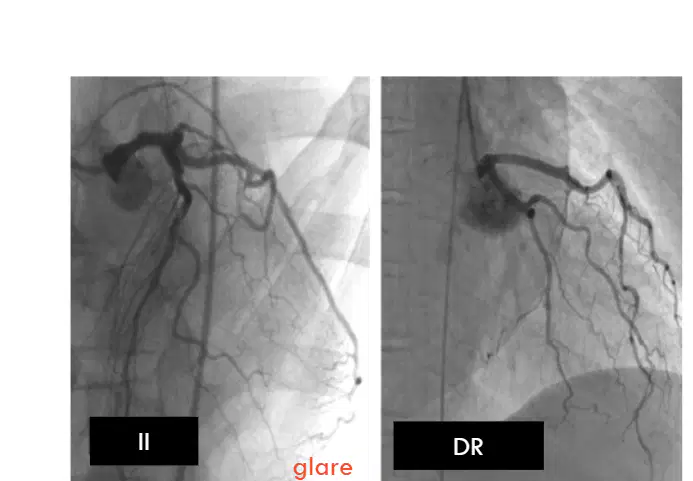
What is an example of glare in medical imaging?
Dark lung field in II image
Bright lung field in II image
Clear lung field in DR image
Blurred lung field in DR image
The next few are the socrative questions
Select me
Don't select me
The image intensifier (II) is a rectangular image receptor
True
False
Which of the following accurately shows the energy sequence within the II?
Light to X‐rays to electrons to light
X‐rays to electrons to light
Electrons to light to electrons
X‐rays to light to electrons to light
The formula for minification gain is:
minification gain = input phosphor diameter / output phosphor diameter
minification gain = output phosphor diameter / input phosphor diameter
minification gain = (input phosphor diameter / output phosphor diameter)2
minification gain = (output phosphor diameter / input phosphor diameter)2
The input phosphor of an II is made of CsI. This material is also used in:
Direct DR detectors
Contrast agents
CR plates
Indirect DR detectors
Magnification of the fluoroscopic image results in improved:
SNR
spatial resolution
contrast
brightness
ABC stands for
Automatic Beam Control
Automatic Beam Contrast
Automatic Brightness Control
Automatic Brightness and Contrast
For an II, the image is much larger at the output phosphor than it is at the input phosphor
True
False
Bones appear black on a fluoroscopy image
True
False
An II has a wider dynamic range than a DR detector
True
False
If the patient entrance skin dose rate is 30 mGy/min, how long would it take to reach the threshold dose for epilation?
10 minutes
100 minutes
1 day
1 week
In interventional radiology, estimation of patient peak skin dose from the displayed CAK requires knowledge of all of the following EXCEPT:
Backscatter factor
Overlap of X‐ray fields
Screening time
X‐ray Source to Skin Distance
Which of the following statements about radiation‐induced skin damage (deterministic effects) is FALSE?
The severity of the effect increases with dose
One example is erythema
They occur many years after exposure
They are not observed below a threshold dose of about 2 Gy
The maximum field of view on an II is 40 cm. Magnification is used and a FOV of 20 cm is selected to improve spatial resolution. How will patient dose change if the equipment is used under ABC? Be as quantitative as possible (i.e. do a calculation!).
Dose will increase by a factor of 8x
Dose will increase by a factor of 2x
Dose will increase by a factor of 4x
Dose will increase by a factor of 16x
The maximum field of view on a DR detector is 40 cm. Magnification is used and a FOV of 20 cm is selected. How will the spatial resolution change?
It will decrease because fewer pixels are used
It will remain the same because detector size is unchanged
It will improve due to increased pixel density over the smaller FOV
It will worsen due to increased geometric unsharpness
Categorise the characteristics
Beam is on at low mA all the time during screening
Only an option on mobile units
Generator delivers pulses at high mA at a pulse rate of 3-30 pulses per second
Pulses delivered at a short pulse width
Only mode for fixed units, but can also be used on mobile
What does temporal resolution refer to in the context of continuous fluoroscopy?
The ability to detect stationary objects
The ability to detect moving objects
The ability to enhance image contrast
The ability to increase image brightness
________ fluoroscopy has better temporal resolution than the other (i.e. little to no motion blur)
Continuous
Pulsed
Longer exposure times will reduce blurring from patient motion in the image
True
False
In pulsed fluoroscopy, each pulse is 3-10ms. This is known as the _____ _____
(a)
In pulsed fluoroscopy, what is adjusted to provide the same X-ray exposure rate as continuous fluoroscopy?
Pulse width
Number of pulses
mA (milliamperes)
X-ray generator type
Assuming all other parameters are the same, changing from 30 pulse/second (p/s) to 15 p/s means the patient dose is halved.
It is therefore safe to assume that the lower the pulse rate, the lower the patient dose.
True
False
What happens if the pulse rate is very low in imaging?
The images become clearer
The display becomes "jerky"
The images become faster
The display becomes smoother
What happens to the displayed image when the monitor frame rate is 30 f/s and the X-ray pulse rate is 15 p/s?
The image flickers on and off in synchronization with the X-ray pulse
The image is displayed continuously at the refresh rate of the monitor
The image is not displayed at all
The image is displayed only when the X-ray pulse is active
____ rate is a measure of how many exposures per second are being made by the x-ray unit.
____ rate is a measure of how many images are displayed per second on the monitor
(a)
If the monitor frame rate is 30 f/s and the pulse rate is 10 p/s, how many times is each x-ray image displayed?
1
2
3
4
Categorise each under the correct stage
To assist with positioning
Fairly noisy images
Low patient dose-rate is important because of time
Used to capture diagnostic-quality images
Patient dose-rate is about 20x higher in acquisition mode
Better image quality is required
What is the primary purpose of using Automatic Exposure Control in fluoroscopy?
To manually select exposure factors
To terminate the exposure
To maintain image quality
To increase patient exposure
How does the ABC maintain a constant image brightness in fluoroscopy?
By adjusting the room lighting
By altering the image resolution
By modulating kV and mA
By modulating the pulse width and rate
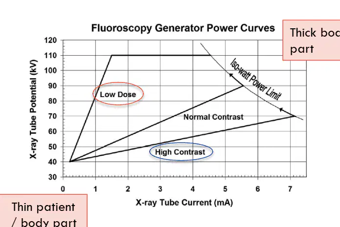
What happens to kV and mA in low dose mode?
Both kV and mA decrease
kV decreases and mA increases
kV increases and mA decreases
Both kV and mA increase
In high contrast mode, how are kV and mA adjusted?
Low kV and high mA
High kV and low mA
Both kV and mA are high
Both kV and mA are low
What is the limitation of the ABC in distinguishing objects?
It cannot distinguish a patient from lead shielding.
It cannot detect any objects.
It can only detect metal objects.
It can distinguish all objects accurately.
How does the ABC respond to the reduction in brightness caused by lead gloves?
It reduces the radiation output
It boosts the radiation output.
It turns off automatically
It does not respond at all.
Which of the following factors affects spatial resolution in X-ray imaging?
Focal spot size of x-ray tube
Patient positioning
Properties of the image receptor
Speed of the x-ray machine
What is an additional factor in fluoroscopy that affects magnification?
Electronic magnification
Optical magnification
Thermal magnification
Aliasing magnification
What determines the largest field of view in magnification?
The physical size of the II or DR Detector
The colour of the image
The type of lens used
The distance from the object
A smaller FOV without magnification can be achieved by bringing in the lead shutters. What is a consequence of doing this?
Spatial resolution improves
Contrast resolution improves
Spatial resolution decreases
Spatial resolution remains the same
Read this & understand pls
In Magnification Mode (B), a higher voltage is applied resulting in a more focussed input electron beam. This causes magnification due to divergence of the output electron beam.
Result? Magnified view of a smaller area of anatomy; better spatial resolution
What happens to the minification gain in magnification mode?
It increases
It decreases
It remains the same
It doubles
To Recap:
In magnification mode:
- The collimators adjust to the smaller FOV
- The data from the smaller FOV are displayed across the entire monitor, thereby achieving (a) magnification
What effect does binning dels have on spatial resolution?
Improves spatial resolution
Has no effect on spatial resolution
Results in poorer spatial resolution
Doubles the spatial resolution
Binned dels have a greater area, meaning they capture more x-ray photons. This also means that for the same patient dose, the image would have better (a)
Mag mode = smaller FOV = better spatial resolution but higher patient dose
True
False
Transient erythema: Threshold dose = (a) mGy, Time to appear = (b)
Temporary epilation: Threshold dose = (c) , Time to appear = (d)
The legal limit for maximum patient ESD rate
150mGy/min
100mGy/min
20mGy/min
1000mGy/min
We use DAP as the starting point to calculate Effective Dose
True
False
What is one use of Cumulative Air Kerma (CAK)?
To indicate potential skin injury
To determine Effective Dose
To measure total body dose
To average the absorbed dose
CAK is used as a starting point to calculate patient ____ ____ ____
(a)
Dosimeter- Absorbed Dose vs KERMA
Input dose from x-ray tube + backscatter dose from patient
Input dose from x-ray tube + free air (i.e. no backscatter)
Air kerma is higher than absorbed dose, because it does not include backscatter
True
False
CAK is
total air kerma
air kerma per projection
What does the Interventional Reference Point (IRP) represent in terms of patient measurement?
The distance from the source to the imaging table
The source to skin distance for an average-sized patient
The distance from the source to the floor
The distance from the source to the ceiling
Why might a patient not exhibit skin effects if CAK is received over multiple projections?
The projections are too weak
The projections are too fast
The projections are not overlapping
The projections are too slow
Peak Skin Dose = Cumulative Air Kerma, corrected by:
Backscatter factor (~1.4) [convert air kerma to dose]
Overlapping projections
Attenuation by table and mattress
Inverse square law [location of actual focus to skin distance compared to IRP]

