
 Worksheets
OBGYN Week 2 pt 1 (transabdominal)
Total questions: 119
Worksheet time: 2hrs 53mins
Pelvic pain is an example of an (a)
Dysmenorrhea and Amenorrhea are examples of
(a)
Menorrhagia and Metrorrhagia are examples of (a)
Prepubertal and postmenopausal bleeding is considered
(a)
Signs/Symptoms of pelvic infection are examples of (a)
Infertility is an (a)
localization of an IUD is an (a)
Screening for malignancy is an
(a)
Pregnancy evaluation/Complications are (a)
Which of the following are contradictions that ONLY apply to transvaginal ultrasounds?
Gravidity
Pediatric age
Premature rupture of membranes
Bleeding associated with known Placenta Previa
Date of last menstrual period (LMP) is considered important (a)
Gravidity and Parity are (a)
Hormone regimen and symptoms are considered
(a)
Family history of cancer, past pelvic surgeries, previous Pap smear/biopsy results, and pelvic examination findings are all (a)
means the number of pregnancies
(a)
means # of deliveries > 20 weeks
(a)
FPAL relates to (a)
Total number of term delivers after 37 weeks
(a)
Total number of preterm deliveries (20-36 weeks)
(a)
Total of abortions/miscarriages (before 20 weeks)
(a)
Total number of living children
(a)
When doing a pelvic ultrasound examination, which of the following are anatomical landmarks?
Rectum
Ovaries
Uterus
Vagina
Which structures does the vagina serve as a landmark for?
Uterus
Cervix
LUS
ovaries
When doing a (a) ultrasound examination you must document:
✓Uterine size, shape, orientation
✓Endometrium
✓Myometrium
✓Cervix
It is where the cervix ends and the LUS (lower uterine segment) begins, and where the uterus bends for positions (anteverted, retroverted, etc)
(a)

The blue arrow is pointing to
(a)
When measuring the length of the uterus in the long axis you should measure from __ to __
Anterior to posterior
Length to width
Fundus to cervix
Depth
When measuring the AP depth of the uterus you should measure from
Uterus to fundus
Anterior to posterior
Lateral to medial
Inferior to superior
Width of the uterus can be measured from __ or __ point of view.
(a)
What is the endometrium analyzed for?
Contour changes
Thickness
Masses
Echogenicity
Which of the following should you evaluate the myometrium for?
Contour changes
thickness
masses
echogenicity
When would you usually measure the cervix?
Pre pregnancy
Pregnancy
Fluid
Nabothian cysts
The (a) region includes the ovaries and fallopian tubes
The ovaries are identified as (a) to the internal iliac arteries (hypogastric artery)
When scanning the adnexa, which of the following should you document?
Size, shape, contour, echogenicity
Position relative to uterus
Relation to the vaginal canal
Ovarian size
Ovarian size determined by measuring length of long axis with AP dimension measured perpendicular to
length
True
False
Ovarian width measured in __ (TA - transabdominally) or __ view (TV - transvaginally)
(a)
Ovarian (a) may be calculated during the examination
Why would you evaluate the Cul - De - Sac?
Ectopic
Free fluid
Cysts
Mass
IF a mass is detected during an examination of the Cul - De - Sac, what would you document?
Size, position, shape
Endometrium size
echographic pattern relationship to ovaries,
and uterus
When examining the cul-de-sac,
Differentiate normal loops of (a) from mass
Usually, US Examination of the female pelvic is performed in two parts: __ and __
(a)
This approach requires distended urinary bladder
(a)
Transducer is carefully scanned across lower abdomen after warm gel is applied to produce the ultrasound images
(a)
What position should the patient be while doing a trans abdominal exam?
Prone
Supine
LLD
RLD
is moving a patient from one flat surface to another (from a bed to a stretcher, from a bed to a wheelchair
(a)
While seemingly intuitive, successful patient transfers rely on (a) each patient's needs while
adhering to evidence-based guidelines
Privacy, relaxation, communication, equipment, support, and lubricants are all ways to make the patients more (a)
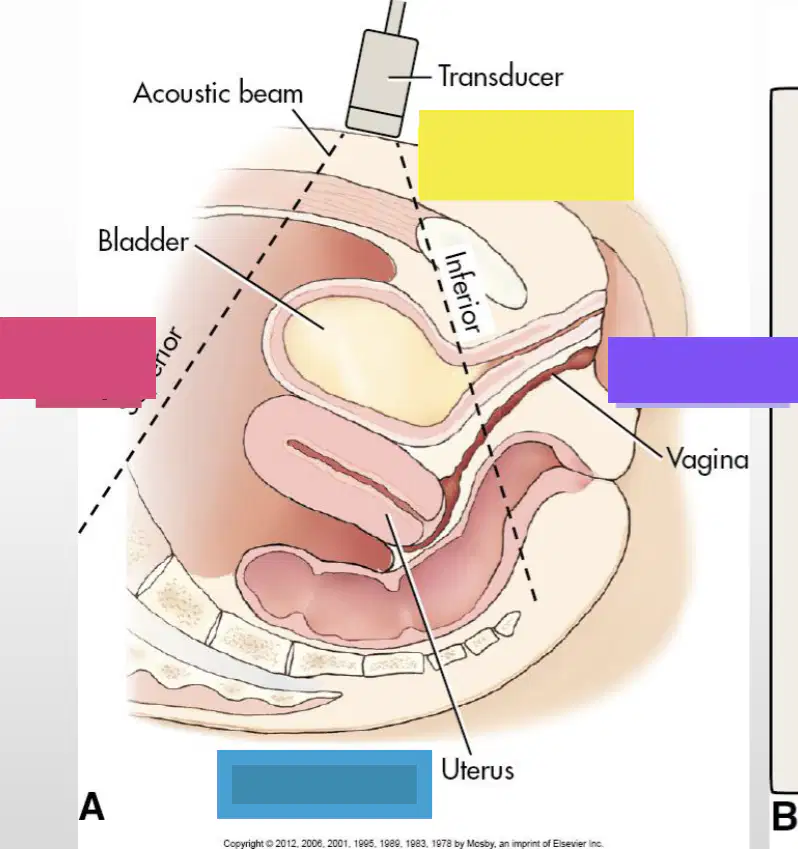
Yellow
(a)
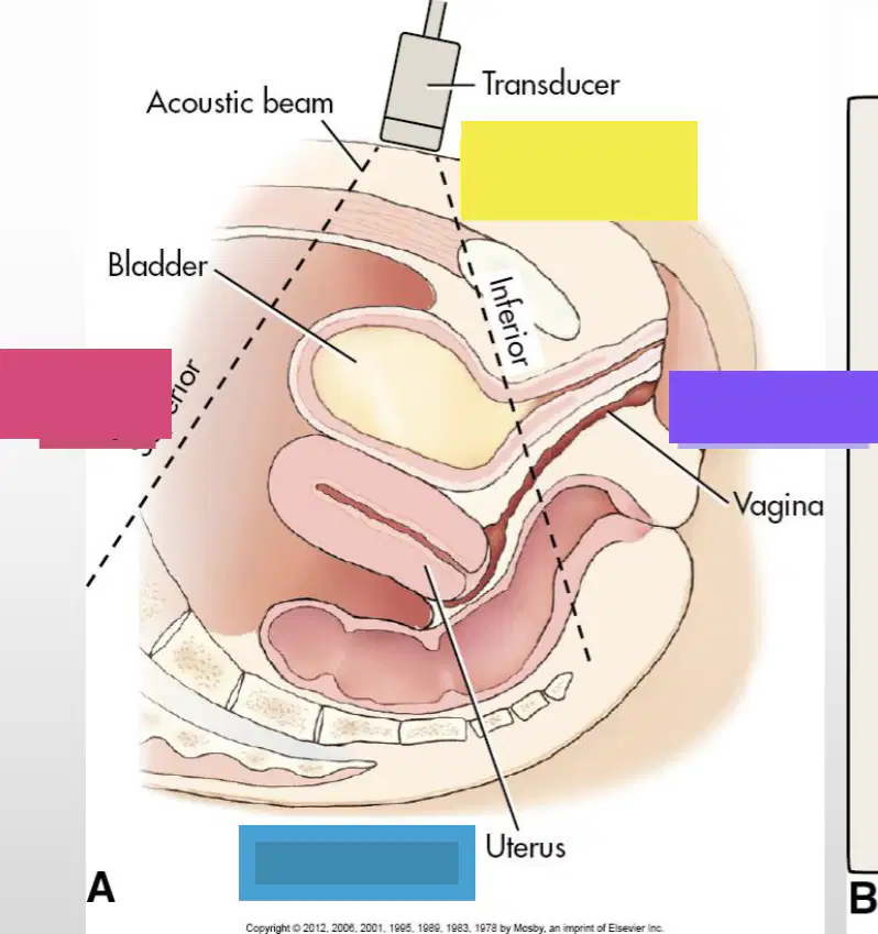
Purple
(a)
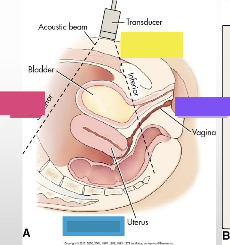
Blue
(a)
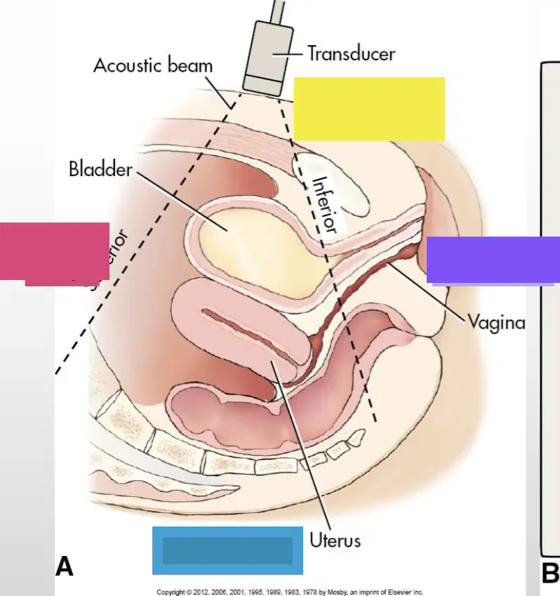
Pink
(a)
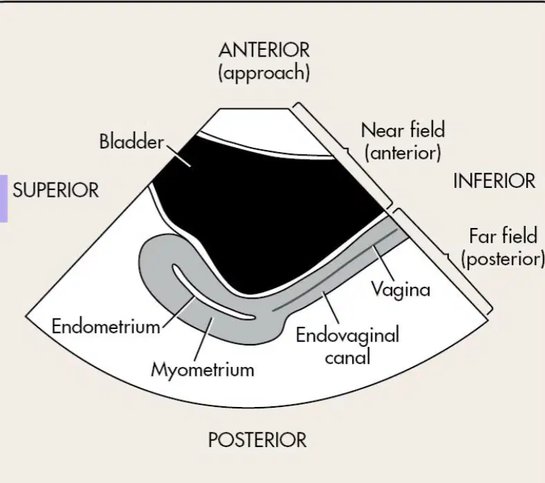
This image is a (a) view of the uterus
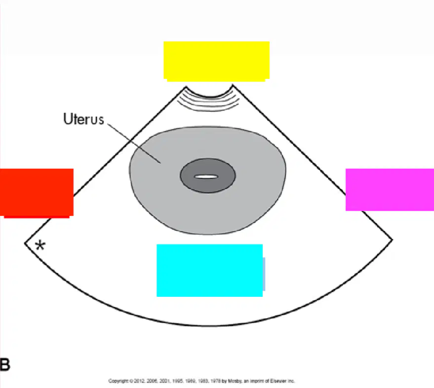
Yellow
(a)
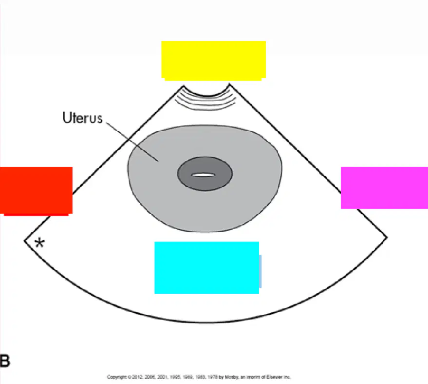
Pink
(a)
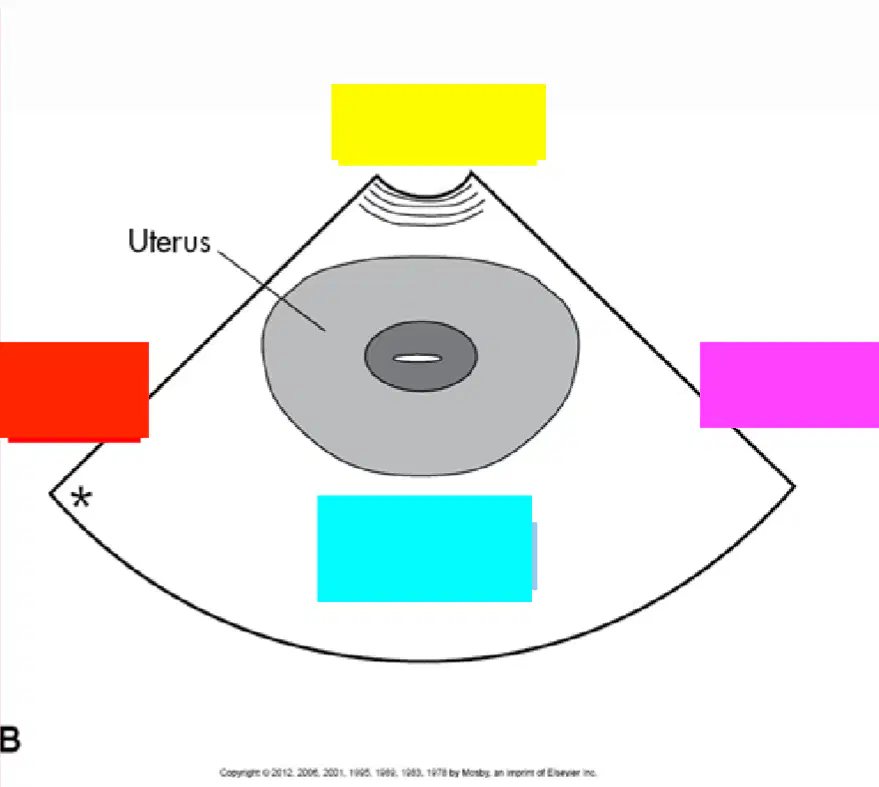
Blue
(a)
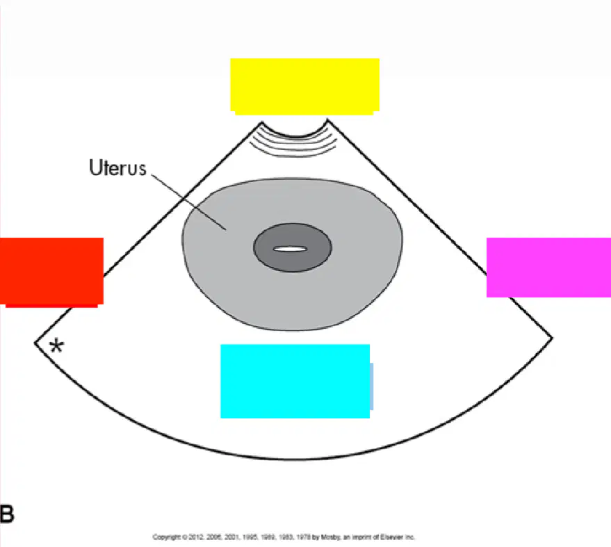
Red
(a)
The right of the patient is located on the (a) side of the image
Midline: Distended urinary bladder, uterus, endometrium, cervix, vagina
(a)
Measure length of uterus from fundus to cervix and A/P perpendicular
(a)
Measure Endo thickness
(a)
Angle Right of midline: Bladder, uterus RT, area of right ovary
(a)
Angle Left of midline: Bladder, uterus LT, area of left ovary
(a)
Look at both right and left adnexal areas in true pelvis (iliacus muscle is lateral border)
(a)
Measure Ovaries
- Sag – length & A/P
- Trans - width
(a)
When mass found on sonography, characterize the following features
1. (a)
When mass found on sonography, characterize the following features
2. (a)
When mass found on sonography, characterize the following features
3. (a)
well-defined, ill-defined, irregular borders
(a)
When mass found on sonography, characterize the following features
4. (a)
cystic, complex, predominantly cystic, complex, predominantly solid, solid
(a)
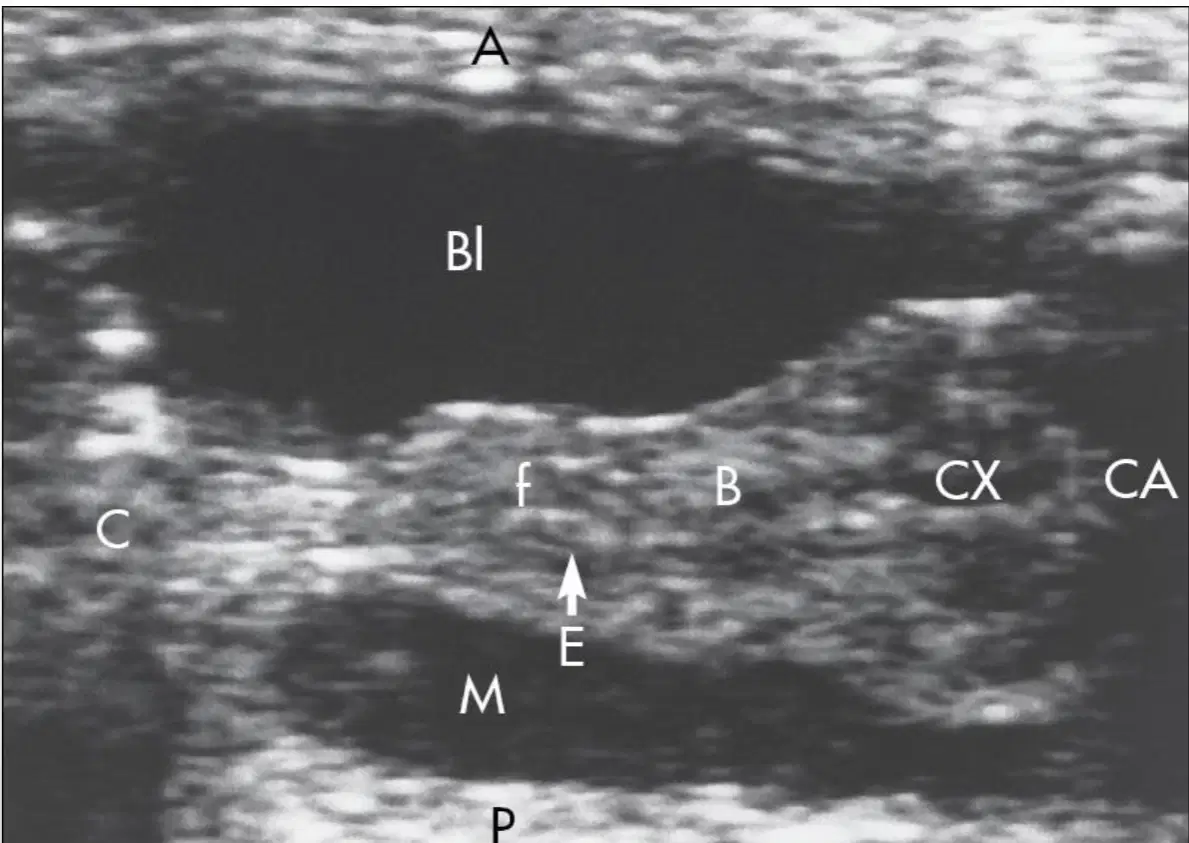
This image shows a (a) uterus
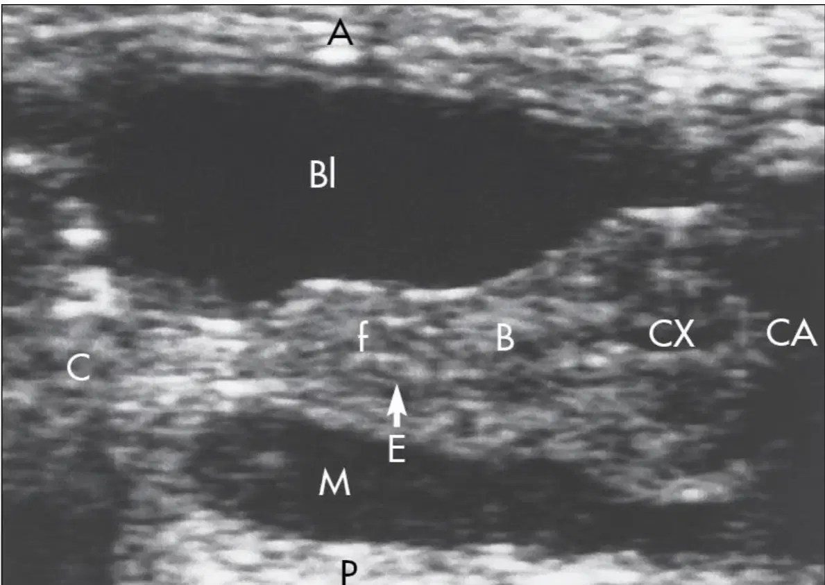
1-day-old neonatal uterus. CX=2/3 entire uterine length due to (a)

This image shows (a) uterus

A 6-year-old. The cervix occupies (a) of the entire uterine length,
but it loses the pear shape of a neonatal uterus

Red arrow
(a)
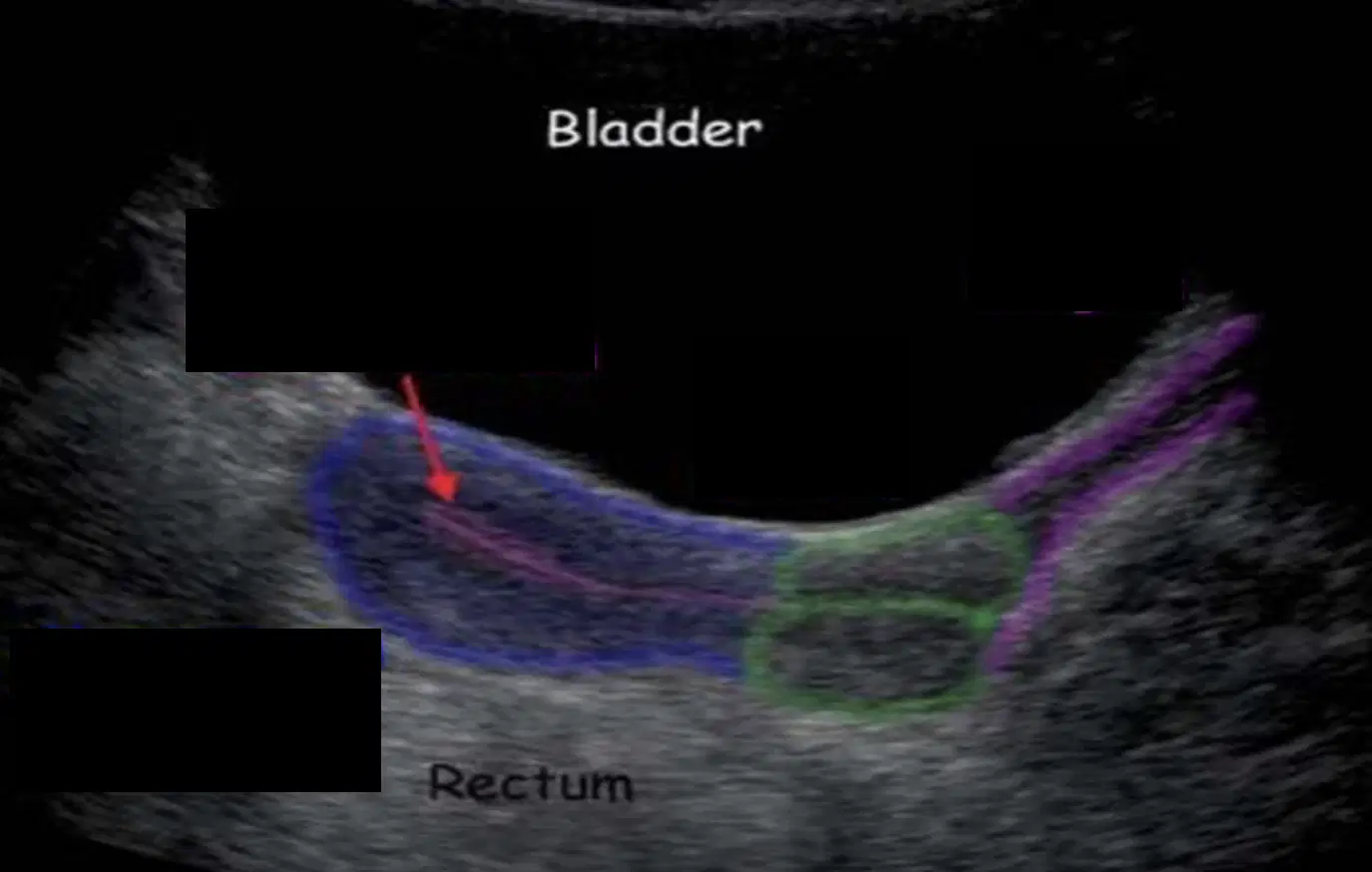
Blue area
(a)

Green area
(a)

Purple area
(a)

The arrow pointing towards the free fluid is where the (a) sits

This image contains a __ __ of the uterus with labeling of cervix, cervical canal, uterine body, fundus, and endometrial line
(a)

Why are we unable to see the uterus in this image?
Bladder is filled
Bladder is not adequately filled
Shadowing from a bowel
Mass is present

This image shows a (a) view of the uterus
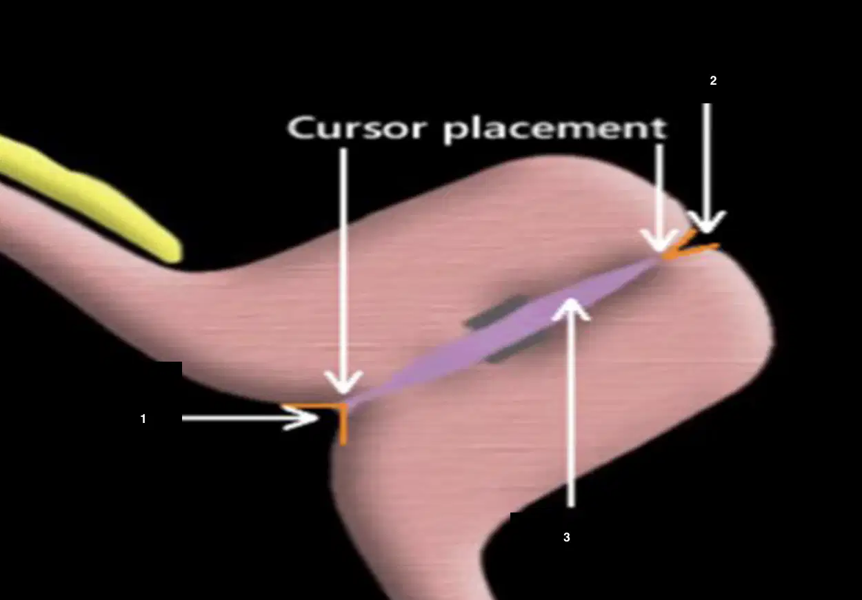
1
(a)

2
(a)

3
(a)
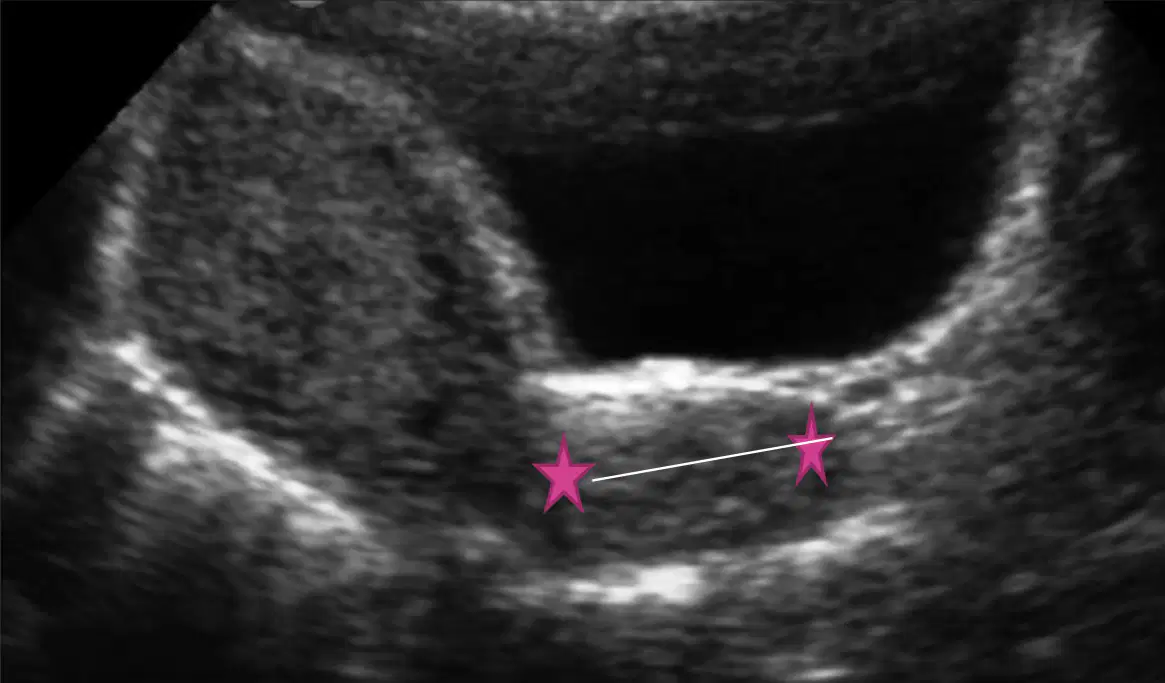
This image shows a sagittal view of (a)
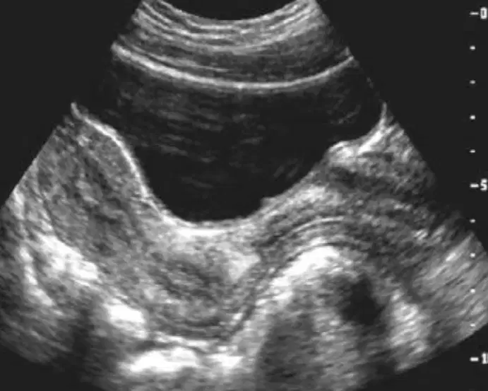
This image shows a (a) uterus
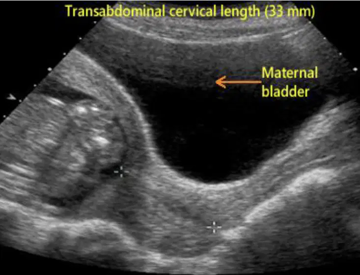
This image shows a (a) uterus
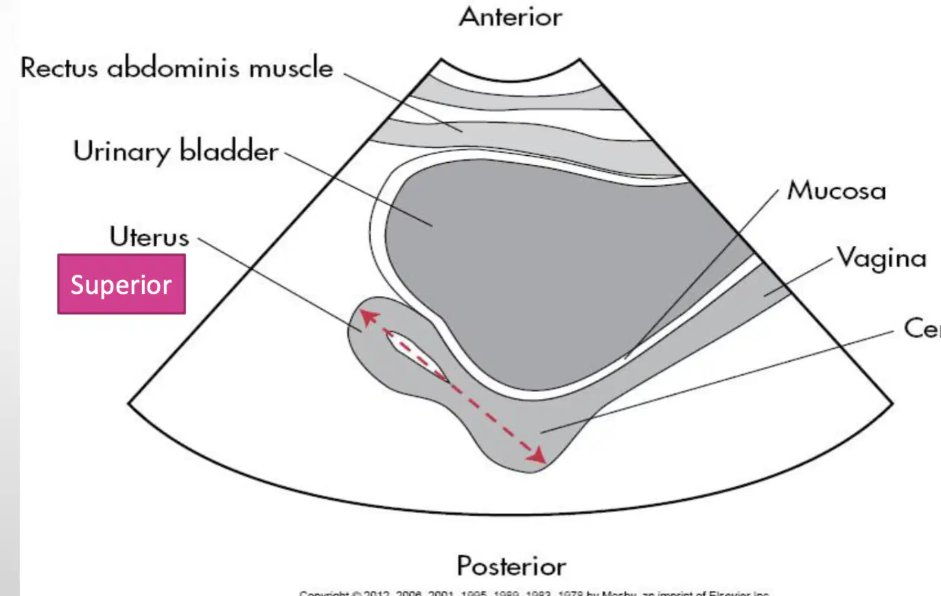
When measuring the length of the uterus in sag you measure from:
Top to bottom
Anterior to posterior
Fundus to cervix
Left to right

This image shows a (a) of the uterus at the ML in sag

1
(a)

2
(a)

The bright echogenic structure in this image is a (a)

These images show a sagittal measurement of the (a)
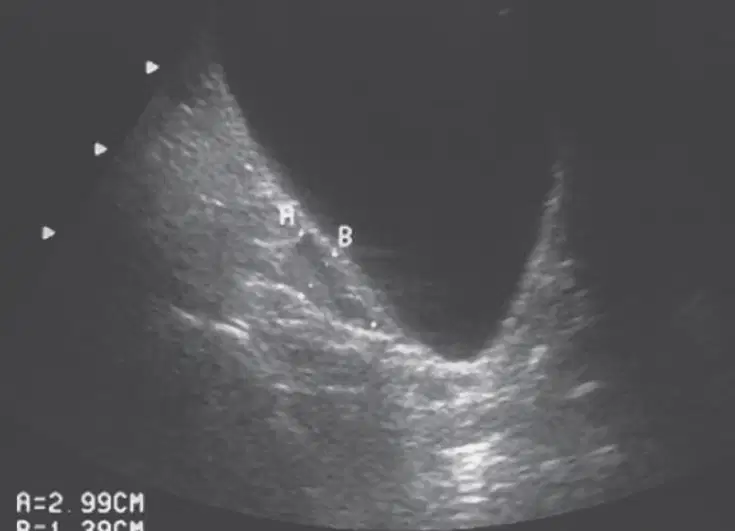
This image shows a (a)
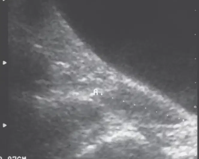
this image shows a (a)
When scanning in a (a) you must sweep from inferior to superior
Low structures in a transverse image:
LUS
Distended bladder
Vagina
Cervix
The Low/Mid structures in a transverse plane
LUS
Cervix
Uterus
Rectum
Mid structures in a transverse plane
LUS
Cervix
Body of uterus
Endometrium
What would you measure at the mid portion of the transverse plane?
Width
Look for ovaries
Length
AP
High portion of the transverse plane
(a)
look for ovaries lateral to cornu of uterus
(a)

1
(a)

2
(a)

3
(a)

The arrow is pointing to the
(a)

blue
(a)
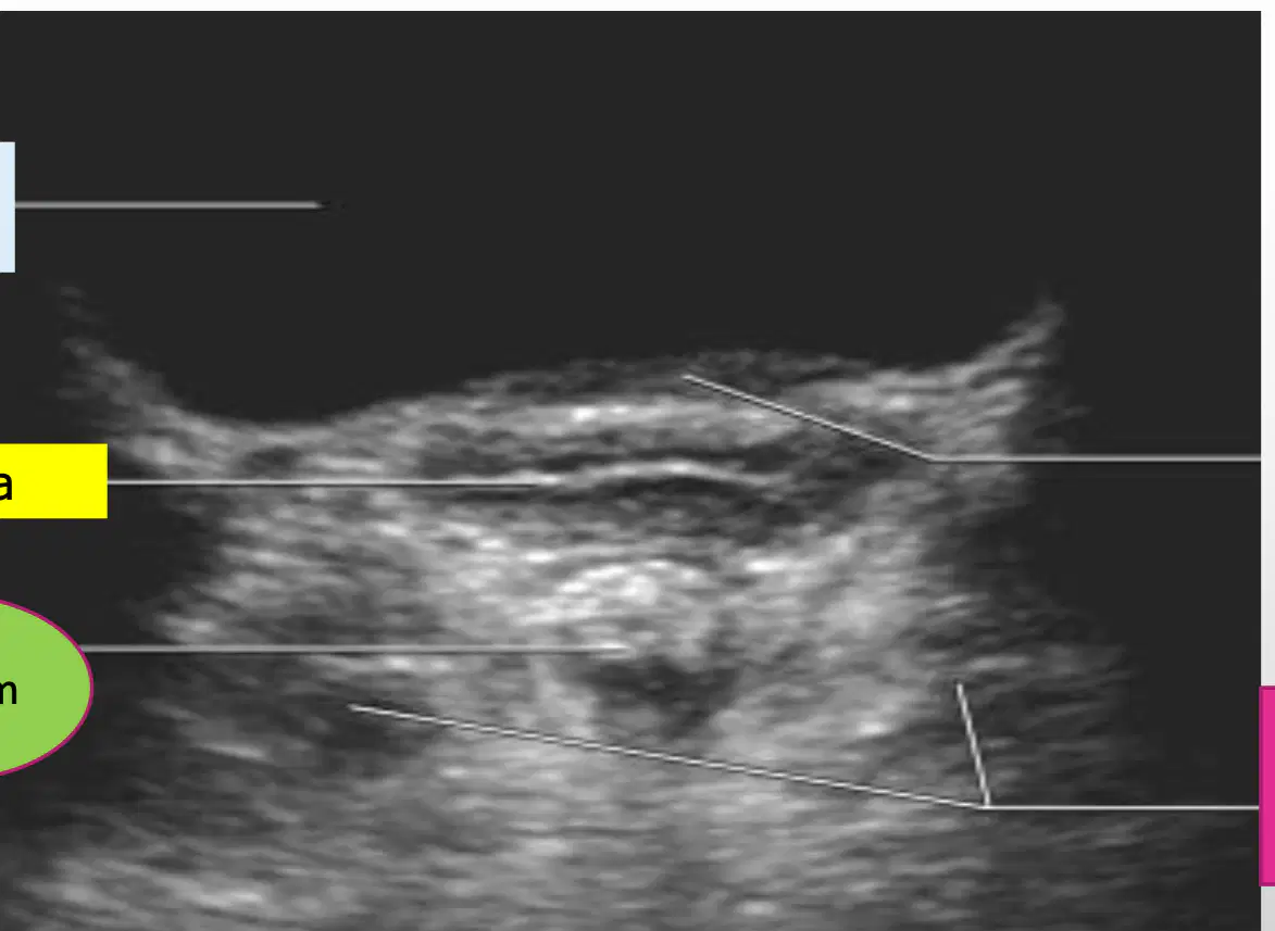
yellow
(a)
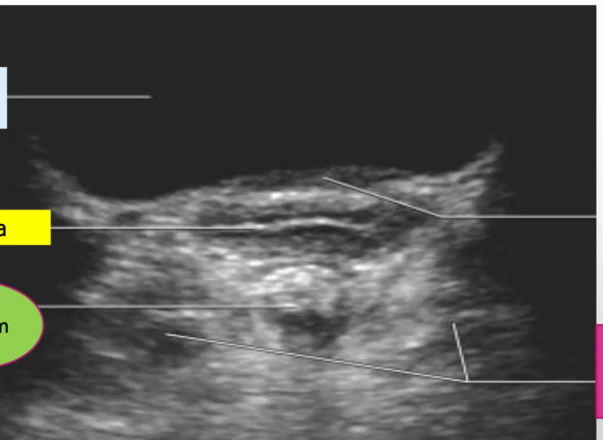
green
(a)
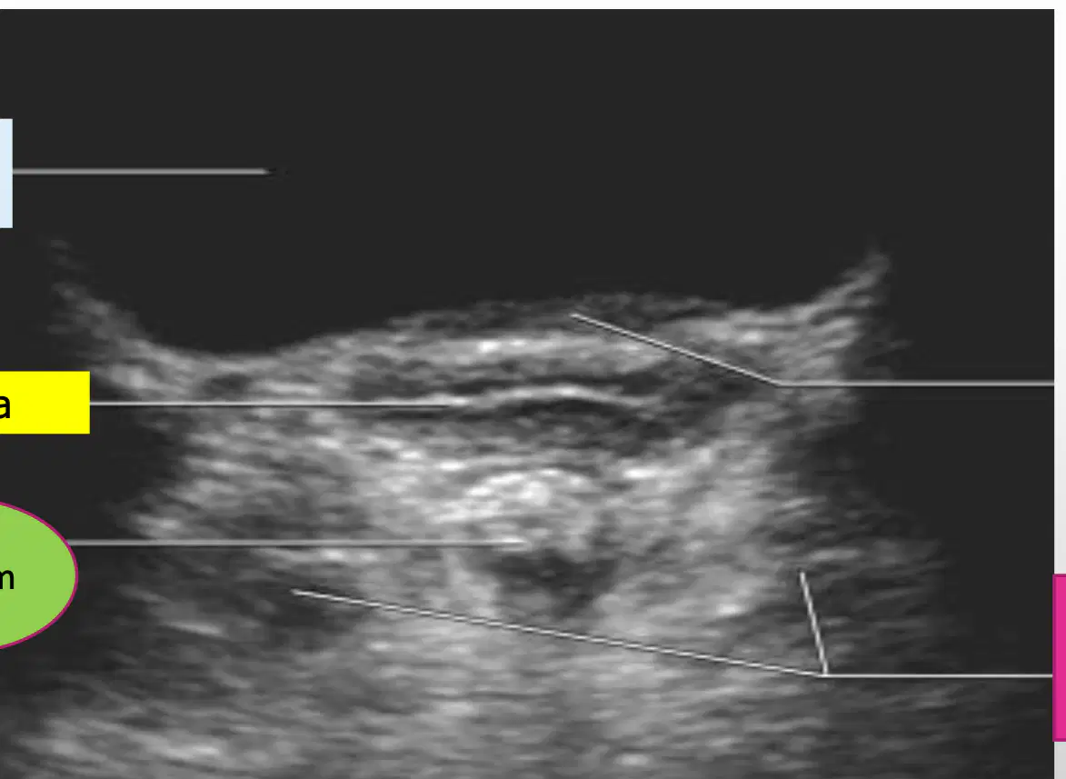
Pink
(a)
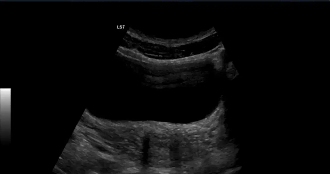
This image shows a trans (a)
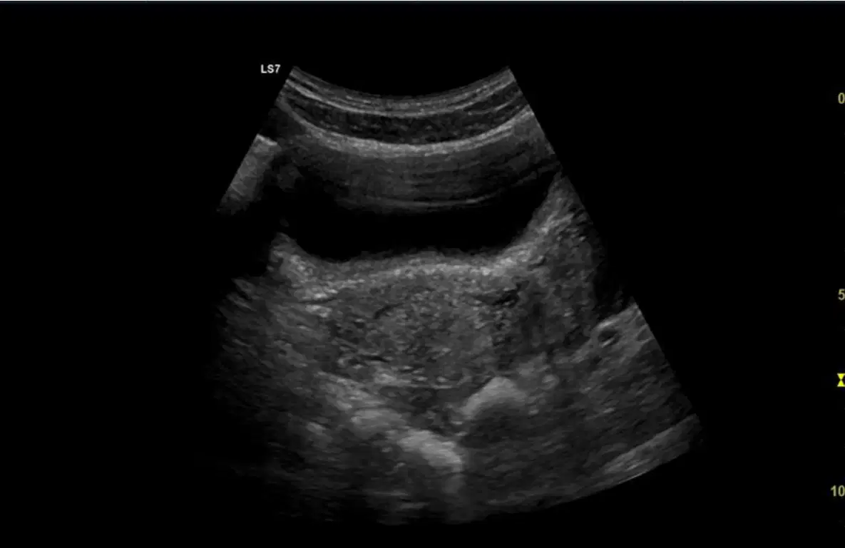
This shows a trans (a)

This shows a trans __ __
(a)

This shows a trans (a) of the uterus

This shows a trans (a) measurement of uterus mid

This shows a (a) view of the uterus mid
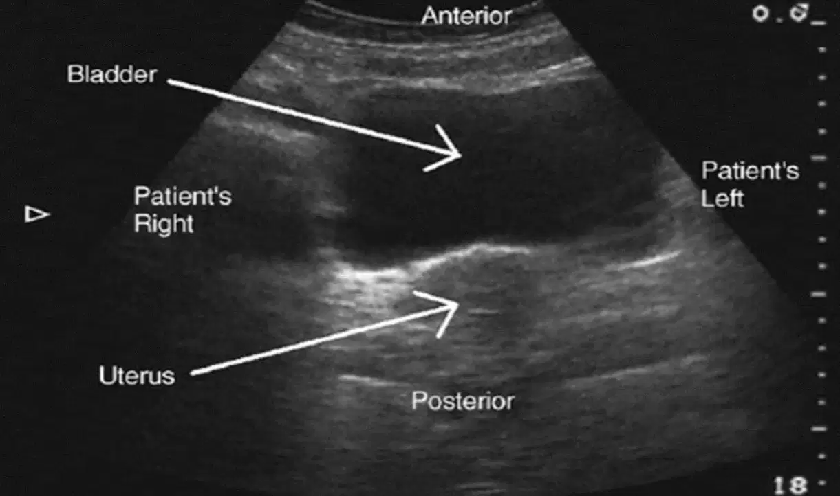
This shows the (a)

