
Font size
 Worksheets
Lesson 2 (BSC208L)
Total questions: 96
Worksheet time: 48hrs 0mins
What is the functional unit of a living organism?
(a)
A group of cells with similar structure and function is called a (a) .
What is formed by the organization of tissues?
(a)
An organ system is formed by a group of related organs performing a common function.
True
False
Why is understanding tissue histology important for understanding organ physiology?
(a)
Fill in the blank: One of the characteristics of epithelial tissue is (a) & specialized contacts (membranes/special junctions).
Fill in the blank: Epithelial tissue shows polarity, which means it has an apical (top) or (a) (bottom) surface.
Fill in the blank: Epithelial tissue is supported by (a) tissue (attached to basement membrane).
Fill in the blank: Epithelial tissue is characterized by avascularity, meaning it has no (a) (nutrients need to diffuse to the epithelial tissue).
Fill in the blank: Epithelial tissue has the ability for (a) , which is the replacement of cells lost due to friction.
According to the 'Classification of Epithelia', what are the two criteria used to classify epithelia?
A) Arrangement (number of layers) and cell shape
B) Color and size
C) Function and location
D) Age and origin
Fill in the blank: Epithelia can be classified based on the number of layers, such as (a) (1 layer) or stratified (multiple layers).
Which of the following is NOT a cell shape mentioned in the 'Classification of Epithelia'?
Squamous (flat)
Cuboidal (cube-shaped)
Columnar (column-shaped)
Spherical (round)
Fill in the blank: Glands are formed by (a) cells.

Which type of epithelial cell is flat and thin in appearance?
Squamous
Cuboidal
Columnar

Refer to the diagram labeled 'Classification of epithelia.' Which type of epithelial cell is cube-shaped?
Squamous
Cuboidal
Columnar

Refer to the diagram labeled 'Classification of epithelia.' Which type of epithelial cell is taller than it is wide?
Squamous
Cuboidal
Columnar

Refer to the diagram labeled 'Classification of epithelia.' Fill in the blank: A single layer of epithelial cells is called (a) epithelium.
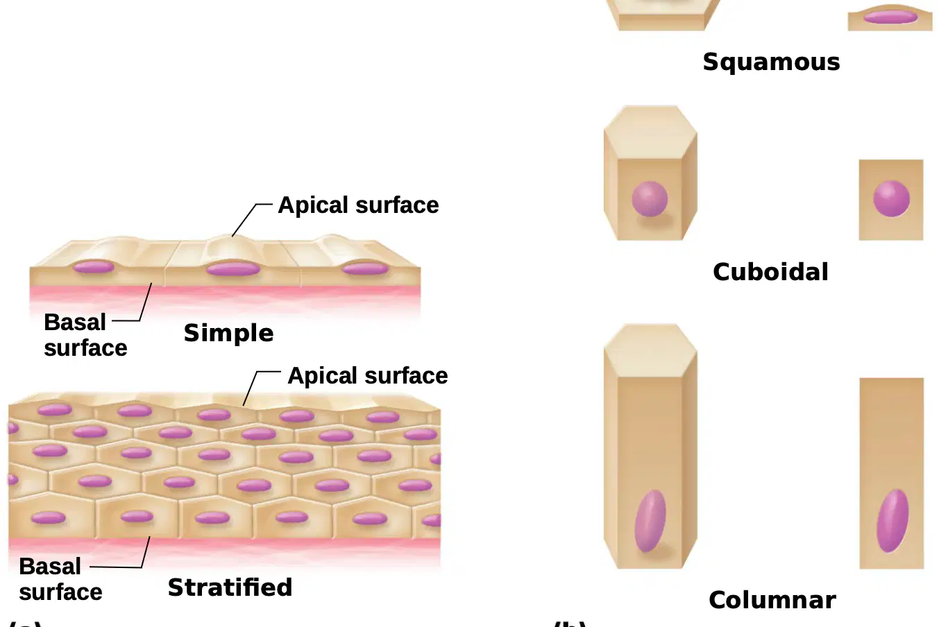
Refer to the diagram labeled 'Classification of epithelia.' Fill in the blank: Multiple layers of epithelial cells are called (a) epithelium.
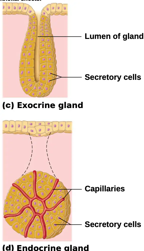
Refer to the diagram labeled (c) Exocrine gland. Which of the following structures is labeled as the 'Lumen of gland'?
The outermost layer
The central cavity
The capillaries
The secretory cells
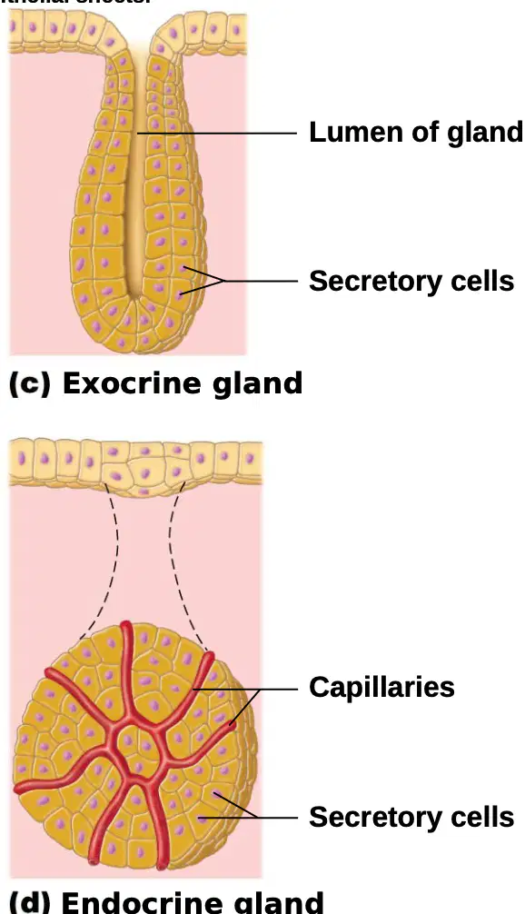
In the diagram labeled (d) Endocrine gland, what is the function of the capillaries?
(a)
Which type of gland releases its secretions into ducts that lead to the surface of an epithelium?
Exocrine gland
Endocrine gland
Fill in the blank: The (a) is the tissue from which both exocrine and endocrine glands are formed, as shown in diagrams (a) and (b).
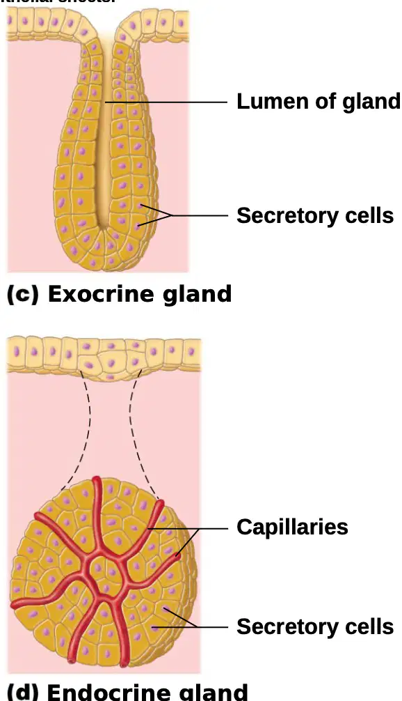
In the diagram labeled (d) Endocrine gland, which cells are responsible for secreting hormones?
(a)
What is the description of simple squamous epithelium?
Single layer of flattened cells with disc-shaped central nuclei and sparse cytoplasm; the simplest of the epithelia.
Multiple layers of cube-shaped cells with dense cytoplasm.
Single layer of columnar cells with elongated nuclei.
Multiple layers of squamous cells with thick cytoplasm.
What is the main function of simple squamous epithelium?
Allows materials to pass by diffusion and filtration in sites where protection is not important; secretes lubricating substances in serosae.
Provides protection against abrasion.
Absorbs nutrients in the intestine.
Produces mucus in respiratory passages.
Where can simple squamous epithelium be found?
Kidney glomeruli; air sacs of lungs; lining of heart, blood vessels, and lymphatic vessels; lining of ventral body cavity (serosae).
Skin surface; lining of mouth.
Intestinal lining.
Sweat glands.
Fill in the blank: The nuclei of squamous epithelial cells are (a) shaped and located centrally.
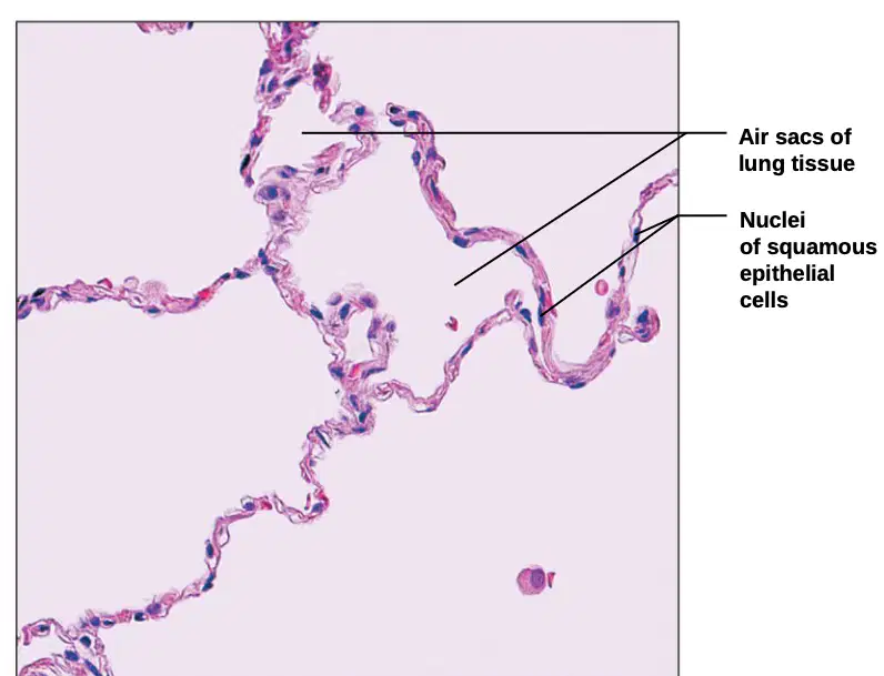
Label the parts shown in the photomicrograph:
(a)
What is the description of simple cuboidal epithelium?
(a)
What is the function of simple cuboidal epithelium?
(a)
Where is simple cuboidal epithelium located?
(a)
What is the description of simple columnar epithelium?
Single layer of flat cells with round nuclei
Single layer of tall cells with round to oval nuclei; some cells bear cilia; layer may contain mucus-secreting unicellular glands (goblet cells).
Multiple layers of cube-shaped cells
Single layer of squamous cells
What is the main function of simple columnar epithelium?
Protection only
Absorption; secretion of mucus, enzymes, and other substances; ciliated type propels mucus (or reproductive cells) by ciliary action.
Filtration
Sensory reception
Where is nonciliated simple columnar epithelium most commonly found?
Skin
Digestive tract (stomach to rectum), gallbladder, and excretory ducts of some glands
Lungs
Heart
Fill in the blank: The layer of simple columnar epithelium may contain mucus-secreting unicellular glands called (a) .
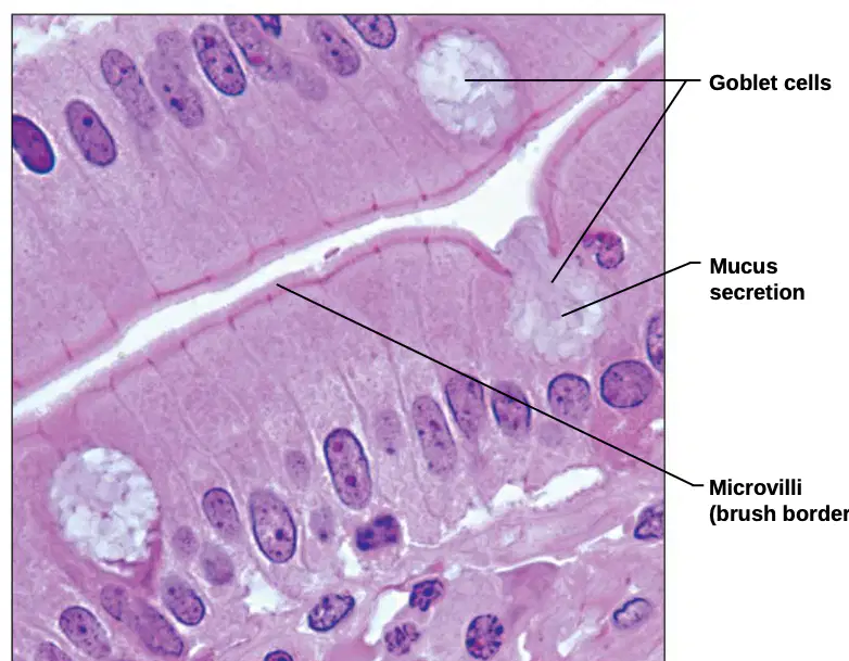
Label the structure indicated in the photomicrograph: The cells responsible for mucus secretion in the simple columnar epithelium are called (a)
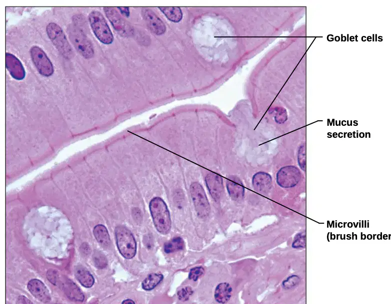
Which structure in the photomicrograph is responsible for increasing surface area for absorption?
Goblet cells
Microvilli (brush border)
Mucus secretion
Cilia
The pseudostratified columnar epithelium may contain mucus-secreting _______ cells and bear _______.
(a)
What is the main function of pseudostratified columnar epithelium?
(a)
Where is the ciliated variety of pseudostratified columnar epithelium commonly found?
(a)
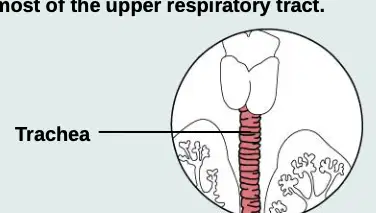
The pseudostratified ciliated columnar epithelium lines the human trachea.
True
False
What is the main function of stratified squamous epithelium?
Absorption
Protection of underlying tissues in areas subjected to abrasion
Secretion
Filtration
Where is the nonkeratinized type of stratified squamous epithelium found?
Lining of the esophagus, mouth, and vagina
Epidermis of the skin
Lining of the stomach
Lining of the intestines
Fill in the blank: The keratinized variety of stratified squamous epithelium forms the (a) of the skin, a dry membrane.
Which of the following best describes the surface cells of stratified squamous epithelium?
Cuboidal
Columnar
Flattened (squamous)
Spherical

Label the following parts in the provided photomicrograph:
(a)
Fill in the blank: The stratified cuboidal epithelium generally consists of two layers of (a) cells.
Fill in the blank: The main function of stratified cuboidal epithelium is (a) .
Fill in the blank: Stratified cuboidal epithelium is found in the largest ducts of sweat glands, (a) glands, and salivary glands.
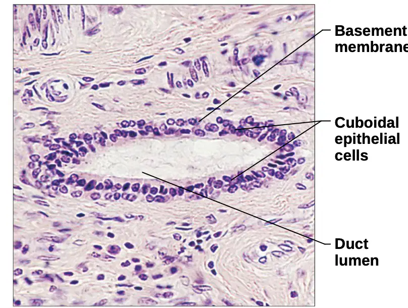
Which of the following is NOT a labeled part of the stratified cuboidal epithelium photomicrograph?
A) Basement membrane
B) Cuboidal epithelial cells
C) Duct lumen
D) Blood vessel
What is the main function of stratified columnar epithelium?
Absorption
Protection and secretion
Filtration
Sensation
Where is stratified columnar epithelium most likely to be found in the human body?
Skin
Lining of the male urethra and large ducts of some glands
Lungs
Stomach lining
Fill in the blank: Stratified columnar epithelium consists of several cell layers; basal cells are usually (a) , and superficial cells are elongated and columnar.
Stratified columnar epithelium is common throughout the human body.
True
False
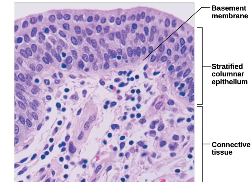
Label the following parts in the photomicrograph:
(a)
Which type of epithelium is being described?
Simple squamous epithelium
Transitional epithelium
Stratified columnar epithelium
Pseudostratified epithelium
The main function of transitional epithelium is to (a) readily and permit distension of urinary organ by contained urine.
Location: Transitional epithelium lines the ureters, urinary bladder, and part of the urethra. Which of the following organs is NOT lined by transitional epithelium?
Ureters
Urinary bladder
Part of the urethra
Esophagus
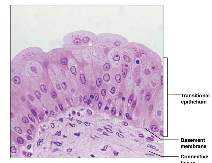
The surface cells of transitional epithelium flatten and become elongated when the bladder is filled with urine.
True
False
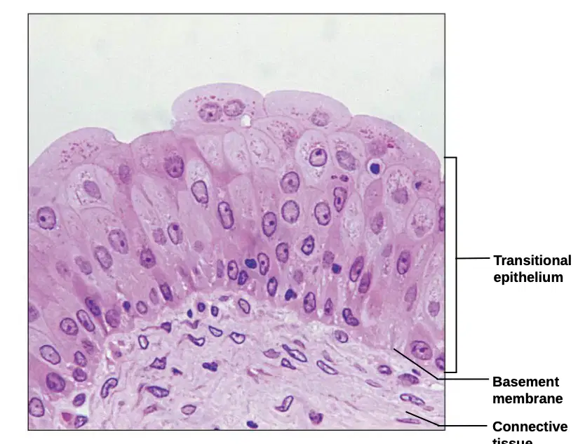
Label the following parts in the provided diagram:
(a)
Connective tissue usually has a rich blood supply.
True
False
Which of the following is NOT a characteristic of connective tissue?
A) Usually has rich blood supply
B) Composed of many cell types
C) Has nonliving material (extracellular matrix) between cells
D) Composed of a single cell type
Fill in the blank: Connective tissue is composed of (a) cell types (unlike epithelia).
What is the nonliving material found between the cells of connective tissue called?
(a)
The matrix of connective tissue is produced by cells and is composed of which two main components?
Ground substance and fibers
Blood and plasma
Epithelia and cartilage
Collagen and bone
Which of the following are types of fibers found in connective tissue?
Collagen, elastic, and reticular fibers
Muscle, nerve, and bone fibers
Plasma, cartilage, and adipose fibers
Epithelial, glandular, and nervous fibers

Identify the cell type labeled as 'Macrophage' in the diagram of areolar connective tissue.
(a)

Identify the cell type labeled as 'Fibroblast' in the diagram of areolar connective tissue.
(a)

Identify the cell type labeled as 'Lymphocyte' in the diagram of areolar connective tissue.
(a)

Identify the cell type labeled as 'Fat cell' in the diagram of areolar connective tissue.
(a)

Identify the cell type labeled as 'Mast cell' in the diagram of areolar connective tissue.
(a)

Identify the cell type labeled as 'Neutrophil' in the diagram of areolar connective tissue.
(a)
Which of the following is NOT a type of fiber found in the extracellular matrix of areolar connective tissue?
Collagen fiber
Elastic fiber
Reticular fiber
Muscle fiber
Which component of the extracellular matrix is responsible for providing a medium for the exchange of nutrients and waste products?
Collagen fiber
Ground substance
Elastic fiber
Reticular fiber
The extracellular matrix of areolar connective tissue contains collagen, elastic, and reticular fibers.
True
False
Fill in the blank: One of the functions of connective tissue is to (a) tissues/organs.
Fill in the blank: Connective tissue functions to support (a) .
Fill in the blank: Connective tissue functions to bind together other (a) .
Which type of connective tissue cushions and protects the body?
Loose connective tissue, areolar
Loose connective tissue, adipose
Loose connective tissue, reticular
Dense connective tissue, elastic
Which type of connective tissue insulates the body and stores food?
Loose connective tissue, areolar
Loose connective tissue, adipose
Loose connective tissue, reticular
Dense connective tissue, elastic
Which type of connective tissue contains fine collagen fibers?
Loose connective tissue, areolar
Loose connective tissue, adipose
Loose connective tissue, reticular
Dense connective tissue, elastic
Which type of connective tissue is found in bronchial tubes and large arteries?
Loose connective tissue, areolar
Loose connective tissue, adipose
Loose connective tissue, reticular
Dense connective tissue, elastic
Dense connective tissue is found in (a) and tendons.
Which type of dense connective tissue is found in tendons and ligaments?
Regular
Irregular
Which type of dense connective tissue is found in the dermis of skin and submucosa of the digestive tract?
Regular
Irregular
What is the description of embryonic connective tissue: mesenchyme?
(a)
What is the function of embryonic connective tissue: mesenchyme?
(a)
Where is embryonic connective tissue: mesenchyme primarily located?
(a)
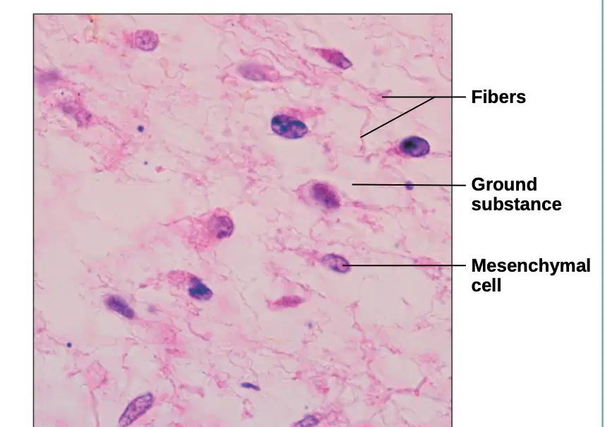
In the photomicrograph of mesenchymal tissue, which label corresponds to the clear-appearing background?
Fibers
Ground substance
Mesenchymal cell
Which of the following is NOT a feature of embryonic connective tissue: mesenchyme?
Star-shaped mesenchymal cells
Gel-like ground substance
Primarily found in adult tissues
Contains fibers
Description: (a) matrix with all three fiber types; cells: fibroblasts, macrophages, mast cells, and some white blood cells. (Fill in the blank)
What is one of the main functions of areolar connective tissue?
Stores fat
Wraps and cushions organs
Transports oxygen
Produces hormones
Location: Areolar connective tissue is widely distributed under (a) of the body, e.g., forms lamina propria of mucous membranes; packages organs; surrounds capillaries. (Fill in the blank)
Which of the following cells are found in areolar connective tissue?
Fibroblasts
Macrophages
Mast cells
All of the above
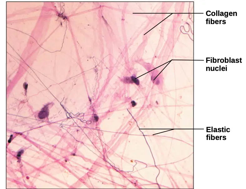
Which fibers are labeled in the photomicrograph of areolar connective tissue?
Collagen fibers
Elastic fibers
Reticular fibers
Both A and B

