
 Worksheets
Blood
Total questions: 100
Worksheet time: 50mins
Observe the prepared microscope slide showing human blood. Based on its appearance and structure, blood is considered a type of ________ tissue.
connective
epithelial
muscular
nervous
Observe the provided blood smear slide under the microscope. Which of the following are the formed elements you can identify in the blood sample?
erythrocytes (red blood cells), leukocytes (white blood cells), and platelets
plasma, serum, and water
hormones, enzymes, and vitamins
glucose, amino acids, and fatty acids
Observe the two test tubes: one contains water and the other contains blood. Based on their appearance and flow, blood is heavier and ______ times thicker than water.
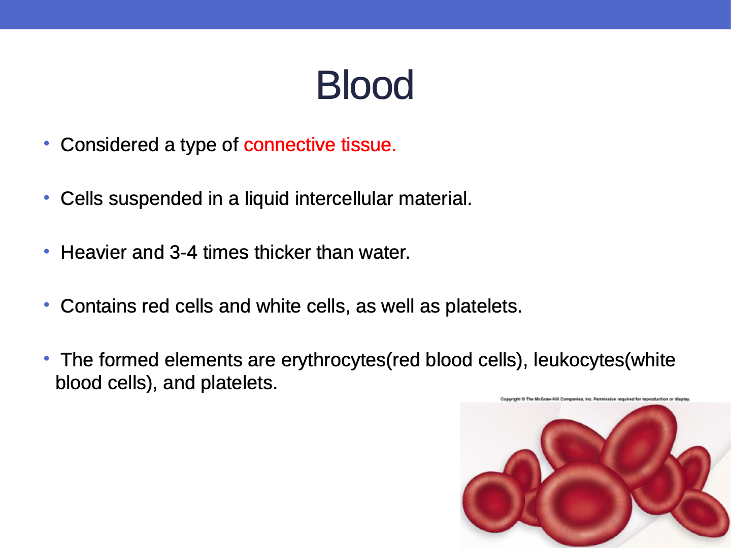
3-4
1-2
5-6
7-8
Observe the provided microscope slides of blood and nerve tissue. Which of the following is NOT visible as a formed element in the blood smear slide?
Erythrocytes
Leukocytes
Platelets
Neurons
Observe the given blood smear image under a microscope. Identify which of the following components you can see: red cells, white cells, and platelets.
All three: red cells, white cells, and platelets
Only red cells and white cells
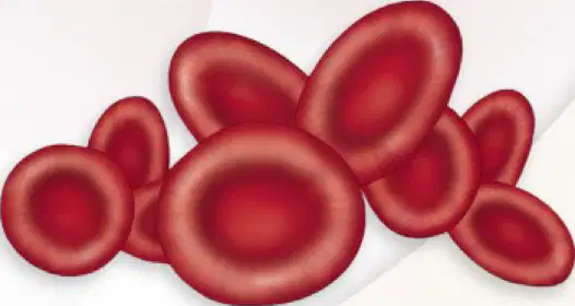
Examine the provided diagram of blood cells. Based on your observation, identify the most abundant type of cell visible in the image by selecting the correct option below.
Red blood cells
White blood cells
Platelets
Plasma cells
Observe the image of a centrifuged blood sample. What does the Hematocrit (HCT) value represent in this visual context?
The percentage of plasma in blood
The percentage of cells by volume in blood, also called Packed Cell Volume (PCV)
The amount of water in blood
The number of white blood cells
Observe the image of a centrifuged blood sample. Based on the visual separation, estimate what percentage of the blood volume is made up of cells (PCV)?
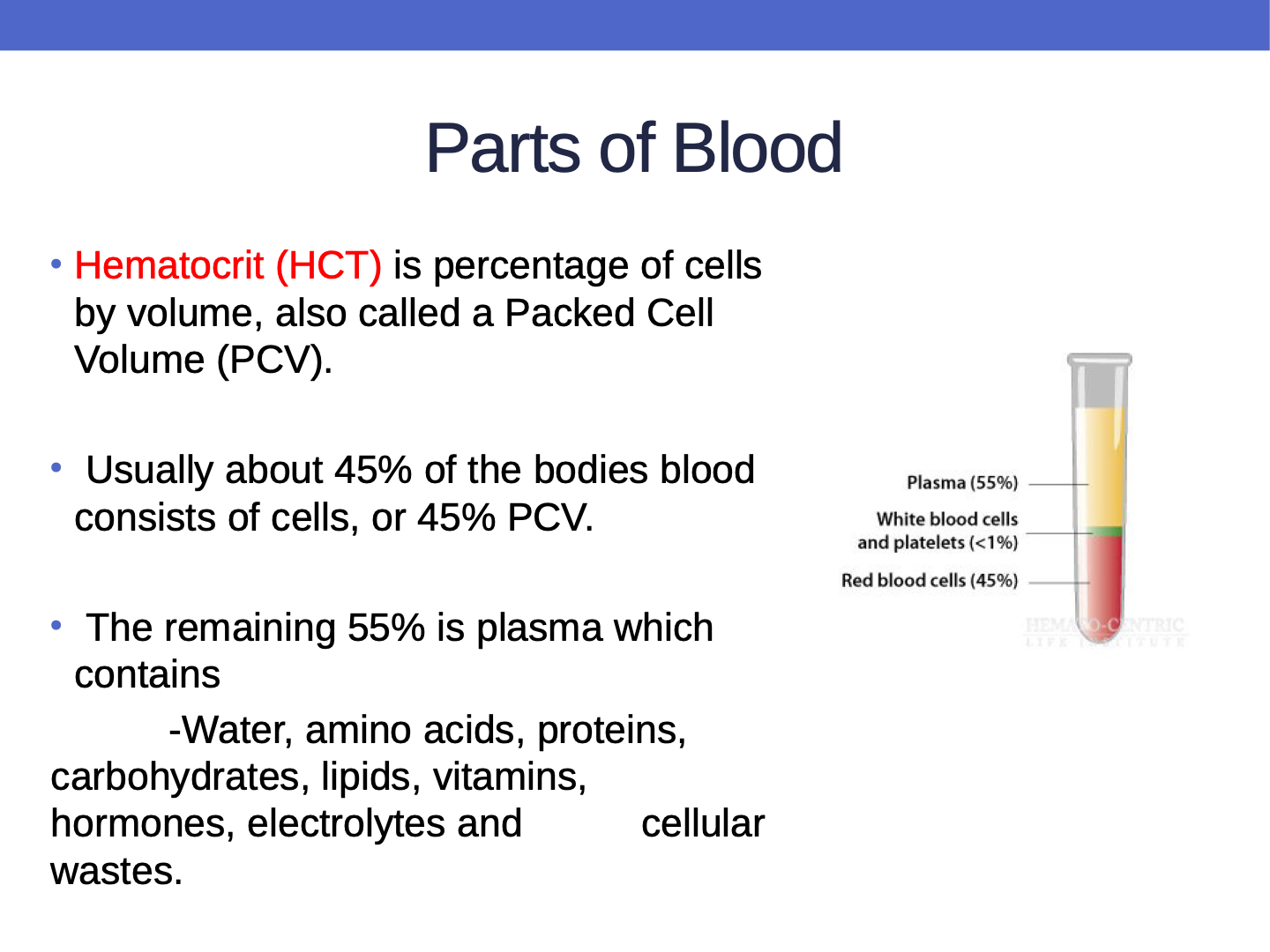
45
25
60
10
Observe the given image of a test tube containing separated blood components. Based on the visual, what percentage of the blood is plasma (the clear, upper layer)? Fill in the blank.
55
25
70
40
Observe the given image of a test tube containing blood after centrifugation. Which of the following components can be visually identified in the plasma layer? Select all that apply.
Proteins
Amino acids
Carbohydrates
All of the above
Water
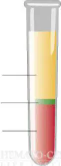
Observe the diagram of the centrifuged blood sample. Using a ruler, measure the height of the red blood cell layer and the total height of the blood sample in the test tube. Calculate and select the percentage of blood made up of red blood cells.
10%
25%
45%
55%
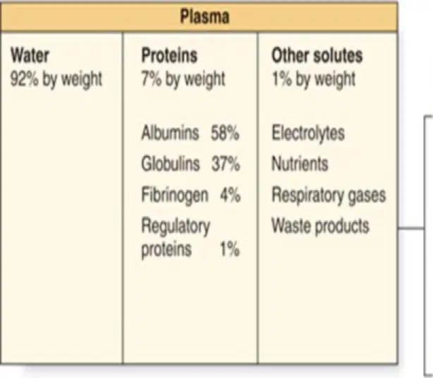
Refer to the diagram showing the composition of plasma. Based on the visual information, what percentage of plasma is made up of water by weight?
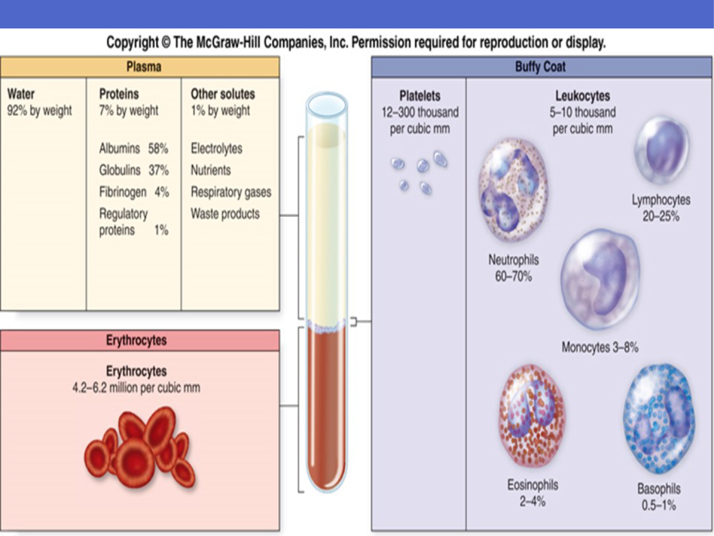
92%
75%
60%
80%
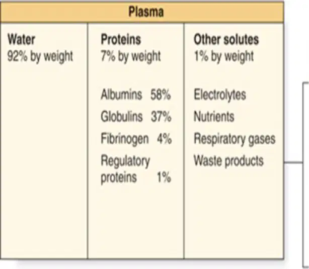
Examine the chart showing the composition of plasma proteins. Based on the visual data, which protein makes up the largest percentage of plasma proteins?
Globulins
Fibrinogen
Albumins
Regulatory proteins
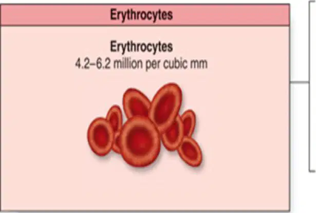
Observe the given image showing erythrocytes and their normal count in blood. Based on the visual, what is the normal range of erythrocytes per cubic mm in blood?
4.2–6.2 million per cubic mm
1.0–2.0 million per cubic mm
7.0–9.0 million per cubic mm
2.5–3.5 million per cubic mm
Match the following leukocytes with their typical percentage in blood:
60–70%
Neutrophils
20–25%
Lymphocytes
3–8%
Monocytes
2–4%
Eosinophils
0.5–1%
Basophils
Observe the provided blood smear slide under the microscope. Based on your observation, estimate the typical range of platelets present per cubic mm of blood.
12–300 thousand per cubic mm
1–5 thousand per cubic mm
500–1000 thousand per cubic mm
300–600 thousand per cubic mm
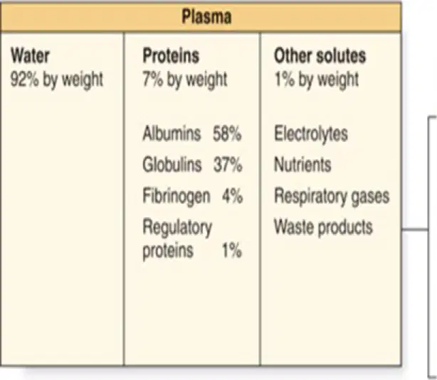
Observe the chart showing the components of plasma. Based on your observation, which of the following is NOT listed as a plasma protein in the chart?
A) Albumins
B) Globulins
C) Fibrinogen
D) Platelets
Observe the provided blood smear image. Identify whether the basophils present make up 20–25% of the leukocytes in this sample.
True
False
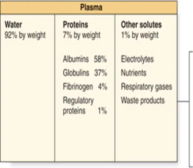
Refer to the diagram of plasma composition provided. If you were to test a plasma sample in the lab, which component under 'Other solutes' would you expect to provide nutrients to cells?
Electrolytes
Nutrients
Respiratory gases
Waste products
Observe the blood smear slide under the microscope. Based on the appearance of the cells labeled as erythrocytes (red blood cells), what is their main function?
Transport nutrients
Carry oxygen from the lungs to body cells and transport carbon dioxide back to the lungs
Secrete hormones
Remove waste products
Observe the labeled diagram of a blood vessel and the GI tract. Which substance, shown moving from the GI tract into the blood plasma, is being transported?
nutrients
hormones
waste products
enzymes
Observe the labeled components in the provided blood sample image. Which of the following is NOT a function of the plasma (the clear liquid part)?
Transporting nutrients
Secreting hormones
Carrying waste products
Producing oxygen
Observe the diagram showing the human circulatory system. Identify the organ labeled as responsible for removing waste products from the blood plasma.
kidneys
lungs
heart
liver
Observe the given blood sample under a microscope. Identify which component (as labeled in the diagram) is responsible for transporting hormones secreted by endocrine organs to their target cells.
Erythrocytes
Blood plasma
Platelets
White blood cells
Observe the thermometer placed next to a beaker of blood in the lab setup. Fill in the blank: Blood helps regulate _______ temperature, as shown by the thermometer reading.
body
room
water
engine
Observe the provided image of a blood sample under a microscope. Based on your observation, which component absorbs and distributes heat throughout the body?
Plasma
Red blood cells
Platelets
White blood cells
Observe the diagram showing blood vessels in the skin under different conditions. Which of the following is happening to help cool the body?
Blood vessels constrict and retain heat
Blood vessels in the dermis dilate and dissipate excess heat through the integument
Plasma absorbs and distributes oxygen
Blood vessels shunt blood to deeper vessels
Observe the diagram showing blood vessels in the skin under different temperature conditions. Based on the image, fill in the blank: When the body needs to conserve heat, the dermal blood vessels _______ and the warm blood is shunted to deeper blood vessels in the body.
constrict
dilate
expand
evaporate
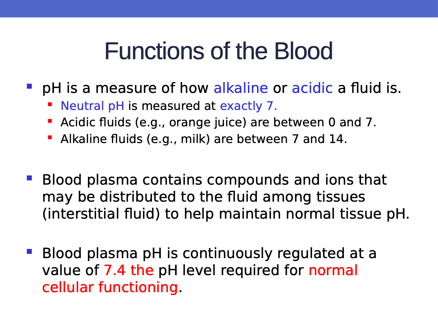
Look at the image of the pH scale provided. What does the pH scale measure in a solution?
pH is a measure of how alkaline or acidic a fluid is.
pH is a measure of temperature of a fluid.
pH is a measure of the density of a fluid.
pH is a measure of the color of a fluid.
Observe the pH scale shown in the image. At what value on the scale is the neutral pH indicated?
Neutral pH is indicated at exactly 7.
Neutral pH is indicated at exactly 5.
Neutral pH is indicated at exactly 9.
Neutral pH is indicated at exactly 3.
Observe the image of different fluids with pH indicators. Which sample (e.g., orange juice) would have a pH between which values, indicating it is acidic?
0 and 7
7 and 14
Exactly 7
1 and 10
Observe the image of a pH scale with various household liquids, including milk. Based on the visual information, between which pH values do alkaline fluids (e.g., milk) fall?
0 and 7
7 and 14
Exactly 7
1 and 10
Observe the color of the blood plasma sample in the provided test tube. Based on the color indicator chart, what is the pH value of the plasma, which is required for normal cellular functioning?
7.4
6.8
8.2
5.5
Observe the diagram of the human body showing different organ systems. Identify which system's fluid levels are maintained by blood and fill in the blank: Blood maintains normal fluid levels in the ________ system.
cardiovascular
digestive
respiratory
nervous
Observe the image of a blood sample with a cut. Based on what you see, blood prevents ________ loss at the site of injury.
fluid
heat
oxygen
protein
Observe the provided microscope slide showing a capillary and surrounding tissues. Based on the visual, fill in the blank: There is a constant exchange of fluid between the blood plasma and the ________ fluid.
interstitial
intracellular
cerebrospinal
synovial
Observe the diagram showing blood vessels before and after excess fluid absorption. Based on the changes, what is the likely result?
Low blood pressure
High blood pressure
No change in blood pressure
Observe a demonstration where fluid is drawn from a model blood vessel into surrounding tissue (using colored water and a sponge to represent tissue). Based on what you see, what happens to blood pressure in the vessel?
Blood pressure rises
Blood pressure drops
Blood pressure stays the same
Observe the microscope slide showing white blood cells. Based on their appearance and function, what do leukocytes help guard against by mounting an immune response if a pathogen or an antigen is found?
infection
digestion
respiration
excretion
Observe the provided microscope slide showing plasma and antigens. Identify which molecule, visible in the plasma, is responsible for immobilizing antigens until a leukocyte can remove them.
antibodies
enzymes
hormones
lipids
Observe the image of a blood smear under a microscope. The platelets and blood proteins you see help protect the body against blood loss by forming ______ on damaged vessels.
blood clots
plasma
antibodies
hormones
Examine the provided diagram showing the process of blood cell formation in the bone marrow. Based on the visual information, which of the following cells would you NOT expect to observe developing from a hematopoietic stem cell in a bone marrow slide under the microscope?
Neuron
Erythrocyte
Neutrophil
Platelets
Examine the diagram showing blood cell development in the bone marrow. The blue arrow in the image points to a specific cell type. Based on the diagram, which cell type does this arrow indicate, that gives rise to B cells and T cells?

common lymphoid progenitor
myeloid progenitor
erythroblast
megakaryocyte
According to the diagram, blood cells are made in the bone marrow from stem cells called hemocytoblasts or hematopoietic stem cells. Match the following progenitor cells to the correct cell types they produce:
Platelets
B
T cell
C
Neutrophil
A

Examine the diagram showing the development of blood cells in the bone marrow. Using the arrow highlighted in blue, identify which cell type is directly produced from an erythroblast by selecting the correct option below.
Platelets
Erythrocyte
Neutrophil
Monocyte
Observe the provided diagram showing blood cell development in the bone marrow. Using the diagram, identify which cell type is the direct precursor to plasma cells by pointing to or labeling it on the diagram. Which of the following is the correct precursor?
B cell
T cell
Monocyte
Erythrocyte
Examine the provided blood smear diagram. Using a marker or pointer, identify and indicate the cell that is a 'Monocyte'.
I have correctly identified the cell in the top left corner as the 'Monocyte'.
I have correctly identified the cell in the bottom right corner as the 'Monocyte'.
I have correctly identified the cell in the center as the 'Monocyte'.
I have correctly identified the cell in the top right corner as the 'Monocyte'.
Examine the provided image of blood cells. Identify and select the cell that is a 'Lymphocyte' by observing its visual characteristics.
The cell in the top row, second from the left, is a 'Lymphocyte'.
The cell in the bottom row, first from the right, is a 'Lymphocyte'.
The cell in the top row, first from the left, is a 'Lymphocyte'.
The cell in the bottom row, second from the left, is a 'Lymphocyte'.
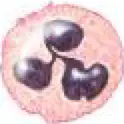
Examine the provided diagram of blood cells. Using a microscope or the image, identify which cell is a 'Neutrophil' by selecting the correct location.
The cell in the top row, third from the left, is a 'Neutrophil'.
The cell in the bottom row, first from the right, is a 'Neutrophil'.
The cell in the top row, first from the left, is a 'Neutrophil'.
The cell in the bottom row, second from the left, is a 'Neutrophil'.
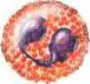
Examine the provided blood cell diagram under the microscope. Using the diagram, identify and point to the cell that is an 'Eosinophil'.
The cell in the top row, fourth from the left, is an 'Eosinophil'.
The cell in the bottom row, first from the right, is an 'Eosinophil'.
The cell in the top row, second from the left, is an 'Eosinophil'.
The cell in the bottom row, third from the left, is an 'Eosinophil'.
Examine the provided diagram of blood cells carefully. Using a marker or pointer, identify and point to the cell labeled as 'Basophil' on the diagram.
The cell in the top row, fifth from the left, is labeled as 'Basophil'.
The cell in the bottom row, second from the right, is labeled as 'Basophil'.
The cell in the top row, first from the left, is labeled as 'Basophil'.
The cell in the bottom row, third from the left, is labeled as 'Basophil'.
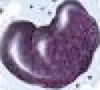
Examine the provided microscope image of a blood cell. Based on its shape and appearance, identify which type of blood cell this is.
Macrophage
Neutrophil
Eosinophil
Lymphocyte
Look at the diagram of blood cells. Which cell is labeled as 'Erythrocyte'?
The cell in the bottom row, second from the left, is labeled as 'Erythrocyte'.
The cell in the top row, first from the right, is labeled as 'Erythrocyte'.
The cell in the middle row, third from the left, is labeled as 'Erythrocyte'.
The cell in the bottom row, last on the right, is labeled as 'Erythrocyte'.
Look at the diagram of blood cells. Which cell is labeled as 'Platelets'?
The cell in the bottom row, third from the left, is labeled as 'Platelets'.
The cell in the top row, first from the right, is labeled as 'Platelets'.
The cell in the middle row, second from the left, is labeled as 'Platelets'.
The cell in the top row, third from the left, is labeled as 'Platelets'.
Which of the following is true about mature erythrocytes?
They have nuclei
They lack nuclei
They can reproduce
They contain mitochondria
Mature erythrocytes lack the ability to ______ or repair themselves.
reproduce
move
digest
divide
What do mature erythrocytes lack that makes them unable to make ATP from the mitochondria?
Mature erythrocytes lack mitochondria.
Mature erythrocytes lack ribosomes.
Mature erythrocytes lack a nucleus.
Mature erythrocytes lack hemoglobin.
Damaged erythrocytes are sent to the ______ or spleen for removal from the bloodstream.
liver
kidney
pancreas
lung
How long do mature erythrocytes live?
10-20 days
30-40 days
100-120 days
200-220 days
Erythrocytes transport oxygen and carbon dioxide to and from the tissues and the lungs with the use of _______.
hemoglobin
insulin
myosin
collagen
Lack of nuclei enables erythrocytes to carry respiratory gases more efficiently.
True
False
Fill in the blank: Every erythrocyte is filled with approximately ______ molecules of a red-pigmented protein called hemoglobin.
280 million
2 million
28 thousand
2.8 billion
What does hemoglobin transport?
A) Oxygen and carbon dioxide
B) Water and salt
C) Glucose and insulin
D) Proteins and fats
Fill in the blank: Hemoglobin is responsible for the characteristic ______ color of arterial blood.
bright red
dark blue
green
yellow
Hemoglobin that contains no oxygen has a deep red color that is perceived as blue through the skin and subcutaneous tissue.
True
False
Which antigen is present on the erythrocytes of Type A blood?
Antigen A
Antigen B
Antigens A and B
Neither antigen A nor B
Which antibodies are found in the plasma of a person with Type B blood?
Anti-A antibodies
Anti-B antibodies
Both anti-A and anti-B antibodies
Neither anti-A nor anti-B antibodies
Type AB blood has which of the following characteristics?
Erythrocytes with both type A and type B surface antigens, and plasma with neither anti-A nor anti-B antibodies
Erythrocytes with type A surface antigens and plasma with anti-B antibodies
Erythrocytes with type B surface antigens and plasma with anti-A antibodies
Erythrocytes with neither type A nor type B surface antigens, but plasma with both anti-A and anti-B antibodies
Fill in the blank: Type O blood has erythrocytes with ________ type A nor type B surface antigens, but plasma with both anti-A and anti-B antibodies.
neither
both
only
either
Match the following blood types with their corresponding plasma antibodies:
Anti-B antibodies
Anti-B antibodies
Anti-A antibodies
Anti-A antibodies
Neither anti-A nor anti-B antibodies
Neither anti-A nor anti-B antibodies
Both anti-A and anti-B antibodies
Both anti-A and anti-B antibodies
Type O blood has both anti-A and anti-B antibodies in its plasma.
True
False
Where are antibodies (agglutinins) to the surface antigens found?
In the plasma
On the red blood cell surface
In the nucleus
In the cytoplasm
Which type of antibodies does blood type A have?
Anti-A
Anti-B
Both anti-A and anti-B
Neither anti-A nor anti-B
Which type of antibodies does blood type B have?
Anti-A
Anti-B
Both anti-A and anti-B
Neither anti-A nor anti-B
Which type of antibodies does blood type AB have?
Anti-A
Anti-B
Both anti-A and anti-B
Neither anti-A nor anti-B
Which type of antibodies does blood type O have?
Anti-A
Anti-B
Both anti-A and anti-B
Neither anti-A nor anti-B
What does Rh stand for?
Rhesus factor
Red blood cells
Rhombus factor
Ribosome factor
The Rh factor is inherited independently of the ABO blood type.
True
False
Fill in the blank: There are two types of Rh factor: ______ and ______.
Rh- and Rh+
A and B
O and AB
Positive and Negative
If a mother is Rh-, there is a risk she will produce Rh+ antibodies. During pregnancy, this could lead to ______ for the fetus.
anemia (lack of healthy blood cells)
increased bone density
improved vision
enhanced muscle growth
What is the function of RhoGAM during pregnancy?
A) Prevents antibody development
B) Increases red blood cells
C) Causes anemia
D) Changes blood type
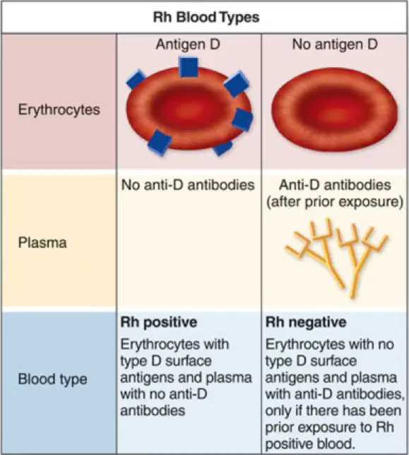
According to the diagram, which blood type has erythrocytes with type D surface antigens and plasma with no anti-D antibodies?
Rh positive
Rh negative
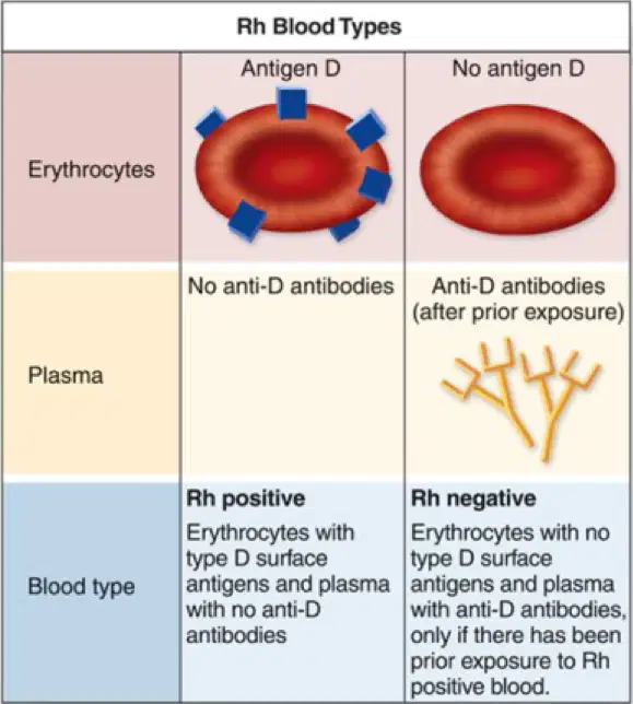
According to the diagram, which blood type has erythrocytes with no type D surface antigens and plasma with anti-D antibodies (after prior exposure)?
Rh positive
Rh negative
Based on the diagram about agglutination, what happens when Type A blood of a donor (has surface antigen A) is given to a recipient with Type A blood (contains anti-B antibodies)?
Agglutination occurs
No agglutination occurs
Hemolysis occurs
Clumping is seen
According to the agglutination diagram, what is the result when Type A blood of a donor (has surface antigen A) is given to a recipient with Type B blood (contains anti-A antibodies)?
No agglutination occurs
Agglutination occurs
No clumping is seen
Successful blood type match
Fill in the blank: In a successful blood type match, ________ is not seen and there is no agglutination.
clumping
swelling
dissolving
shrinking
According to the Red Blood Cell Compatibility Table, can a person with O- blood receive red blood cells from an A+ donor?
True
False
According to the Red Blood Cell Compatibility Table, which of the following blood types can donate to an AB+ recipient?
O-
A+
B+
All of the above
A person with B- blood can receive red blood cells from (a) .
O-
B-
AB+
Both A and B
According to the Red Blood Cell Compatibility Table, can a person with AB- blood receive red blood cells from an O+ donor?
True
False
According to the Red Blood Cell Compatibility Table, which blood type is known as the universal donor?
O-
A+
B+
AB+
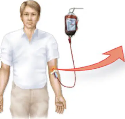
Refer to the diagram showing a blood transfusion from a type A donor to a type B recipient. What happens when type A blood is given to a type B recipient?
The blood is accepted without any reaction
The recipient's anti-A antibodies attack the donor erythrocytes
The donor's blood attacks the recipient's cells
No antibodies are involved
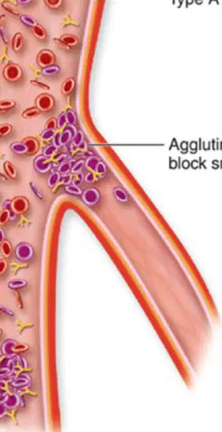
In the diagram, what is the result of agglutinated erythrocytes in the blood vessels?
They help transport oxygen
They block small vessels
They increase blood flow
They produce more antibodies
Fill in the blank: In the diagram, the type B blood recipient has ______ antibodies in plasma.
anti-A
anti-B
anti-O
no
What is the main function of leukocytes? (Fill in the blank)
Protect against disease.
Transport oxygen.
Clot blood.
Store energy.
How are leukocytes transported to sites of infection? (Fill in the blank)
In the blood.
By nerve impulses.
Through bone marrow only.
Via lymphatic vessels only.
How many types of leukocytes can be found in the blood?
Five.
Three.
Six.
Four.
Leukocytes are distinguished by which of the following?
Size, cytoplasm, and shape of nucleus
Color only
Location in the body
Function only
Leukocytes with granules in the cytoplasm are called _________.
Granulocytes.
Agranulocytes.
Erythrocytes.
Thrombocytes.
Leukocytes with non-granular cytoplasm are called _________.
Agranulocytes
Granulocytes
Erythrocytes
Thrombocytes

