
Font size
 Worksheets
Anatomy of the Heart
Total questions: 102
Worksheet time: 51mins
In which cavity is the heart located?
Abdominal cavity
Thoracic cavity
Pelvic cavity
Cranial cavity
The heart rests on the ________.
diaphragm
liver
stomach
lungs
The heart is bordered laterally by the ________.
lungs
liver
stomach
diaphragm
Approximately how much does the heart weigh?
100 to 200 grams
250 to 350 grams
400 to 500 grams
600 to 700 grams
The heart is located left of the body midline posterior to the sternum in the ________ mediastinum.
middle
anterior
superior
posterior
The right side or border of the heart (right atrium and ventricle) is located more ________, while its left side or border (left atrium and ventricle) is located more ________.
anteriorly, posteriorly
posteriorly, anteriorly
superiorly, inferiorly
inferiorly, superiorly
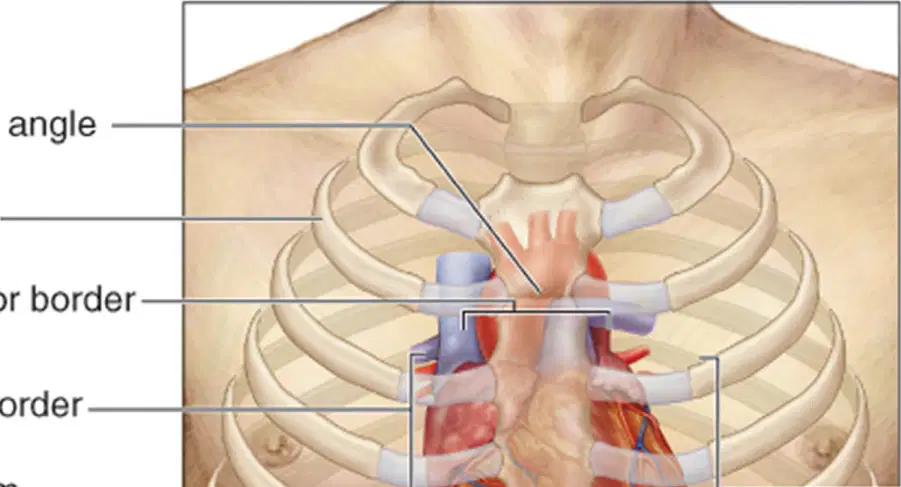
Identify the anatomical structure labeled as 'Sternal angle' in the anterior view of the thorax diagram.
Sternal angle
Xiphoid process
Manubrium
Clavicle
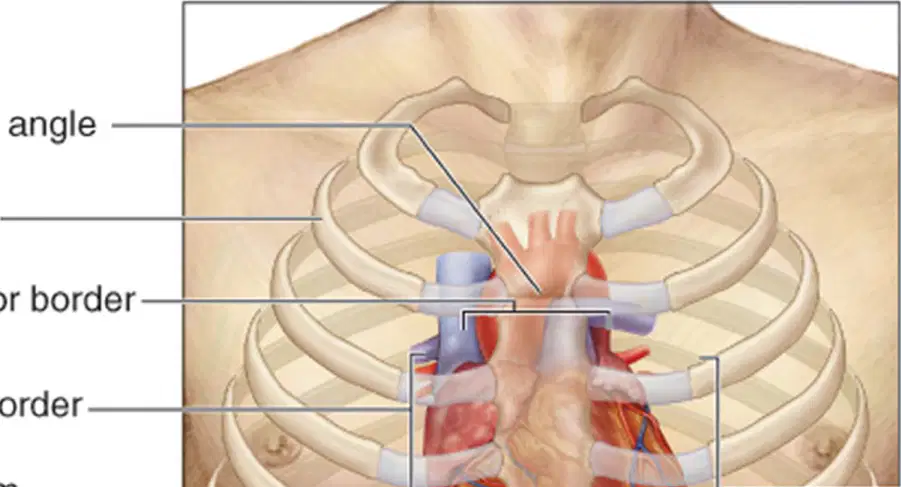
Identify the anatomical structure labeled as '2nd rib' in the anterior view of the thorax diagram.
2nd rib
Clavicle
Sternum
5th rib
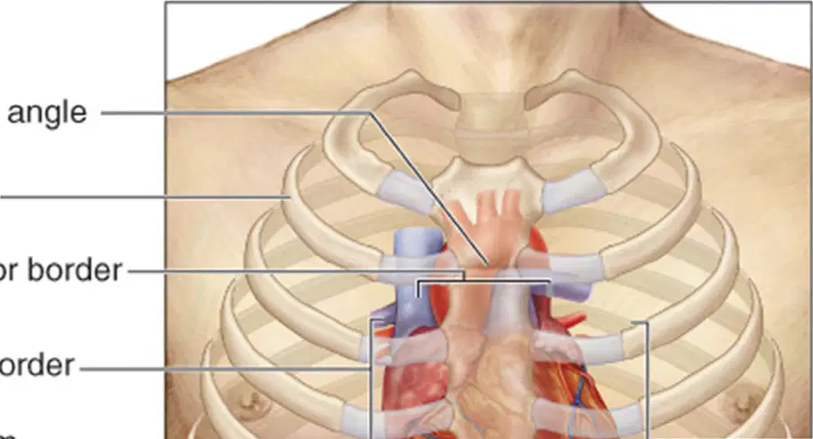
Identify the anatomical structure labeled as 'Superior border' in the anterior view of the thorax diagram.
Superior border
Inferior border
Lateral border
Costal margin
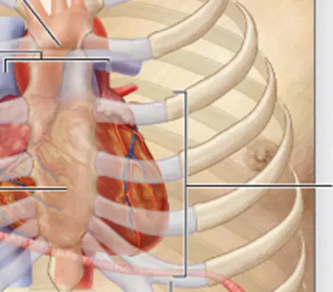
Identify the anatomical structure labeled as 'Right border' in the anterior view of the thorax diagram.
Right border
Left border
Apex
Base
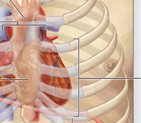
Identify the anatomical structure labeled as 'Sternum' in the anterior view of the thorax diagram.
Sternum
Clavicle
Scapula
Humerus
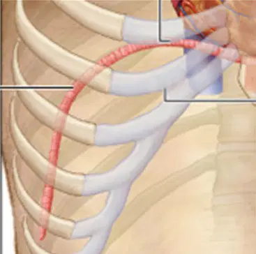
Identify the anatomical structure labeled as 'Diaphragm' in the anterior view of the thorax diagram.
Diaphragm
Trachea
Clavicle
Sternum
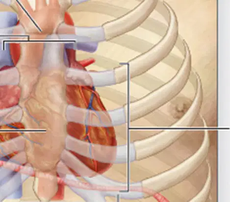
Identify the anatomical structure labeled as 'Left border' in the anterior view of the thorax diagram.
Left border
Right border
Apex
Base
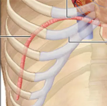
Identify the anatomical structure labeled as 'Inferior border' in the anterior view of the thorax diagram.
Inferior border
Manubrium
Xiphoid process
Costal cartilage
What is the outer portion of the pericardium called?
Serous pericardium
Fibrous pericardium
Parietal layer
Visceral layer
The outer portion of the pericardium is attached to both the ______ and the diaphragm.
sternum
clavicle
scapula
vertebra
What type of tissue makes up the outer portion of the pericardium?
Tough, dense connective tissue
Smooth muscle tissue
Simple squamous epithelium
Loose areolar connective tissue
The inner portion of the pericardium is called the ______ pericardium.
serous
fibrous
parietal
visceral
Which of the following are layers of the serous pericardium?
Parietal layer and fibrous layer
Visceral layer and parietal layer
Fibrous layer and visceral layer
Outer layer and inner layer
What does the outer wall correspond to?
Visceral pericardium
Parietal pericardium
Pericardial cavity
Diaphragm
Which layer of the heart wall is also known as the visceral layer of serous pericardium?
A) Epicardium
B) Myocardium
C) Endocardium
The ________ is the outermost heart layer.
Epicardium
Myocardium
Endocardium
Pericardium
Which heart wall layer is composed chiefly of cardiac muscle tissue?
Epicardium
Myocardium
Endocardium
The thickest of the three heart wall layers is the ________.
Myocardium
Endocardium
Epicardium
Pericardium
Which layer of the heart wall covers the internal surface of the heart and the external surfaces of the heart valves?
Epicardium
Myocardium
Endocardium
The endocardium is made up of ________.
thin endothelium
thick connective tissue
smooth muscle fibers
adipose tissue
The epicardium consists of simple squamous epithelium underlined by fat.
True
False
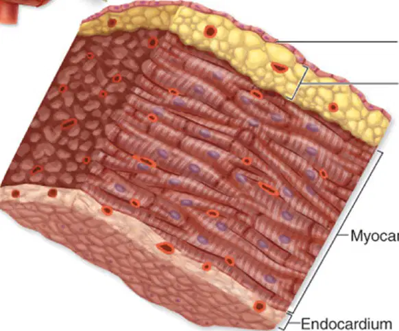
Which layer of the heart wall is composed of cardiac muscle?
Epicardium
Myocardium
Endocardium
Pericardium
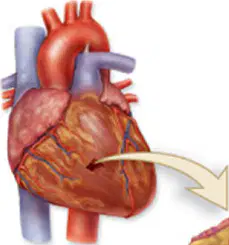
The outermost layer of the heart wall is called the _________.
Epicardium
Myocardium
Endocardium
Pericardium
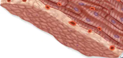
Which layer of the heart wall contains areolar connective tissue and endothelium?
Epicardium
Myocardium
Endocardium
Pericardium
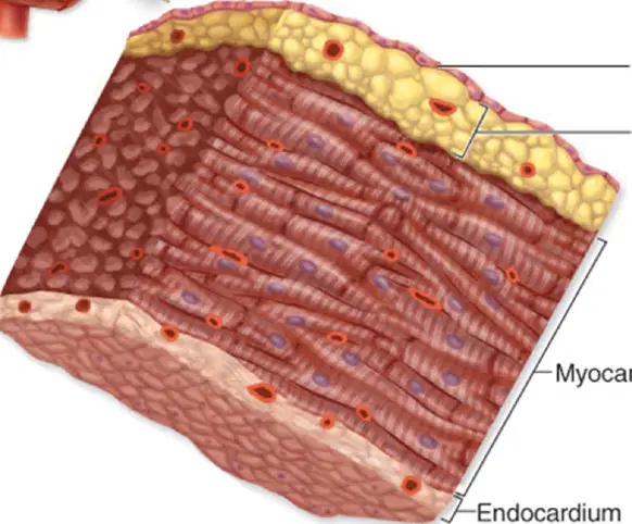
The epicardium is also known as the ________ layer of the serous pericardium.
visceral
parietal
fibrous
myocardial
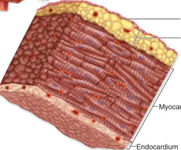
Which of the following is NOT a component of the epicardium?
A) Simple squamous epithelium
B) Areolar connective tissue and fat
C) Cardiac muscle
D) Visceral layer of serous pericardium
Label the part of the heart wall indicated in the diagram: ________
Fibrous pericardium
Myocardium
Endocardium
Epicardium
Label the part of the heart wall indicated in the diagram: ________
Parietal layer of serous pericardium
Epicardium
Myocardium
Endocardium
Label the space between the parietal and visceral layers of the serous pericardium: ________
Pericardial cavity
Pleural cavity
Peritoneal cavity
Mediastinum
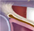
Label the part of the heart wall indicated in the diagram: ________
Visceral layer of serous pericardium (epicardium)
Parietal layer of serous pericardium
Myocardium
Endocardium
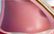
Label the muscular layer of the heart wall indicated in the diagram: ________
Myocardium
Endocardium
Epicardium
Pericardium
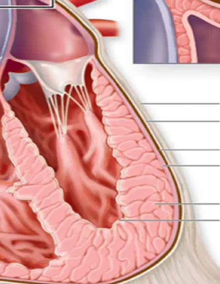
Label the innermost layer of the heart wall indicated in the diagram: ________
Endocardium
Myocardium
Epicardium
Pericardium
How many hollow chambers does the heart have?
Four
Two
Six
Five
Which chambers of the heart are smaller: atria or ventricles?
Atria
Ventricles
Septum
Valves
Are the atria thin-walled and located superiorly?
True
False
Which atrium receives blood from the systemic circuit?
Right atrium
Left atrium
Right ventricle
Left ventricle
Which atrium receives blood from the pulmonary circuit?
Left atrium
Right atrium
Left ventricle
Right ventricle
Blood that enters an atrium is passed to the ______ on the same side of the heart.
ventricle
artery
vein
auricle
The ventricles are the ______ chambers of the heart.
inferior
superior
anterior
posterior
Which two large arteries exit the heart at the basal surface?
Pulmonary trunk and aorta
Pulmonary vein and vena cava
Aorta and carotid artery
Pulmonary trunk and subclavian artery
The pulmonary trunk carries blood from the ______ into the pulmonary circuit.
right ventricle
left atrium
left ventricle
right atrium
The aorta conducts blood from the left ventricle into the ______ circuit.
systemic
pulmonary
coronary
lymphatic
Refer to the diagram (anterior view of the human heart). What is the name of the part labeled as 'Aortic arch'?
Aortic arch
Pulmonary vein
Left atrium
Right ventricle
What is the name of the part labeled as 'Ligamentum arteriosum'?
Ligamentum arteriosum
Ductus arteriosus
Coronary ligament
Falciform ligament
Refer to the diagram (anterior view of the human heart). What is the name of the part labeled as 'Branches of the right pulmonary artery'?
Branches of the right pulmonary artery
Left atrium
Right ventricle
Aorta
What is the name of the part labeled as 'Ascending aorta'?
Ascending aorta
Descending aorta
Pulmonary artery
Left atrium
What is the name of the part labeled as 'Right pulmonary vein'?
Right pulmonary vein
Left atrium
Aorta
Inferior vena cava
What is the name of the part labeled as 'Left pulmonary vein'?
Left pulmonary vein
Right atrium
Aorta
Inferior vena cava
What is the name of the part labeled as 'Pulmonary trunk'?
Pulmonary trunk
Aorta
Superior vena cava
Left atrium
What is the name of the part labeled as 'Auricle of right atrium'?
Auricle of right atrium
Left ventricle
Pulmonary trunk
Interventricular septum
What is the name of the part labeled as 'Auricle of left atrium'?
Auricle of left atrium
Left ventricle
Right atrium
Interventricular septum
What is the name of the part labeled as 'Right atrium'?
Right atrium
Left ventricle
Pulmonary artery
Left atrium
What is the name of the part labeled as 'Left coronary artery'?
Left coronary artery
Right coronary artery
Pulmonary artery
Aorta
What is the name of the part labeled as 'Right coronary artery (in coronary sulcus)'?
Right coronary artery (in coronary sulcus)
Left coronary artery
Pulmonary trunk
Aortic arch
Refer to the diagram (anterior view of the human heart). What is the name of the part labeled as 'Marginal artery'?
Marginal artery
Circumflex artery
Great cardiac vein
Left coronary artery
What is the name of the part labeled as 'Anterior interventricular artery'?
Anterior interventricular artery
Circumflex artery
Right marginal artery
Posterior interventricular artery
What is the name of the part labeled as 'Right ventricle'?
Right ventricle
Left atrium
Pulmonary artery
Aorta
What is the name of the part labeled as 'Left ventricle'?
Left ventricle
Right atrium
Pulmonary artery
Right ventricle
Refer to the diagram (anterior view of the human heart). What is the name of the part labeled as 'Apex of heart'?
Apex of heart
Left atrium
Right ventricle
Pulmonary artery
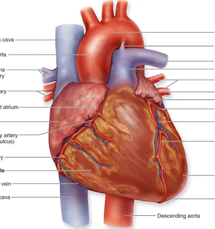
What is the name of the vein labeled at the top left of the heart diagram?
Superior vena cava
Inferior vena cava
Pulmonary vein
Aorta
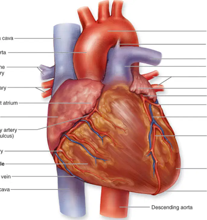
Which chamber of the heart is labeled on the left side of the diagram as 'Right atrium'?
Right atrium
Left ventricle
Left atrium
Right ventricle
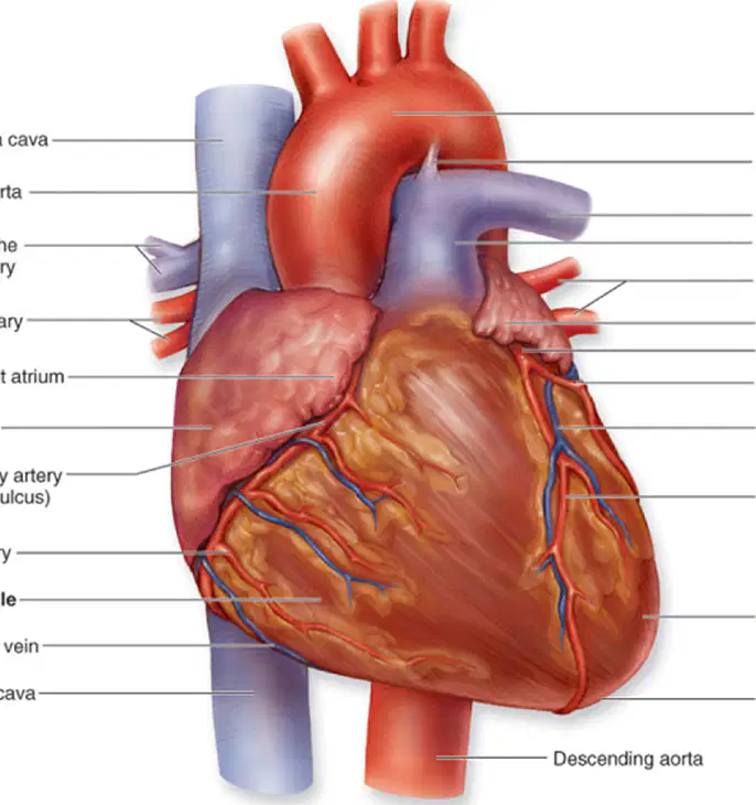
Which chamber of the heart is labeled at the bottom left of the diagram as 'Right ventricle'?
Right ventricle
Left atrium
Left ventricle
Right atrium
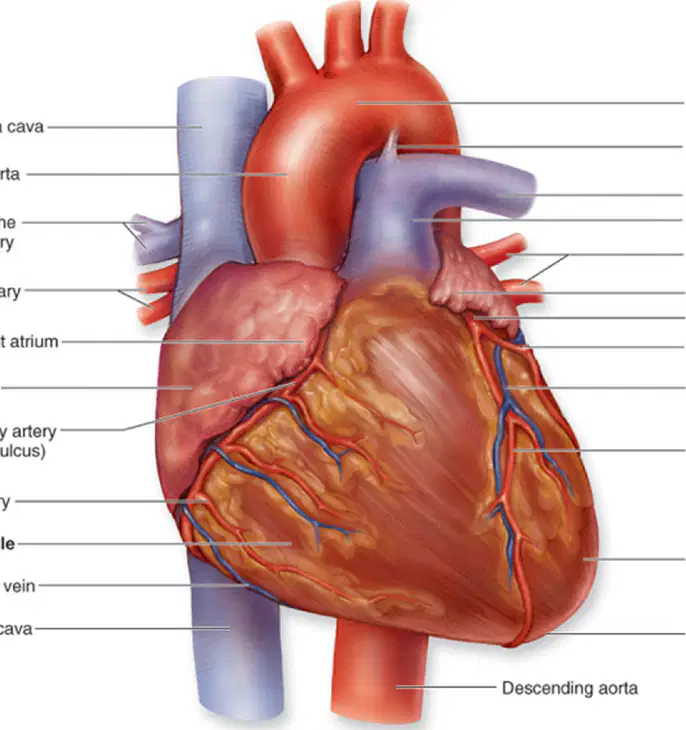
What is the name of the artery labeled at the top right of the heart diagram?
Aortic arch
Pulmonary vein
Inferior vena cava
Right atrium
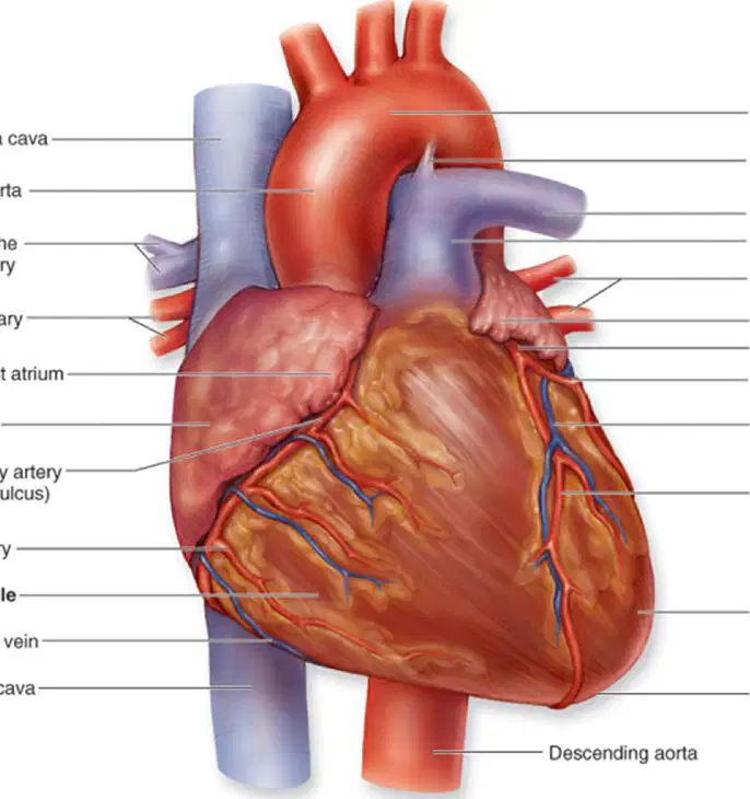
Which chamber of the heart is labeled on the right side of the diagram as 'Left atrium'?
Left atrium
Right atrium
Left ventricle
Right ventricle

Which chamber of the heart is labeled at the bottom right of the diagram as 'Left ventricle'?
Left ventricle
Right atrium
Right ventricle
Left atrium
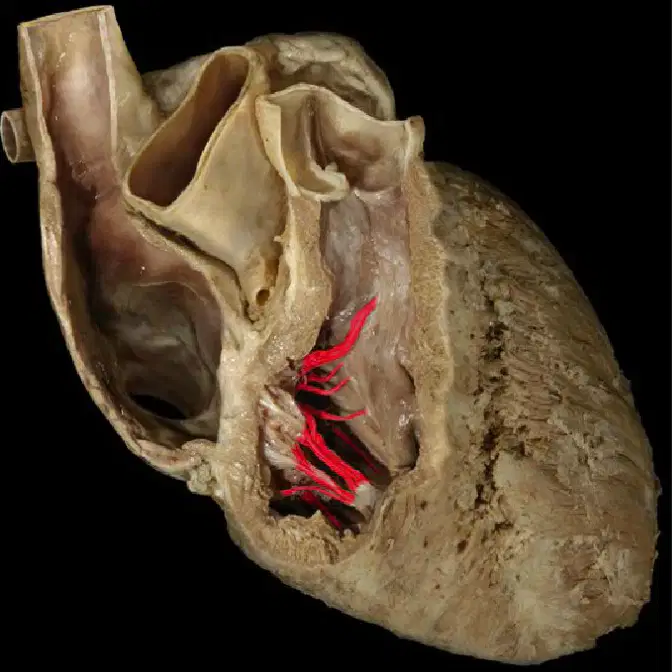
Name the ventricle & the valve shown here. ________ (ventricle), ________ (valve)
Right ventricle, tricuspid valve
Left ventricle, mitral valve
Right atrium, pulmonary valve
Left atrium, aortic valve
The right atrium receives ______ blood from the systemic circuit and from the heart muscle itself.
venous
arterial
oxygenated
nutrient-rich
Which of the following vessels does NOT empty into the right atrium?
Superior vena cava (SVC)
Inferior vena cava (IVC)
Pulmonary vein
Coronary sinus
The superior vena cava (SVC) drains blood from which regions?
Lower limbs and trunk
Head, upper limbs, and superior regions of the trunk
Heart wall
Lungs
The inferior vena cava (IVC) drains blood from the ______.
lower limbs and trunk
head and neck
lungs
upper limbs
The coronary sinus drains blood from the ______.
heart wall
lungs
liver
kidneys
The interatrial septum forms a wall between the right and left ______.
atria
ventricles
auricles
arteries
What does the right atrioventricular valve separate?
The left atrium from the left ventricle
The right atrium from the right ventricle
The right ventricle from the left ventricle
The left atrium from the right atrium
What is another name for the right atrioventricular valve?
Bicuspid valve
Tricuspid valve
Pulmonary valve
Aortic valve
How many triangular flaps does the tricuspid valve have?
Three
Two
Four
Five
Venous blood flows from the right atrium, through the valve into the ________.
right ventricle
left atrium
left ventricle
aorta
The right atrioventricular valve is forced closed when the right ventricle begins to contract.
True
False
What does the right atrioventricular valve prevent?
Blood flow into the left atrium
Blood backflow into the right atrium
Blood flow into the left ventricle
Blood backflow into the left ventricle
The Right Ventricle: Receives ________ venous blood from the right atrium.
deoxygenated
oxygenated
nutrient-rich
filtered
The Right Ventricle: An ________ forms a wall between the right and left ventricles.
interventricular septum
atrioventricular valve
pulmonary artery
chordae tendineae
The Right Ventricle: Where are papillary muscles located?
On the external wall surface
On the internal wall surface
In the left atrium
In the aorta
The Right Ventricle: What is the shape of papillary muscles?
Spherical
Cone-shaped
Flat
Cylindrical
The Right Ventricle: Papillary muscles anchor ________.
chordae tendineae
pulmonary veins
aortic valve
interventricular septum
What marks the end of the right ventricle and the entrance into the pulmonary trunk?
Pulmonary semilunar valve
Mitral valve
Aortic valve
Tricuspid valve
The pulmonary trunk divides shortly into ______ and left pulmonary arteries.
right
anterior
superior
inferior
What do the right and left pulmonary arteries carry to the lungs?
Oxygenated blood
Deoxygenated blood
Nutrients
Carbon dioxide only
Where are semilunar valves located?
Within the walls of both ventricles, immediately before the connection of the ventricle to the pulmonary trunk and aorta.
Between the atria and ventricles of the heart.
Inside the left atrium and right atrium.
At the entrance of the superior and inferior vena cava.
How many cusps are semilunar valves composed of?
Three thin, pocket-like semilunar cusps.
Two thick, flat cusps.
Four round, muscular cusps.
Five irregular, pointed cusps.
What happens to the semilunar valves as blood is pumped into the arterial trunks?
It pushes against the cusps, forcing the valves open.
They close tightly to prevent blood from entering the arteries.
They become flaccid and collapse inward.
They detach from the arterial walls.
What prevents blood from flowing back into the ventricles when ventricular contraction ceases?
The cusps 'inflate' and meet at the artery center, effectively blocking blood backflow.
The ventricles contract again to push the blood forward.
The atria absorb the excess blood.
The valves open wider to allow blood to flow back.
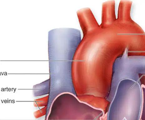
Refer to the diagram of the human heart. What is the name of the part labeled as 'Ascending aorta'?
Ascending aorta
Pulmonary vein
Left atrium
Inferior vena cava
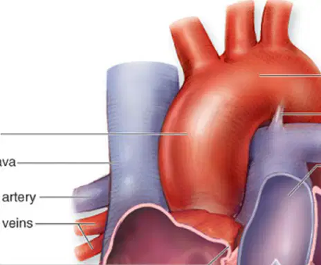
What is the name of the part labeled as 'Superior vena cava'?
Superior vena cava
Inferior vena cava
Pulmonary artery
Aorta
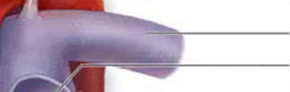
What is the name of the part labeled as 'Right pulmonary artery'?
Right pulmonary artery
Left pulmonary vein
Aorta
Inferior vena cava
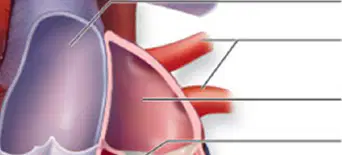
What is the name of the part labeled as 'Right pulmonary veins'?
Right pulmonary veins
Left atrium
Superior vena cava
Right ventricle

interventricular septum
vena cava
pulmonary artery
apex
coronary artery

The lower chambers of the heart which pump blood out of the heart, are known as what?
Pericardium
Atrium / Atria
Ventricle / Ventricles
Endocardium

