
 Worksheets
zoollab 2
Total questions: 133
Worksheet time: 2hrs 12mins
is made out of stratified squamous epithelial tissue of flattened cells.
(a)
consists of a living and growing layer of stratified epithelial tissue.
(a)
outer sublayer, where the body of cutaneous glands is situated, thus giving it a spongy appearance.
(a)
consists of layers of white fibrous connective tissue, which are compactly arranged.
(a)
At the cross-section of the epidermis and dermis are the chromatophores, specialized connective tissue that contains pigments.
(a)
chromatophores are a specialized connective tissue that contains pigments. It contains a row of black pigment-containing cells called (a)
Where the mucous comes out when a frog feels threatened. a milky, toxic substance, like the bufotoxin
(a)
The slimy material comes from the (a) to keep frogs from being dry.
closes when the frog is in the water. covers the eyes
(a)
equivalent of the ears for the frogs
(a)
WHY WAS THERE A PIECE OF WOOD OPENING DANNY’S MOUTH?
(a)
exit of air
(a)
open to allow air to move into or out of the middle ear, equalizing the pressure on both sides of the eardrum.
(a)
enable breathing, protect the airway during swallowing, and produce sound
(a)
opening of the esophagus
(a)
Why are male frogs' thumbs swollen? It is for (a)
dense layer from the outside of the bone.
(a)
spongelike meshwork consisting of trabeculae. The spaces are continuous and occupied by marrow and blood vessels.
(a)
The trabecula contain the (a)
The lines radiating from the lacunae is the (a)
A compact bone is Compact without any spaces. The whole section is called (a)
move a part towards the main
axis of the body
(a)
move a part away from the main axis of the body
(a)
move jointed parts away from
each other
(a)
SUBMENTALIS: Origin
(a)
SUBMENTALIS: Insertion
(a)
SUBMENTALIS: Action
(a)
MEDIAN RAPHE: Origin
(a)
MEDIAN RAPHE: Insertion
(a)
MEDIAN RAPHE: Action
(a)
MYLOHYOID: Origin
(a)
MYLOHYOID: Insertion
(a)
MYLOHYOID: Action
(a)
GENIOHYOID: Origin
(a)
GENIOHYOID: Insertion
(a)
GENIOHYOID: Action
(a)
DELTOID: Action
(a)
DELTOID: Insertion
(a)
DELTOID: Origin
(a)
STERNORADIALIS: Origin
(a)
STERNORADIALIS: Insertion
(a)
STERNORADIALIS: Action
(a)
ANTERIOR PECTORALIS: Action
(a)
ANTERIOR PECTORALIS: Insertion
(a)
ANTERIOR PECTORALIS: Origin
(a)
MIDDLE PECTORALIS: Origin
(a)
MIDDLE PECTORALIS: Insertion
(a)
MIDDLE PECTORALIS: Action
(a)
POSTERIOR PECTORALIS: Action
(a)
POSTERIOR PECTORALIS: Insertion
(a)
POSTERIOR PECTORALIS: Origin
(a)
EXTERNAL OBLIQUE: Origin
(a)
EXTERNAL OBLIQUE: Insertion
(a)
EXTERNAL OBLIQUE: Action
(a)
Why was it named EXTERNAL OBLIQUE?
(a)
RECTUS ABDOMINIS: Origin
(a)
RECTUS ABDOMINIS: Insertion
(a)
Why was it named RECTUS ABDOMINIS
(a)
TRICEP FEMORIS: dorsally situated
(a)
TRICEP FEMORIS: medially located
(a)
TRICEP FEMORIS: ventrally located
(a)
TRICEP FEMORIS: Insertion
(a)
TRICEP FEMORIS: Action
(a)
ADDUCTOR LONGUS: Origin
(a)
ADDUCTOR LONGUS: Insertion
(a)
ADDUCTOR LONGUS: Action
(a)
SARTORIUS: Origin
(a)
ADDUCTOR MAGNUS: Origin
(a)
GRACILIS MAJOR: Origin
(a)
GRACILIS MAJOR: Insertion
(a)
GRACILIS MAJOR: Action
(a)
EXTERNAL CRURIS: Action
(a)
EXTERNAL CRURIS: Insertion
(a)
EXTERNAL CRURIS: Action
(a)
TIBIALIS POSTICUS: Action
(a)
TIBIALIS POSTICUS: Insertion
(a)
TIBIALIS POSTICUS: Origin
(a)
GASTROCNEMIUS: Origin
(a)
GASTROCNEMIUS: Insertion
(a)
GASTROCNEMIUS: Action
(a)
TEMPORALIS: Action
(a)
TEMPORALIS: Insertion
(a)
TEMPORALIS: Origin
(a)
CUCCULARIS: Origin
(a)
CUCCULARIS: Insertion
(a)
CUCCULARIS: Action
(a)
DORSALIS SCAPULAE: Action
(a)
DORSALIS SCAPULAE: Insertion
(a)
DORSALIS SCAPULAE: Origin
(a)
LONGISSIMUS DORSI: Origin
(a)
LONGISSIMUS DORSI: insertion
(a)
LONGISSIMUS DORSI: Action
(a)
LATISSIMUS DORSI: Action
(a)
LATISSIMUS DORSI: Insertion
(a)
LATISSIMUS DORSI: Origin
(a)
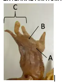
Based on the picture, identify the WHOLE structure:
(a)
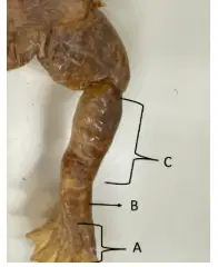
Based on the picture, identify the WHOLE structure:
(a)
Based on the picture, identify STRUCTURE A
(a)
Based on the picture, identify STRUCTURE B
(a)
Based on the picture, identify STRUCTURE C
(a)
Based on the picture, identify STRUCTURE A
(a)
Based on the picture, identify STRUCTURE B
(a)
Based on the picture, identify STRUCTURE C
(a)
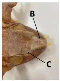
Identify structure B
(a)
What is the structure postero-lateral to C?
(a)
What is the structure posterior to your answer in number 2?:
(a)
Going back to patrotid glands, what substances is found in the structure?
(a)
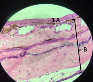
1-2 Identify the sublayers that make up A: top-most layer:
(a)
Identify B:
(a)
Identify the sublayer of B that contains the poison glands:
(a)
Chromatophores contain rows of black pigments that give the toad its color. What is the specific term for these black pigments?:
(a)
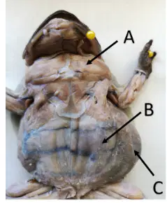
identify A
(a)
identify B
(a)
identify C
(a)
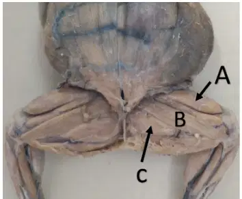
identify A
(a)
identify B
(a)
identify C
(a)
What muscle must be cut for the adductor longus to be visible?
(a)
origin of C
(a)
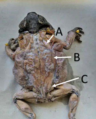
choose the letter (from the picture) the best describes the muscle: A
large, broad muscle underneath the depressor mandibulae and can be visible by cutting the depressor mandibulae
small but thick muscle found anterior to the vastus externus and medial to the rectus femoris.
muscle lateral to the longissimus dorsi
choose the letter (from the picture) the best describes the muscle: B
large, broad muscle underneath the depressor mandibulae and can be visible by cutting the depressor mandibulae
small but thick muscle found anterior to the vastus externus and medial to the rectus femoris.
muscle lateral to the longissimus dorsi
choose the letter (from the picture) the best describes the muscle: C
large, broad muscle underneath the depressor mandibulae and can be visible by cutting the depressor mandibulae
small but thick muscle found anterior to the vastus externus and medial to the rectus femoris.
muscle lateral to the longissimus dorsi
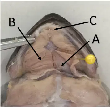
identify A
(a)
identify B
(a)
identify C
(a)
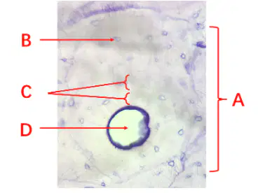
Structure A
(a)
Structure B
(a)
Structure C
(a)
Structure D
(a)
What is the specialized cell found in Structure B?
(a)
Based on the picture, what specific type of bone is this?
(a)
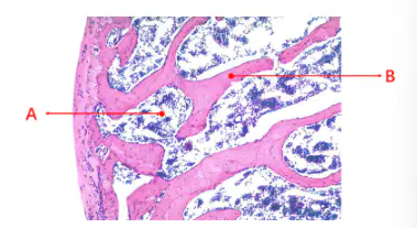
Structure A
(a)
Structure B
(a)

