
 Worksheets
Study Guide Muscular System HSFD 25
Total questions: 117
Worksheet time: 56mins

plantar flexion of the ankle and knee flexion

Trunk flexion
Trapezius
Buccinator
Flatus abdominis
Rectus abdominis

Adducts, medially rotates and extends the arm at the shoulder joint.
Trapezius
External Obliques
Latissmus dorsi
Rectus abdominis

Elevates and retracts the scapula
Latissimius Dorsi
Trapezius
Triceps
Erectors

As a group these muscles extend the knee
Hamstring
Quadriceps
Gastrocnemius
Sartorius

Adduction, flexion and internal rotation of the arm. Stabilizes the shoulder and assist in breathing
Pectoralis Major
Tricep
Bicep
Deltoid
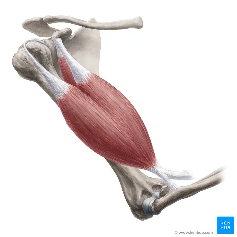
Flexion of the elbow
Triceps Brachii
Biceps Brachii

trunk rotation, lateral flexion, abdominal compression, stabilization, assist in forced expiration
Pectoralis Major
External Oblique
Erectors
Quadriceps

abduction, flexion, ,internal and external rotation, and stabilizes the shoulder to help prevent dislocations
Deltoid
Triceps
Latissimus dorsi
Trapezius
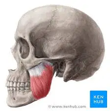
Elevates the mandible closing the mouth
sartorius
masseter
deltoid
bicep
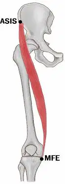
flexes the hip and knee
sartorius
quadriceps
tibialis anterior
deltoid
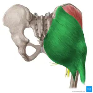
extends the hip joint
hamstrings
gluteus maximus
gastrocnemius
deltoid
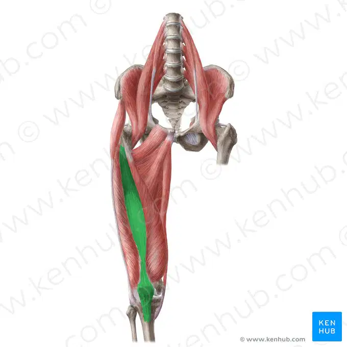
Knee extension
vastus intermedius
vastus medialis
vastus lateralis
rectus femoris
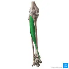
Dorsiflexion and inversion of the ankle joint
Tibialis Anterior
Hamstrings
Gastrocnemius
Rectus Femoris

Which muscle group flexes the knee joint?
Deltoid
Hamstrings
Trapezius
Quadriceps
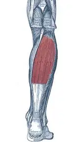
Plantarflexion of the ankle
vastus medialis
semitendinosis
soleus
tibialis anterior
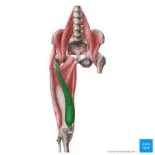
Knee extension, patella tracking, adduction
vastus lateralis
vastus medialis
semimembranosus
tibialis anterior
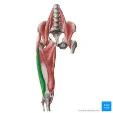
Knee extension, hip flexion, patella stabilization
bicep femoris
vastus intermedius
vastus medialis
vastus lateralis
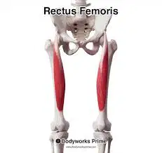
Knee extension, hip flexion
bicep femoris
rectus femoris
vastus medialis
deltoid
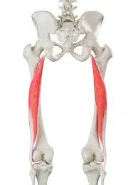
Knee flexion, hip extension
bicep femoris
rectus femoris
vastus medialis
tibialis anterior
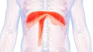
Enables breathing by contracting and relaxing rhythmically
deltoid
diaphragm
rectus femoris
biceps brachii
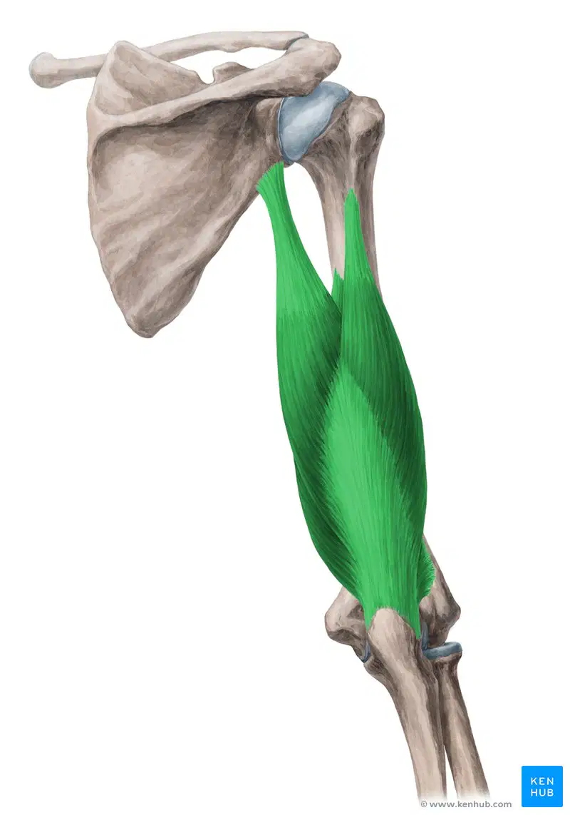
Extension of the elbow
biceps brachii
triceps brachii
deltoid
external oblique
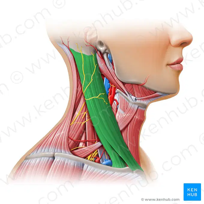
Flexion and rotation of the Neck
triceps brachii
sternocleidomastoid
masseter
deltoid
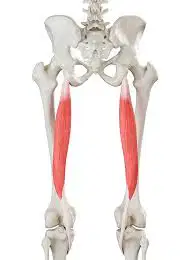
Knee flexion hip extension
gastrocnemius
vastus medialis
semitendinosus
tibialis anterior
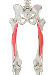
knee extension, hip flexion
deltoid
rectus femoris
rectus abdominus
gastrocnemius
Skeletal muscle is:
involuntary
voluntary
Skeletal Muscle has ______nuclei.
No
One
many
Skeletal muscle is:
striated
unstriated
Has a centrally located nucleus
smooth and skeletal
cardiac and skeletal
skeletal and smooth
smooth and cardiac
Found inside organs
smooth
cardiac
skeletal
This muscle contracts slowly
smooth
cardiac
skeletal
Which myofilament shortens when a sarcomere contracts?
Actin
Myosin
Both
Neither
Which myofilament is thick?
Actin
Myosin
Both
Neither
Which myofilament has many heads to grab the other one?
Actin
Myosin
Both
Neither
Which myofilament is attached to the Z line?
Actin
Myosin
Both
Neither
Which myofilament has tropomyosin on it?
Actin
Myosin
Both
Neither
Which myofilament has troponin on it?
Actin
Myosin
Both
Neither
Which myofilament is powered by ATP?
Actin
Myosin
Both
Neither
Which myofilament has two regulatory proteins on it?
Actin
Myosin
Both
Neither
Which regulatory protein blocks myosin-binding sites on actin?
Troponin
Tropomyosin
Neither
Which regulatory protein blocks actin-binding sites on myosin?
Troponin
Tropomyosin
Neither
Which regulatory protein has a binding site for calcium ions?
Troponin
Tropomyosin
Neither
Which regulatory protein stiffens actin so that it can resist tension?
Troponin
Tropomyosin
Neither
Which regulatory protein has 3 binding sites?
Troponin
Tropomyosin
Neither
Which regulatory protein can move the other one?
Troponin
Tropomyosin
Neither
What happens when calcium binds to troponin?
It moves tropomyosin on top of the myosin-binding sites
It moves tropomyosin off of the myosin-binding sites
It moves tropomyosin to cover the actin-binding sites
It moves tropomyosin off of the actin-binding sites
Once the myosin-binding sites are uncovered, what does myosin need in order to power it to cock up and bind?
Calcium
Acetylcholine
ATP
Actin
With ATP and calcium present, what is able to happen?
Myosin can grab actin, shortening the sarcomere
Actin can grab myosin, shortening the sarcomere
Actin and myosin can shorten
With ATP and calcium absent, what happens?
Actin relaxes
Actin contracts
Myosin relaxes
The sarcomere relaxes
Why does the sarcomere relax when calcium is absent?
The myosin heads release and go back down
The actin heads release
Troponin covers the myosin-binding sites
Tropomyosin covers the myosin-binding sites
What is the smallest unit of a muscle to contract?
Sarcomere
Myofibril
Muscle fiber
Myofilaments
Which myofilament is thin?
Collagen
Elastin
Actin
Myosin
Which myofilament is thick?
Myosin
Collagen
Elastin
Acetylcholine
Which part of a sarcomere contains only thin filaments?
I band
A band
H zone
M zone
Which part of a sarcomere is the wall between two different sarcomeres?
M line
Z line
Which part of a sarcomere contains BOTH actin and myosin myofilaments?
A band
I band
H zone
M line
Which part of the sarcomere is the line down the middle of the myosin?
Z line
I band
M line
Which myofilament pulls the other one when a muscle contracts?
Actin pulls myosin
Myosin pulls actin
Connects muscle to bone
ligament
tendon
cartilage
none of these
Connective tissue sheath that surrounds the entire muscle
perimysium
epimysium
endomysium
fascicle
Bundle of muscle fibers
fascicle
epimysium
perimysium
tendon
Connective tissue sheath that surrounds each fascicle
tendon
endomysium
perimysium
ligament
Connective tissue sheath that surrounds each muscle fiber
endomysium
perimysium
ectomysium
sarcolemma
Plasma membrane that around each muscle fiber
actin
myosin
sarcolemma
myofilament
Long proteins that make up each muscle fiber
fascicle
myofibril
tendon
calcium
Contacting unit of the muscle
sarcomere
tendon
sarcolemma
epimysium
What is a crossbridge?
Where myosin binds to actin
Where the calcium crosses into the cytoplasm
Where calcium binds with troponin
Where titin and myosin are connected
What does calcium do to the actin?
Covers myosin binding sites
Uncovers myosin binding sites
Covers actin binding sites
Uncovers actin binding sites
What is the name of the protein that covers the binding sites on actin?
Myosin
Titin
Troponin
Tropomyosin
What does the sarcoplasmic reticulum do?
Stores sodium
Stores ATP
Releases sodium
Stores calcium
What is the name of the place where a nerve meets a muscle fiber
Nerve junction
Neuromuscular junction
Muscle junction
Crossbridge
How does the sarcomere shorten (contract) ?
The myosin filaments contract
The actin filaments contract
The myosin is pulled by the actin
The actin is pulled by the myosin
What two chemicals are necessary for muscle contraction to occur?
Niacin and Potassium
Calcium and ATP
Sodium and Potassium
Acetylcholine and Potassium
Skeletal Muscle contraction is initiated when the ________ sends a message to the muscle cell.
Muscle cell
Gland
Neuron
None of the above
The neurotransmitter that signals muscle contraction is...
Calcium
Actin
Myosin
Ach (acetylcholine)
Which of the following statements correctly explains the sliding filament model?
Actin head attaches to a binding site on a myosin filament, forming a cross bridge.
Myosin head attaches to a binding site on an actin filament, forming a neuromuscular junction.
Actin head attaches to a binding site on a myosin filament, forming a neuromuscular junction.
Myosin head attaches to a binding site on an actin filament, forming a cross bridge.
Abduction
movement of a limb in a circle
movement of a limb toward the midline
movement of a limb away from the midline
turning a limb upward
Movement of a limb toward the midline
adduction
abduction
circumduction
rotation
Lack of muscle coordination during voluntary movements
ataxia
dyskinesia
atonic
atrophy
Circular movement at the distal end of a limb
dorsiflexion
pronation
circumduction
ataxia
Moving the foot up toward the ankle
plantarflexion
dorsiflexion
rotation
pronation
Turning the palm or sole of the foot downward
pronation
supination
dorsiflexion
adduction
Abnormally increased muscle function or activity
ataxia
hyperkinesia
dystonia
subluxation
Partial contraction of a muscle; firmness
atrophy
tonus
flaccid
dyskinesia
Greater than normal growth
atrophy
hypertrophy
hypotrophy
ataxia
Limp or without muscle tone
origin
prime mover
flaccid
myalgic
A loss of muscle bulk in one or more muscles
strain
dyskinesia
atrophy
hypertrophy
A general term for muscle disease
strain
myopathy
sprain
flaccid
Cordlike, band of nonelastic fibrous connective tissue that attaches a muscle to a bone
tendon
ligament
neuron
antagonist
Fibrous band that connects bone to bone
prime mover
tendon
synergist
ligament
Sheet of connective tissue that acts as a muscle attachment
neuron
tonus
aponeurosis
synergist
Inflammation of a tendon
synergist
strain
tendinitis
sprain
Chronic injury to a tendon
tendinosis
tendinitis
sprain
strain
Pain in one or more muscles
ataxia
tendinosis
myalgia
dyskinesia
Pain located at specific points in the body; along with widespread pain in the body
myasthenia gravis
fibromyalgia
muscular dystrophy
shin splints
The most important muscle that causes a movement; prime mover
origin
insertion
agonist
fixator
Muscle attachment at the immovable bone
insertion
origin
Muscle attachment to the movable bone
insertion
origin
Cooperating action of certain muscles
agonist
insertion
synergistic
origin
Opposite motion of the agonist - example biceps flex and the triceps extend
antagonist
agonist
prime mover
synergist
A lack of coordination of a muscle during movement, particularly gait(how you walk)
synergistic
ataxia
agonist
atagonist
Bending a joint to decrease the angle of a joint
extension
flexion
rotation
adduction
Straightening a joint to increase the angle of a joint
flexion
extension
rotation
circumduction
Moving a body part toward the midline of the body
adduction
abduction
extension
rotation
The rotation is a single spinning movement around a fixed axis
rotation
flexion
extension
abduction
A progressive, genetic muscle disorder that primarily affects boys
duchenne's Muscular dystrophy
fibromyalgia
myasthenia gravis
ataxia
A common over use injury of the lower extremity commonly called shin splints
multiple sclerosis
medial tibial stress syndrome
fibromyalgia
sprain
An injury to a ligament
strain
sprain
An injury to a muscle
strain
sprain
Characterized by stretched ligaments and mild pain
grade 1 sprain
grade 2 sprain
grade 3 sprain
Partial tear of ligaments, Moderate pain and swelling, Significant limitation of range of motion
grade 1 sprain
grade 2 sprain
grade 3 sprain
Complete tear of ligaments, Severe pain and swelling, Inability to bear weight, and May require surgery.
grade 1 sprain
grade 2 sprain
grade 3 sprain
Minimal pain and swelling, Stretching of muscle fibers without tearing, and Full range of motion with slight discomfort.
grade 1 strain
grade 2 strain
grade 3 strain
Moderate pain and swelling, Partial tearing of muscle fibers, and Decreased range of motion and strength.
grade 1 strain
grade 2 strain
grade 3 strain
Severe pain and swelling, Complete rupture of muscle fibers, Significant loss of range of motion and strength, and May require surgery.
grade 1 strain
grade 2 strain
grade 3 strain
Which statement best describes one main function of the muscular system?
It allows the body to move.
It makes bones grow faster.
It produces blood cells.
It stores vitamins for later use.
Which of the following is a function of the muscular system?
Movement
Support
Protection
Heat Generation
All of the above

