
Font size
 Worksheets
Musculoskeletal System - RAW Flawed File
Total questions: 99
Worksheet time: 50mins
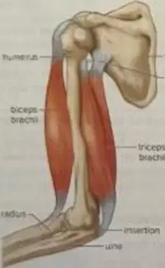
What is the name of the muscle labeled as the 'biceps brachii'?
biceps brachii
triceps brachii
deltoid
pectoralis major
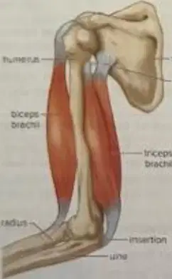
What is the name of the bone labeled as the 'humerus'?
humerus
femur
radius
tibia
Which of the following is NOT a type of muscle found in the human body?
Smooth muscle
Cardiac muscle
Skeletal muscle
Elastic muscle
The musculoskeletal system recognizes the connection between the skeletal and muscular systems.
True
False
Match the following muscle types to their significant features:
Found in the heart
Found in the heart
Found in internal organs
Found in internal organs
Attached to bones
Attached to bones
What is the function of the biceps brachii muscle during the contraction of the arm?
Contraction of the biceps brachii causes the bones of the forearm to be pulled upward.
Contraction of the biceps brachii causes the arm to rotate outward.
Contraction of the biceps brachii causes the fingers to spread apart.
Contraction of the biceps brachii causes the shoulder to move downward.
What is the function of a hydrostatic skeleton in segmented worms?
It provides a framework, structural support, and assists movement by applying muscular contraction pressure to fluid-filled body compartments.
It helps in digestion by breaking down food particles mechanically.
It acts as a respiratory organ for gas exchange.
It stores nutrients for long-term energy use.
Identify the series of concentric rings in compact bone.
Lamellae
Canaliculi
Osteons
Lacunae
What are cavities within the lamellae that contain bone cells called?
Lacunae
Canaliculi
Osteons
Trabeculae
Which specialized substance produces blood cells and is present in spongy bone?
Bone marrow
Cartilage
Ligament
Tendon
What type of marrow is a fat storage tissue present in the medullary cavity?
Yellow marrow
Red marrow
Spongy marrow
Compact marrow
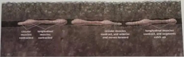
Refer to the diagram of the earthworm. Which muscles contract to move the segments forward?
Longitudinal muscles
Circular muscles
Oblique muscles
Radial muscles
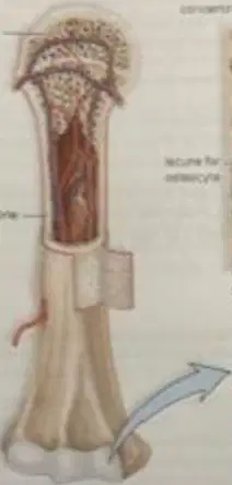
What is the function of the central canal in bone tissue?
The central canal is the canal in the center of each osteon.
It stores fat for the bone tissue.
It produces red blood cells.
It connects bone to muscle.

What are canaliculi in bone tissue?
Canaliculi are tiny tubules that allow nutrients to pass between the osteocytes.
Canaliculi are large cavities that store fat in bone tissue.
Canaliculi are thick fibers that provide strength to bone tissue.
Canaliculi are cells responsible for bone formation.

What is the matrix in bone tissue composed of?
The matrix is the nonliving material maintained by osteocytes. It contains mineral salts (notably calcium salts) and protein.
It is composed mainly of living cells and water.
It consists only of collagen fibers and no minerals.
It is made up entirely of blood vessels and nerves.
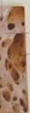
Examine a prepared slide of spongy bone (Fig. 32.3b and c), and identify the numerous bony bars and plates separated by irregular spaces. What are these spaces often filled with?
These spaces are often filled with red marrow.
These spaces are often filled with yellow marrow.
These spaces are often filled with cartilage.
These spaces are often filled with synovial fluid.
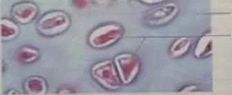
What do lacunae contain?
Lacunae contain chondrocytes.
Lacunae contain osteoblasts.
Lacunae contain fibroblasts.
Lacunae contain erythrocytes.
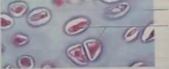
What is the matrix of hyaline cartilage primarily composed of?
The matrix of hyaline cartilage is primarily composed of water and protein.
The matrix of hyaline cartilage is primarily composed of nucleic acids and lipids.
The matrix of hyaline cartilage is primarily composed of bone marrow and calcium.
The matrix of hyaline cartilage is primarily composed of muscle fibers and glycogen.
The shape of the treated femur compared with that of the untreated femur is:
identical to the untreated femur
more irregular than the untreated femur
smoother than the untreated femur
shorter than the untreated femur
The treated bone is more flexible or less flexible than the untreated bone.
More flexible
Less flexible
Equally flexible
Not flexible at all
The treated bone is able to withstand more direct downward compression or less direct downward compression than the untreated bone.
More direct downward compression
Less direct downward compression
The same amount of direct downward compression
Cannot withstand any compression
The effect of this treatment on bone strength is:
It increases bone strength.
It decreases bone strength.
It has no effect on bone strength.
It weakens bone structure.

Label the following bones on the human skeleton diagram: skull, clavicle, scapula, humerus, radius, ulna, femur, tibia, fibula, pelvis, vertebral column.
Skull, clavicle, scapula, humerus, radius, ulna, femur, tibia, fibula, pelvis, vertebral column (as shown in the diagram).
Skull, mandible, sternum, humerus, radius, ulna, femur, tibia, fibula, pelvis, vertebral column.
Skull, clavicle, scapula, humerus, radius, ulna, femur, tibia, fibula, patella, vertebral column.
Skull, clavicle, scapula, humerus, radius, ulna, femur, tibia, fibula, pelvis, coccyx.
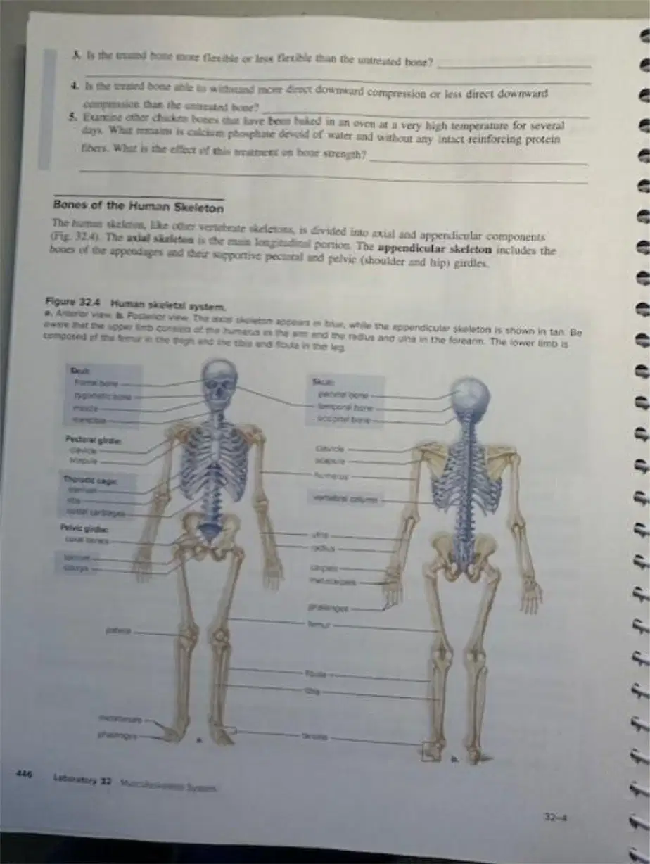
Match the following bones to their correct location in the human skeleton:
Skull
Head
Clavicle
Shoulder
Scapula
Upper arm
Humerus
Forearm (thumb side)
Radius
Forearm (little finger side)
Examine a human skeleton, and with the help of Figure 32.4, identify the bone that is composed of many small bones fused together, enclosing the brain and major sense organs. What is the name of this bone?
Skull
Femur
Rib
Pelvis
Which bone is the only bone of the skull not immovably fused to another?
Mandible
Maxilla
Frontal bone
Parietal bone
What is the bone that provides support and also protects the spinal cord?
Vertebral column
Femur
Rib cage
Skull
The vertebral column is divided into five series. Name the series that forms the neck region.
Cervical vertebrae
Thoracic vertebrae
Lumbar vertebrae
Sacral vertebrae
The vertebral column is divided into five series. Name the series with which the ribs articulate.
Thoracic vertebrae
Cervical vertebrae
Lumbar vertebrae
Sacral vertebrae
The vertebral column is divided into five series. Name the series in the abdominal region.
Lumbar vertebrae
Cervical vertebrae
Thoracic vertebrae
Sacral vertebrae
The vertebral column is divided into five series. Name the series called the sacrum.
Sacral vertebrae
Cervical vertebrae
Thoracic vertebrae
Lumbar vertebrae
The vertebral column is divided into five series. Name the series that forms the coccyx.
Caudal vertebrae
Cervical vertebrae
Thoracic vertebrae
Lumbar vertebrae
What are ribs that do not have sternal attachment called?
Floating ribs
True ribs
False ribs
Costal ribs
Examine a human skeleton, and with the help of Figure 32.4, identify the bone that supports the upper limb and is composed of the clavicles.
Pectoral girdle
Pelvic girdle
Femur
Scapula
Which bone is known as the shoulder blade?
Scapula
Femur
Tibia
Clavicle
Which bone is the large long bone of the upper arm?
Humerus
Femur
Tibia
Radius
Which bone is the long bone of the forearm with a pivot joint at the elbow that allows rotational motion?
Radius
Ulna
Humerus
Femur
Which bone is the other long bone of the forearm, with a hinge joint at the elbow that allows motion in only one plane?
Ulna
Femur
Tibia
Scapula
Name the group of small bones forming the wrist.
Carpals
Tarsals
Metacarpals
Phalanges
Name the slender bones forming the palms and back of the hand.
Metacarpals
Phalanges
Carpals
Tarsals
Name the bones of the fingers.
Phalanges
Metacarpals
Carpals
Tarsals
What forms the basal support for the lower limbs and is composed of the coxal (hip) bone?
Pelvic girdle
Pectoral girdle
Femur
Vertebral column
Which bone is the long bone of the thigh?
Femur
Tibia
Humerus
Radius
Which bone is the larger of the two long bones of the leg?
Tibia
Fibula
Femur
Humerus
Which bone is the smaller of the two long bones of the leg?
Fibula
Tibia
Femur
Humerus
Name the group of small bones forming the ankle.
Tarsals
Metatarsals
Phalanges
Carpals
Name the slender anterior bones of the foot.
Metatarsals
Tarsals
Phalanges
Calcaneus
Name the bones of the toes.
Phalanges
Metatarsals
Carpals
Tarsals
What are structures that are similar because they are inherited from a common ancestor called?
Homologous structures
Analogous structures
Vestigial structures
Convergent structures
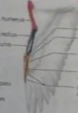
A bird uses its forelimb for ________.
flying.
swimming.
digging.
running.

2. Keeping function in mind, compare the forelimb bones in the animals listed in Table 32.1 to those of a human. Where a bone is noted, compare the humerus; where it is noted, compare the radius and ulna; and where it is noted, compare the carpals, metacarpals, and phalanges as a whole. Are the bones fused, longer, stronger, or more delicate than those of a human?
The forelimb bones in some animals are fused, longer, stronger, or more delicate compared to those of a human, depending on their function.
The forelimb bones in all animals are identical to those of a human.
The forelimb bones in animals are always shorter and weaker than those of a human.
The forelimb bones in animals are never fused and are always more robust than those of a human.

Give examples to show that the forelimbs of these animals are modified to suit their means of locomotion.
Wings in birds for flying, flippers in whales for swimming, and forelegs in horses for running.
Forelimbs in all animals are used only for walking.
Forelimbs in birds are used for digging.
Forelimbs in whales are used for climbing trees.
Only the phalanges in the hindlimb of a frog, bird, and cat ever touch the ground, whereas the entire foot of a human touches the ground. This difference exists because:
Humans have a plantigrade posture, while frogs, birds, and cats have digitigrade posture.
Humans have more phalanges than other animals.
Frogs, birds, and cats walk on their heels, while humans walk on their toes.
The bones in the human foot are shorter than those in frogs, birds, and cats.
Using Figure 32.6, label the bones of the forelimb in these animals.
Humerus, radius, ulna, carpals, metacarpals, phalanges
Femur, tibia, fibula, tarsals, metatarsals, phalanges
Scapula, clavicle, sternum, ribs, vertebrae
Pelvis, sacrum, coccyx, femur, patella, tibia
In Table 32.2, the structure of the hindlimb bones compared to the forelimb bones in a frog, bird, cat, and human differs primarily due to which factor?
The specific locomotor function of the hindlimb
The color of the bones
The age of the animal
The diet of the animal
Fill in the differences in hindlimb and forelimb bones and explanation for the frog.
Hindlimbs are longer and stronger for hopping; forelimbs are shorter and used for landing.
Hindlimbs are shorter and weaker; forelimbs are longer and used for swimming.
Both hindlimbs and forelimbs are of equal length and strength.
Forelimbs are longer and stronger for jumping; hindlimbs are shorter and used for walking.
Fill in the differences in hindlimb and forelimb bones and explanation for the bird.
Hindlimbs are adapted for perching or walking; forelimbs are modified as wings for flying.
Hindlimbs are used for flying; forelimbs are adapted for swimming.
Hindlimbs are modified as wings for flying; forelimbs are adapted for perching.
Hindlimbs and forelimbs are both adapted for running only.
Fill in the differences in hindlimb and forelimb bones and explanation for the cat.
Hindlimbs are powerful for leaping; forelimbs are used for landing and walking.
Hindlimbs are used for grasping; forelimbs are used for swimming.
Hindlimbs are shorter and weaker; forelimbs are longer and stronger for running.
Hindlimbs are adapted for flying; forelimbs are adapted for digging.
Fill in the differences in hindlimb and forelimb bones and explanation for the human.
Hindlimbs (legs) are adapted for bipedal walking and support; forelimbs (arms) are adapted for manipulation and tool use.
Hindlimbs (legs) are adapted for flying; forelimbs (arms) are adapted for swimming.
Hindlimbs (legs) are adapted for grasping; forelimbs (arms) are adapted for running.
Hindlimbs (legs) are adapted for climbing; forelimbs (arms) are adapted for digging.
Observation: Muscular Tissues Smooth Muscle Smooth muscle is located in the walls of hollow internal organs, and its involuntary contraction moves substances through an organ. Smooth muscle fibers are spindle-shaped cells, each with a single nucleus. The cells are usually arranged in parallel lines, forming sheets. Smooth muscle does not have striations (bands of light and dark) as seen in cardiac and skeletal muscle. Although smooth muscle is slower to contract than skeletal muscle, it can maintain prolonged contractions and does not fatigue easily. Examine the slide of smooth muscle (Figure 32.7, 227), and identify the spindle-shaped fibers and the oval-shaped nucleus found in the center of the cytoplasm. Which of the following best describes the appearance of smooth muscle fibers?
Spindle-shaped fibers with a single, oval-shaped nucleus in the center
Branched fibers with multiple nuclei and striations
Cylindrical fibers with striations and peripheral nuclei
Irregularly shaped fibers with no visible nucleus
Which of the following types of muscle tissue is under voluntary control?
Smooth muscle
Cardiac muscle
Skeletal muscle
None of the above
Fill in the blank: ________ muscle tissue is found in the heart and contains alternating light and dark bands.
Cardiac
Smooth
Skeletal
Connective
Smooth muscle tissue has striations (alternating light and dark bands).
True
False
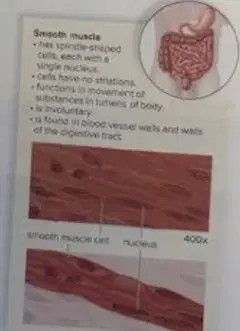
Identify the structures labeled in the diagram:
Smooth muscle cell, nucleus
Cardiac muscle cell, mitochondria
Neuron, axon
Epithelial cell, cytoplasm
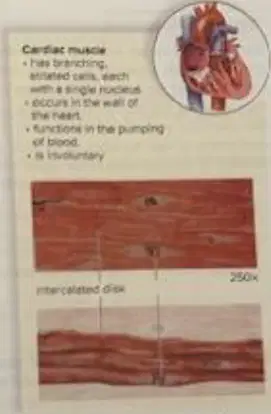
Cardiac Muscle: Examine a prepared slide of cardiac muscle (Figure 32.8), and identify the features such as a single nucleus per cell, branched fibers joined at intercalated disks, and striations under high power. Which of the following best describes what you see?
Branched fibers with intercalated disks and striations, single nucleus per cell
Multiple nuclei per cell, smooth fibers, no intercalated disks
Unbranched fibers, no striations, multiple nuclei per cell
Branched fibers, no intercalated disks, no striations
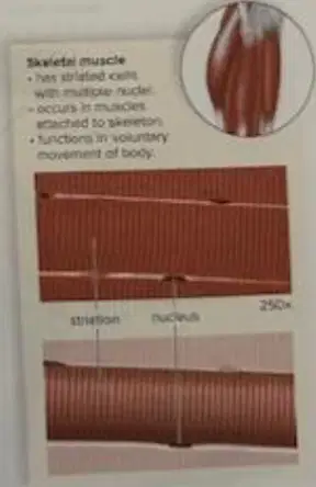
Skeletal Muscle: Examine a prepared slide of skeletal muscle (Figure 32.9), and identify the following features: long fibers arranged in a parallel fashion, multinuclei per fiber, and striations. Which of the following best describes what you see?
Long fibers arranged in parallel, multinuclei per fiber, and striations
Short fibers with single nucleus and no striations
Branched fibers with intercalated discs and single nucleus
Spindle-shaped fibers with central nucleus and no striations
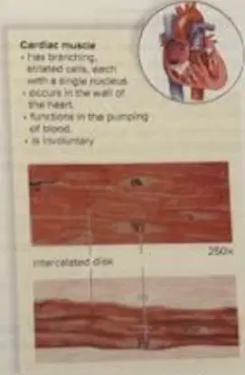
Which of the following is true about cardiac muscle?
It is voluntary and has multiple nuclei per cell.
It is striated, involuntary, and has a single nucleus per cell.
It is not striated and is voluntary.
It is attached to bones and is voluntary.
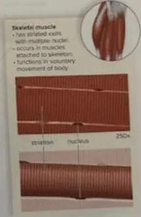
Which of the following is true about skeletal muscle?
It is striated, voluntary, and has multiple nuclei per cell.
It is involuntary and has a single nucleus per cell.
It is not striated and is involuntary.
It is branched and joined at intercalated disks.
What is the term for the movement of joint parts toward each other?
Flexion
Extension
Abduction
Rotation
What is the term for the movement of joint parts away from each other?
Extension
Flexion
Rotation
Adduction
What is the term for the movement of a part toward the midline of the body?
Adduction
Abduction
Extension
Flexion
What is the term for the movement of a part away from the midline of the body?
Abduction
Adduction
Flexion
Extension
Match the following terms to their definitions:
Movement of joint parts toward each other
Flexion
Movement of joint parts away from each other
Extension
Movement of a part toward the midline of the body
Adduction
Movement of a part away from the midline of the body
Abduction
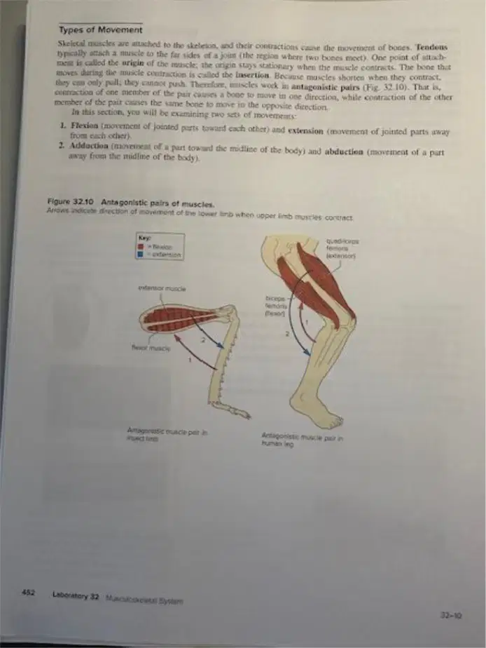
Which muscle pair in the human leg is shown as antagonistic?
A. Quadriceps and biceps
B. Quadriceps and hamstrings
C. Flexor and extensor muscles
D. Adductor and abductor muscles
What is the action caused by the gastrocnemius when the thigh is rigid?
Extension of the shank (leg) at the knee joint.
Flexion of the thigh at the hip joint.
Abduction of the thigh.
Rotation of the shank (leg) externally.
When the shank is rigid what is the action caused by contraction of the gastrocnemius?
Flexion of the thigh at the knee joint.
Extension of the thigh at the hip joint.
Abduction of the thigh at the hip joint.
Rotation of the thigh at the knee joint.
Locate the frog's triceps femoris, a very large ventral muscle of the thigh. It is seen best in the dorsal view. This muscle has two origins on the pelvic girdle and inserts into the femur by way of the broad, white tendon that passes over the anterior surface of the knee. Hold the thigh firmly, and pull on the triceps. Is the triceps femoris a shank extensor or flexor?
Shank extensor.
Shank flexor.
Thigh abductor.
Thigh adductor.
Identify the following muscles in the diagram labeled 'Dorsal view of hindleg muscles in frogs (Figure 32.11)'.
gluteus, pyriformis, triceps femoris, biceps femoris, semimembranosus, peroneus, rectus anticus femoris, vastus externus, vastus internus, rectus internus, iliacus internus, anal sphincter, gastrocnemius, Achilles tendon (all labeled in the diagram).
pectoralis, deltoid, trapezius, latissimus dorsi, serratus anterior, biceps brachii, triceps brachii, flexor carpi ulnaris, extensor digitorum, palmaris longus, pronator teres, supinator, abductor pollicis longus, extensor pollicis brevis, flexor digitorum profundus (not present in frog hindleg).
masseter, temporalis, digastric, mylohyoid, sternohyoid, omohyoid, sternocleidomastoid, platysma, levator scapulae, scalenes, splenius capitis, semispinalis capitis, longus colli, rectus capitis anterior, rectus capitis lateralis (muscles of the head and neck).
external oblique, internal oblique, transversus abdominis, rectus abdominis, pyramidalis, quadratus lumborum, iliopsoas, psoas major, psoas minor, iliacus, sartorius, tensor fasciae latae, adductor longus, gracilis (muscles of the abdomen and thigh).
Where does the biceps femoris insert?
The biceps femoris inserts on the tibia and fibula.
The biceps femoris inserts on the patella.
The biceps femoris inserts on the femur.
The biceps femoris inserts on the calcaneus.
What action does the biceps femoris cause?
The biceps femoris causes flexion of the leg at the knee.
The biceps femoris causes extension of the arm at the elbow.
The biceps femoris causes abduction of the thigh at the hip.
The biceps femoris causes rotation of the wrist.
Is the biceps brachii or the triceps brachii located on the ventral surface of the arm?
The biceps brachii is located on the ventral surface of the arm.
The triceps brachii is located on the ventral surface of the arm.
Both are located on the ventral surface of the arm.
Neither is located on the ventral surface of the arm.
What change did you notice in the firmness of this muscle as it contracted?
The muscle became firmer as it contracted.
The muscle became softer as it contracted.
The muscle did not change in firmness as it contracted.
The muscle disappeared as it contracted.
Did your hand or forearm move as you pushed up against the table?
No, the hand or forearm did not move.
Yes, the hand moved slightly.
Yes, the forearm moved upward.
Both the hand and forearm moved.
Given your answer to question 3, did this muscle's fibers shorten as you pushed up against the tabletop?
No, the muscle's fibers did not shorten.
Yes, the muscle's fibers shortened significantly.
The muscle's fibers lengthened instead of shortening.
The muscle's fibers remained unchanged in length.
What makes this contraction isotonic rather than isometric?
The muscle fibers shorten and the length of the muscle changes, making the contraction isotonic rather than isometric.
The muscle fibers remain the same length and the muscle does not change shape.
The contraction occurs without any movement of the muscle.
The muscle fibers lengthen and the muscle stretches during contraction.
Contraction of Muscle Fibers: The current model for muscle contraction states that as muscle contraction occurs, actin filaments ______ past myosin filaments. This causes ______ to shorten.
slide; sarcomeres
bind; mitochondria
contract; nuclei
move; ribosomes
According to the passage, ATP serves as the immediate energy source for ______ contraction.
sarcomere
neuron
epidermal
alveolar

What is the microscopic structure shown in the diagram?
Cardiac muscle fiber
Skeletal muscle fiber
Smooth muscle fiber
Nerve fiber
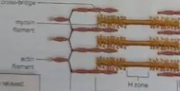
In the diagram, which structure is labeled as the site where actin and myosin filaments interact?
Sarcomere
Mitochondrion
Nucleus
T tubule

Match the following substances with their correct function.
Provides energy for muscle contraction
Myosin filaments
Interacts with myosin for contraction
Actin filaments
Interacts with actin for contraction
K+/Mg2+ salt solution
Provides necessary ions for contraction
ATP

Fill in the blank: According to Table 32.3, the solution that causes the greatest change in muscle fiber length is ________.
Both salt solution and ATP
Salt solution only
ATP only
Distilled water
Fill in the blank: In the experimental procedure, ATP solution is added to slide 1 to observe any change in ________.
strand length
temperature
pH level
color
Based on the experimental procedure, explain why both the K+/Mg2+ salt solution and ATP are required to observe muscle contraction.
Both K+/Mg2+ salt solution and ATP are necessary because they provide essential ions and energy for muscle contraction.
Only ATP is needed for muscle contraction; K+/Mg2+ salt solution is not required.
K+/Mg2+ salt solution alone can induce muscle contraction without ATP.
Neither K+/Mg2+ salt solution nor ATP are required for muscle contraction.
Where is the pressure of muscle contraction applied in a hydrostatic skeleton?
The pressure is applied to the fluid within the body cavity.
The pressure is applied to the bones.
The pressure is applied to the skin surface.
The pressure is applied to the muscles themselves.
What are the characteristics of an exoskeleton?
An exoskeleton is a hard external structure that provides protection and support, and must be shed for growth.
An exoskeleton is a soft internal structure that helps in digestion.
An exoskeleton is a flexible membrane that allows for rapid movement.
An exoskeleton is a liquid layer that keeps the body moist.
What are the characteristics of an endoskeleton?
An endoskeleton is an internal support structure made of bone or cartilage, which grows with the organism.
An endoskeleton is an external shell that protects the body from outside harm.
An endoskeleton is a hard outer covering that does not grow with the organism.
An endoskeleton is a fluid-filled cavity that provides support to the body.
What is produced by the red marrow of the spongy bone?
Red blood cells are produced by the red marrow of the spongy bone.
White blood cells are produced by the red marrow of the spongy bone.
Platelets are produced by the red marrow of the spongy bone.
Fat cells are produced by the red marrow of the spongy bone.
Which mineral provides rigidity to bone?
Calcium provides rigidity to bone.
Iron provides rigidity to bone.
Potassium provides rigidity to bone.
Sodium provides rigidity to bone.
What are osteocytes?
Osteocytes are mature bone cells that maintain bone tissue.
Osteocytes are cells that produce cartilage in joints.
Osteocytes are immature cells that form blood vessels.
Osteocytes are cells responsible for breaking down bone tissue.

