
Font size
 Worksheets
The Nervous System and Senses - Raw and Flawed
Total questions: 99
Worksheet time: 50mins
Identify the parts of the sheep brain, and state the functions of each part.
Parts of the sheep brain include the cerebrum, cerebellum, and brainstem. Functions: Cerebrum - higher brain functions; Cerebellum - coordination; Brainstem - basic life functions.
Parts of the sheep brain include the medulla, thalamus, and hypothalamus. Functions: Medulla - memory; Thalamus - movement; Hypothalamus - vision.
Parts of the sheep brain include the cortex, pons, and amygdala. Functions: Cortex - digestion; Pons - respiration; Amygdala - balance.
Parts of the sheep brain include the occipital lobe, spinal cord, and pituitary gland. Functions: Occipital lobe - hearing; Spinal cord - taste; Pituitary gland - smell.
Label a diagram of the human brain, and give examples to show that the parts of the brain work together.
Label: cerebrum, cerebellum, brainstem. Example: Coordination of movement involves cerebrum (planning), cerebellum (execution), and brainstem (relay).
Label: cerebrum, medulla, spinal cord. Example: Coordination of movement involves cerebrum (planning), medulla (execution), and spinal cord (relay).
Label: cerebellum, thalamus, hypothalamus. Example: Coordination of movement involves cerebellum (planning), thalamus (execution), and hypothalamus (relay).
Label: frontal lobe, occipital lobe, temporal lobe. Example: Coordination of movement involves frontal lobe (planning), occipital lobe (execution), and temporal lobe (relay).
Describe the anatomy of the spinal cord, and tell how the cord functions as a relay station.
The spinal cord consists of nerve fibers that transmit signals between the brain and body. It acts as a relay station for sensory and motor information.
The spinal cord is made up of bone tissue and stores nutrients for the body. It acts as a storage center for vitamins.
The spinal cord is a muscle that helps in digestion and absorption of food. It acts as a digestive organ.
The spinal cord is a gland that secretes hormones to regulate sleep. It acts as a hormonal control center.
Distinguish between cranial nerves and spinal nerves on the basis of location and function.
Cranial nerves originate from the brain and control head/neck functions. Spinal nerves originate from the spinal cord and control body functions.
Cranial nerves originate from the spinal cord and control body functions. Spinal nerves originate from the brain and control head/neck functions.
Cranial nerves and spinal nerves both originate from the brain and control only head functions.
Cranial nerves and spinal nerves both originate from the spinal cord and control only body functions.
Describe the anatomy and physiology of a spinal reflex arc.
A spinal reflex arc involves sensory neurons, interneurons in the spinal cord, and motor neurons, allowing quick responses without brain involvement.
A spinal reflex arc only involves the brain and motor neurons, bypassing the spinal cord entirely.
A spinal reflex arc consists solely of sensory neurons and does not include motor or interneurons.
A spinal reflex arc is responsible for voluntary movements controlled by the cerebral cortex.
Compare the eyes of various invertebrates, particularly those of an arthropod and a squid.
Arthropod eyes are compound, made of many lenses; squid eyes are camera-like, similar to vertebrate eyes.
Both arthropod and squid eyes are compound, made of many lenses.
Arthropod eyes are camera-like, similar to vertebrate eyes; squid eyes are compound.
Both arthropod and squid eyes lack lenses and are simple light-sensitive spots.
Identify the parts of the human eye, and state a function for each part.
Cornea - focuses light; Lens - adjusts focus; Retina - detects light; Iris - controls light entry.
Cornea - detects light; Lens - controls light entry; Retina - adjusts focus; Iris - focuses light.
Cornea - controls light entry; Lens - detects light; Retina - focuses light; Iris - adjusts focus.
Cornea - adjusts focus; Lens - focuses light; Retina - controls light entry; Iris - detects light.
Explain how to test for accommodation and the blind spot in the human eye.
Accommodation: Focus on near and far objects. Blind spot: Close one eye and move a dot in peripheral vision until it disappears.
Accommodation: Use colored filters. Blind spot: Look directly at a bright light.
Accommodation: Blink rapidly. Blind spot: Cover both eyes and try to see.
Accommodation: Read text in dim light. Blind spot: Move your head quickly from side to side.
Compare the tympanum of grasshoppers and the lateral line of fishes to the human ear.
Grasshopper tympanum detects sound vibrations; fish lateral line detects water movement; human ear detects sound waves.
Grasshopper tympanum detects water movement; fish lateral line detects sound waves; human ear detects vibrations.
Grasshopper tympanum detects light; fish lateral line detects sound; human ear detects water movement.
Grasshopper tympanum detects air pressure; fish lateral line detects light; human ear detects temperature.
Identify the parts of the ear, and state a function for each part.
Outer ear - collects sound; Middle ear - transmits vibrations; Inner ear - converts vibrations to nerve signals.
Outer ear - transmits vibrations; Middle ear - collects sound; Inner ear - converts vibrations to nerve signals.
Outer ear - converts vibrations to nerve signals; Middle ear - collects sound; Inner ear - transmits vibrations.
Outer ear - collects sound; Middle ear - converts vibrations to nerve signals; Inner ear - transmits vibrations.
Describe the anatomy of human skin, and explain the distribution and function of sensory receptors.
Human skin has layers: epidermis, dermis, hypodermis. Sensory receptors detect touch, temperature, pain, and pressure.
Human skin consists of a single layer called the epidermis, which contains all sensory receptors for vision and hearing.
Human skin is made up of bone and muscle, and sensory receptors only detect movement.
Human skin has no sensory receptors and is only for protection against sunlight.
Relate the ability to distinguish foods to the senses of smell and taste.
Smell and taste work together to identify and distinguish flavors in food.
Only the sense of sight is used to distinguish flavors in food.
The ability to distinguish foods depends solely on texture.
Taste alone is responsible for identifying all flavors in food.
What is the function of the ventricles in the brain?
Produce and serve as a reservoir for cerebrospinal fluid
Control voluntary muscle movements
Receive sensory information from the skin
Coordinate equilibrium and motor activity
Which part of the brain is responsible for higher mental capabilities and is divided into right and left cerebral hemispheres?
Cerebrum
Cerebellum
Medulla oblongata
Thalamus
Which lobe of the cerebrum controls motor functions and permits voluntary muscle control?
Frontal lobe
Parietal lobe
Temporal lobe
Occipital lobe
The parietal lobe receives information from sensory receptors located in the skin and taste receptors in the mouth.
True
False
Which lobe of the cerebrum integrates visual input and combines visual images with other sensory experiences?
Frontal lobe
Parietal lobe
Occipital lobe
Temporal lobe
The temporal lobe has sensory areas for hearing and smelling.
True
False
Which portion of the brain contains the thalamus and hypothalamus and is located where the third ventricle is found?
Diencephalon
Cerebellum
Medulla oblongata
Cerebrum
The thalamus acts as a relay station that processes sensory impulses before sending them to the cerebrum.
True
False
Which part of the brain coordinates equilibrium and motor activity to produce smooth movements?
Cerebellum
Medulla oblongata
Thalamus
Hypothalamus
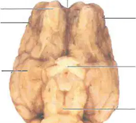
In the ventral view of the sheep brain diagram, identify the structure labeled as 'olfactory bulb'.
olfactory bulb
optic chiasm
cerebellum
medulla oblongata
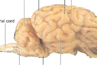
In the lateral view of the sheep brain diagram, which structure is labeled as 'cerebellum'?
cerebellum
optic chiasm
olfactory bulb
medulla oblongata
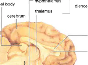
In the longitudinal section of the sheep brain, what is the name of the structure labeled as 'corpus callosum'?
corpus callosum
thalamus
cerebellum
medulla oblongata
Match the following brain structures to their correct location in the sheep brain diagram:
Located at the base of the brainstem
A-1
Located above the medulla oblongata
B-2
Located where the optic nerves cross
C-3
Located at the back of the brain, below the cerebrum
D-4
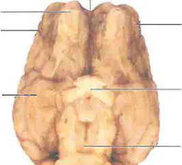
In the ventral view of the sheep brain, which structure is found between the right and left cerebral hemispheres?
longitudinal fissure
optic chiasma
cerebellum
olfactory bulb
Based on your knowledge of the sheep brain, complete Table 31.1 by stating the major functions for each part of the brain listed. Also label Figure 31.3. Table 31.1 Summary of Brain Functions Part: Cerebrum Major Functions: ____________
Higher brain functions such as thought and action
Regulation of heart rate and breathing
Coordination of balance and movement
Control of hormone secretion
Based on your knowledge of the sheep brain, complete Table 31.1 by stating the major functions for each part of the brain listed. Also label Figure 31.3. Table 31.1 Summary of Brain Functions Part: Cerebellum Major Functions: ____________
Coordination of voluntary movements, balance, and posture
Regulation of heart rate and breathing
Processing visual information
Control of hormone secretion
Based on your knowledge of the sheep brain, complete Table 31.1 by stating the major functions for each part of the brain listed. Also label Figure 31.3.
Relay station for sensory impulses to the cerebral cortex
Controls voluntary muscle movements
Regulates heartbeat and breathing
Responsible for vision processing
Based on your knowledge of the sheep brain, complete Table 31.1 by stating the major functions for each part of the brain listed. Also label Figure 31.3. Table 31.1 Summary of Brain Functions Part: Hypothalamus Major Functions: ____________
Regulation of body temperature, hunger, thirst, and hormone secretion
Control of voluntary muscle movements and balance
Processing of visual and auditory information
Coordination of heart rate and breathing
Based on your knowledge of the sheep brain, complete Table 31.1 by stating the major functions for each part of the brain listed. Also label Figure 31.3. Table 31.1 Summary of Brain Functions Part: Midbrain Major Functions: ____________
Relay station for sensory input and motor output; reflex center for eye muscles
Regulates heartbeat and breathing
Controls balance and coordination
Responsible for higher cognitive functions
Based on your knowledge of the sheep brain, complete Table 31.1 by stating the major functions for each part of the brain listed. Also label Figure 31.3. Table 31.1 Summary of Brain Functions Part: Pons Major Functions: ____________
Passageway for nerve impulses running between the medulla and higher brain regions
Regulates visual processing and interpretation
Controls voluntary muscle movements in the limbs
Responsible for hormone secretion and regulation
Based on your knowledge of the sheep brain, complete Table 31.1 by stating the major functions for each part of the brain listed. Also label Figure 31.3. Table 31.1 Summary of Brain Functions Part: Medulla oblongata Major Functions: ____________
Controls internal organs; blood pressure, cardiac, and breathing control centers
Responsible for vision and visual processing
Regulates voluntary muscle movements and balance
Processes auditory information
Which parts of the brain work together to achieve good eye-hand coordination?
Cerebellum and cerebrum
Medulla and pons
Thalamus and hypothalamus
Frontal lobe and occipital lobe
Which parts of the brain work together to achieve the following? 2. Concentrating on homework when TV is playing
Cerebrum and thalamus
Cerebellum and medulla
Hypothalamus and pons
Amygdala and hippocampus
Which parts of the brain work together to achieve the following? 3. Avoiding dark alleys while walking home at night
Cerebrum and midbrain
Cerebellum and medulla
Hypothalamus and pons
Thalamus and cerebellum
Which parts of the brain work together to achieve the following? 4. Keeping the blood pressure within the normal range
Medulla oblongata and hypothalamus
Cerebellum and thalamus
Frontal lobe and occipital lobe
Pons and amygdala
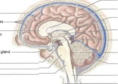
Figure 31.3 The human brain (longitudinal section). The cerebrum is larger in humans than in sheep. Label where indicated. 1. __________________
skull
cerebellum
spinal cord
medulla oblongata
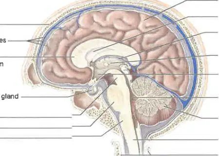
Label where indicated.
meninges
cerebrum
spinal cord
ventricle
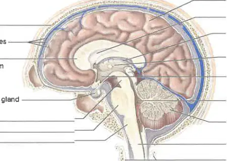
Label where indicated. 3. __________________
corpus callosum
thalamus
cerebellum
medulla oblongata
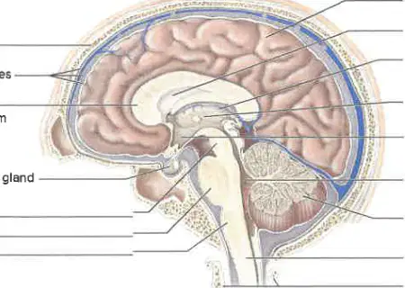
Label where indicated.
pituitary gland
thyroid gland
adrenal gland
pancreas

Figure 31.3 The human brain (longitudinal section). The cerebrum is larger in humans than in sheep. Label where indicated. 5. __________________
lateral ventricle
thalamus
cerebellum
medulla oblongata
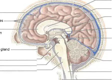
Figure 31.3 The human brain (longitudinal section). The cerebrum is larger in humans than in sheep. Label where indicated. 6. __________________
third ventricle
corpus callosum
cerebellum
optic chiasma
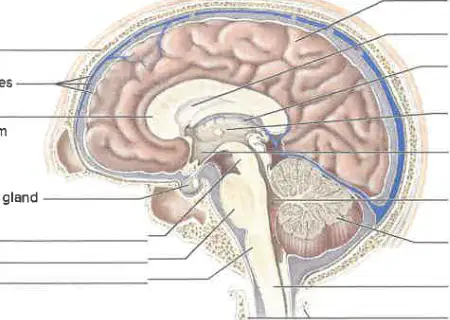
Label where indicated. 7. __________________
fourth ventricle
third ventricle
lateral ventricle
cerebral aqueduct

Figure 31.3 The human brain (longitudinal section). The cerebrum is larger in humans than in sheep. Label where indicated. 8. __________________
spinal cord
cerebellum
medulla oblongata
thalamus
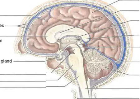
Figure 31.3 The human brain (longitudinal section). The cerebrum is larger in humans than in sheep. Label where indicated. 9. __________________
vertebra
cerebellum
medulla oblongata
thalamus
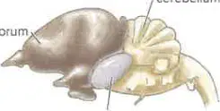
Observation: Comparison of Vertebrate Brains. 1. Examine the brain of a fish, amphibian, bird, and mammal (Fig. 31.4). Which of the following groups is being compared in this observation?
Fish, amphibian, bird, and mammal
Reptile, insect, bird, and mammal
Fish, reptile, amphibian, and insect
Mammal, reptile, amphibian, and insect
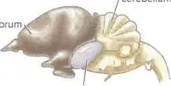
Observation: Comparison of Vertebrate Brains. 2. Which of the following correctly compares the sizes of the forebrain, midbrain, and hindbrain in vertebrates? a. Forebrain: Contains olfactory bulb and cerebrum. b. Midbrain: Contains optic lobe (and other structures). c. Hindbrain: Contains cerebellum (and other structures).
The forebrain is generally larger than the midbrain and hindbrain.
The midbrain is the largest among the three regions.
The hindbrain is always larger than the forebrain and midbrain.
All three regions are typically the same size.
Record your observations for the following vertebrates: Fish (shark): __________
The midbrain is the most prominent region; cerebrum has an olfactory function.
The cerebellum is the largest part; cerebrum controls vision.
The cerebrum is highly developed; midbrain is reduced.
The spinal cord is the most prominent region; cerebrum controls movement.
Record your observations for the following vertebrates: Amphibian (frog): __________
The midbrain is the most prominent region; cerebrum has an olfactory function.
The cerebellum is the largest part; olfactory lobes are absent.
The forebrain is highly developed; midbrain is reduced.
The spinal cord is the most prominent region; cerebrum is absent.
Record your observations for the following vertebrates: Bird (goose): __________
Cerebrum becomes increasingly complex; well-developed cerebellum associated with agility.
Cerebrum is underdeveloped; cerebellum is absent.
Cerebrum and cerebellum are both poorly developed.
Cerebrum is highly convoluted; cerebellum is rudimentary.
Record your observations for the following vertebrates: Mammal (horse): __________
Cerebrum becomes increasingly complex; well-developed cerebellum associated with agility.
Cerebrum is absent; only brainstem is present.
Cerebellum is underdeveloped; limited coordination.
No nervous system; movement is purely reflexive.
Identify the following with the help of Figure 31.5: Gray matter: __________
A central, butterfly-shaped area composed of masses of short nerve fibers, interneurons, and motor neuron cell bodies.
A peripheral layer containing only sensory neuron axons.
A region filled with cerebrospinal fluid and blood vessels.
An outer layer made up of connective tissue and fat.
Identify the following with the help of Figure 31.5: White matter: __________
Masses of long fibers that lie outside the gray matter and carry impulses up and down the spinal cord. In animals, white matter appears white because an insulating myelin sheath surrounds long fibers.
Clusters of neuron cell bodies that process sensory information.
Thin layers of connective tissue that protect the spinal cord.
Regions containing only blood vessels and no nerve fibers.
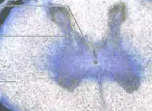
Refer to Figure 31.5 The spinal cord. Which part of the spinal cord is labeled as the central canal?
A) The outermost region
B) The butterfly-shaped area
C) The small central area
D) The white matter
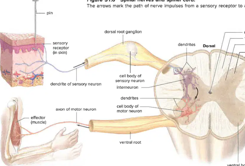
Suppose you were walking barefoot and stepped on a locust thorn. Which of the following best describes the pathway of information, starting with the pain receptor in your foot, that would allow you to both feel and respond to this painful stimulus?
Pain receptor → sensory neuron → spinal cord → interneuron → motor neuron → muscle
Pain receptor → motor neuron → spinal cord → sensory neuron → muscle
Pain receptor → spinal cord → sensory neuron → interneuron → muscle
Pain receptor → interneuron → sensory neuron → spinal cord → muscle
It is important for the legs to hang freely during the knee-jerk reflex test because:
it allows the reflex to occur without resistance.
it makes the muscles contract more forcefully.
it prevents the reflex from happening.
it helps the patient relax completely.
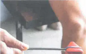
The expected response when the patellar tendon is tapped is:
Flexion of the knee
Extension of the knee
No response
Dorsiflexion of the foot
In this relaxed state, does the leg flex (move toward the buttocks) or extend (move away from the buttocks)?
The leg extends (moves away from the buttocks).
The leg flexes (moves toward the buttocks).
The leg rotates inward.
The leg remains stationary.
The function of each ommatidium in the compound eye is:
to detect a part of the visual field
to produce sound
to digest food
to regulate temperature
The function of the single lens in a squid's eye is:
to focus light onto the retina
to detect color changes
to produce light for camouflage
to filter out harmful UV rays
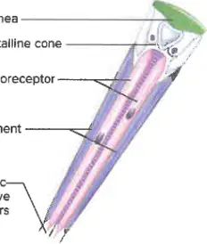
Which part of the compound eye of a fly focuses light onto photoreceptor cells?
Cornea
Crystalline cone
Pigment cells
Optic nerve fibers
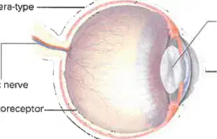
Refer to Figure 31.9. The squid has a camera-type eye similar to those of vertebrates. Which structure in both the squid and vertebrate eye is responsible for transmitting nerve impulses to the brain?
Lens
Cornea
Optic nerve
Photoreceptor cells
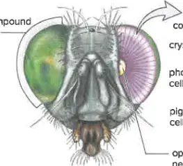
Fill in the blank: In the compound eye of a fly, the ________ cells generate nerve impulses transmitted to the brain, where interpretation produces a mosaic image.
photoreceptor
epidermal
muscle
gland
Observation: Invertebrate Eyes
1. Examine the demonstration slide of a compound eye set up under a stereomicroscope or examine a model of a compound eye.
What type of eye structure is being observed in this demonstration?
Compound eye
Simple eye
Camera-type eye
Pinhole eye
Observation: Invertebrate Eyes
2. Examine the eyes of any other invertebrates on display. What is a common feature of invertebrate eyes?
They often have compound eyes.
They have eyelids.
They can see in complete darkness.
They are always blue in color.
Anatomy of the Human Eye
Observation: The Human Eye
Which of the following is a structure you would identify when examining a human eye model, as listed in Table 31.3 and depicted in Figure 31.10?
Cornea
Femur
Patella
Humerus
Trace the path of light from outside the eye to the retina.
Cornea → Aqueous humor → Pupil → Lens → Vitreous humor → Retina
Cornea → Lens → Aqueous humor → Pupil → Vitreous humor → Retina
Cornea → Pupil → Lens → Aqueous humor → Vitreous humor → Retina
Cornea → Vitreous humor → Lens → Pupil → Aqueous humor → Retina
During accommodation, the lens rounds up to aid in viewing near objects or flattens to aid in viewing distant objects. Which structure holds the lens and is involved in accommodation?
Ciliary body (or ciliary muscle)
Retina
Cornea
Optic nerve
Refraction is the bending of light rays so that they can be brought to a single focus. Which of the structures listed in Table 31.3 aid in refracting and focusing light rays?
Cornea and lens
Retina and optic nerve
Sclera and choroid
Vitreous humor and aqueous humor
Specifically, what are the sensory receptors for sight, and where are they located in the eye?
Rods and cones; located in the retina
Hair cells; located in the cochlea
Olfactory bulbs; located in the nasal cavity
Taste buds; located on the tongue
What structure takes nerve impulses to the brain from the rod cells and cone cells?
Optic nerve
Retina
Cornea
Lens
Which cerebral lobe processes nerve impulses from an eye?
Occipital lobe
Frontal lobe
Temporal lobe
Parietal lobe
What is the location of the Sclera in the human eye?
Outer layer of eye
Inner layer of eye
Lens of eye
Retina of eye
What is the function of the Sclera in the human eye?
Protects and supports eyeball
Regulates the amount of light entering the eye
Focuses light onto the retina
Produces tears
What is the location of the Cornea in the human eye?
Transparent portion of sclera
Retina
Lens
Optic nerve
What is the function of the Cornea in the human eye?
Refracts light rays
Produces tears
Controls eye movement
Detects color
What is the location of the Choroid in the human eye?
Middle layer of eye
Outer layer of eye
Inner layer of eye
Lens of eye
What is the function of the Choroid in the human eye?
Absorbs stray light rays
Regulates the size of the pupil
Focuses light onto the retina
Produces aqueous humor
What is the location of the Retina in the human eye?
Inner layer of eye
Outer layer of eye
Lens of eye
Cornea of eye
What is the function of the Retina in the human eye?
Contains receptors for sight
Produces tears
Controls the shape of the lens
Regulates the amount of light entering the eye
What is the location of Rod cells in the human eye?
In retina
In cornea
In lens
In iris
What is the function of Rod cells in the human eye?
Make black-and-white vision possible
Detect color vision
Control the size of the pupil
Produce tears
What is the location of Cone cells in the human eye?
Concentrated in fovea centralis
Scattered throughout the sclera
Located only in the optic nerve
Found mainly in the cornea
What is the function of Cone cells in the human eye?
Make color vision possible
Help in hearing
Aid in digestion
Regulate body temperature
Match the following parts of the human eye with their functions:
Protects and supports eyeball
Sclera
Refracts light rays
Cornea
Absorbs stray light rays
Choroid
Contains receptors for sight
Retina
Make black-and-white vision possible
Rod cells
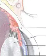
Which part of the human eye is responsible for focusing light onto the retina?
Lens
Sclera
Iris
Choroid
With your partner's help, measure the distance from your eye to the paper when the circle first disappeared. Left eye: ________ cm
(The answer will be the measured distance in cm, which varies for each student.)
10 cm
50 cm
100 cm
With your partner's help, measure the distance from your eye to the paper when the cross first disappeared. Right eye: ________ cm
(The answer will be the measured distance in cm, which varies for each student.)
10 cm
100 cm
0 cm
You are unaware of a blind spot under normal conditions because:
your brain fills in the missing information from the surrounding area
the blind spot is too small to notice
your eyes move constantly to cover the blind spot
light never falls on the blind spot
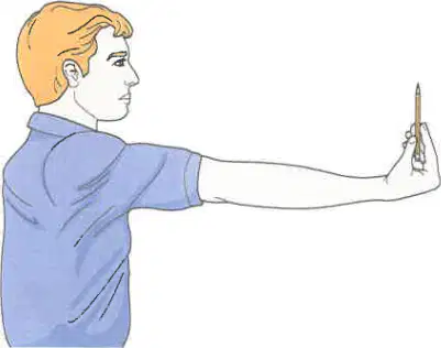
Experimental Procedure: Accommodation of the Eye Step 5: Move the pencil toward you until the end is out of focus. Measure the distance (in centimeters) between the pencil and your eye: _______ cm
10 cm
5 cm
20 cm
Student should fill in the measured distance in cm
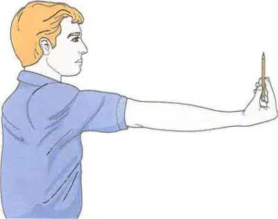
At what distance can your eye no longer accommodate for distance?
Student should fill in the measured distance in cm
10 cm
100 cm
1 cm
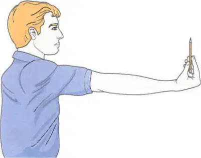
If you wear glasses, repeat this experiment without your glasses, and note the accommodation distance of your eye without glasses: _______ cm
(Student should fill in the measured distance in cm or write 'contact lens' if applicable)
10 cm
100 cm
0.1 cm
How 'old' is the eye you tested? (Use the measured near point distance and match it to the age in the table below)
(Student should match their measured near point to the closest age in the table)
20 years old
50 years old
70 years old

The function of the tympanum in the grasshopper is:
to detect sound vibrations
to help in digestion
to aid in flying
to assist in reproduction
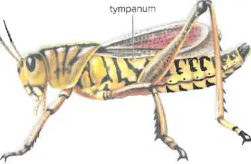
Which of the following animals has a tympanum for detecting sound waves?
Fish
Grasshopper
Human
Spider
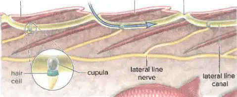
Fill in the blank: The lateral line system in fishes contains hair cells with cilia embedded in a _________?
gelatinous cupula
bony capsule
fibrous sheath
fatty layer
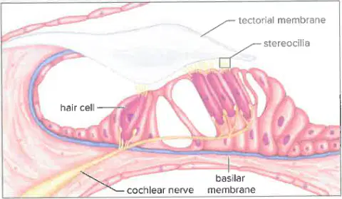
Fill in the blank: In the human ear, hair cells have stereocilia embedded in the ________ membrane.
tectorial
basilar
Reissner's
tympanic
Match the parts of the human ear with their correct function:
Collects sound waves
Pinna
Filters air
Auditory canal
Amplifies sound waves
Tympanic membrane and ossicles
Equalizes air pressure
Auditory tube
Rotational equilibrium
Semicircular canals

