
NEW
Font size
 Worksheets
Figure 12.1 Head and neck muscles
Total questions: 70
Worksheet time: 35mins
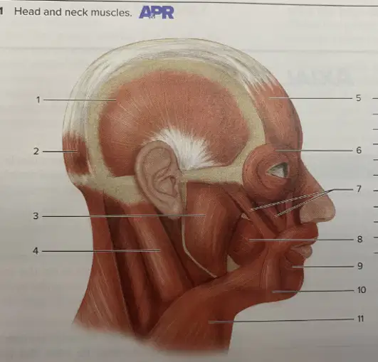
Name the structure identified with number 10.
Depressor anguli oris
Masseter
Zygomaticus major
Orbicularis oris
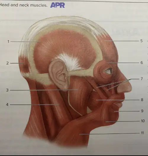
Name the structure identified with number 5.
Frontalis
Occipitalis
Temporalis
Masseter
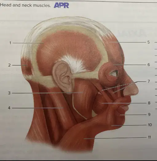
Name the structure identified with number 3.
Masseter
Temporalis
Buccinator
Sternocleidomastoid
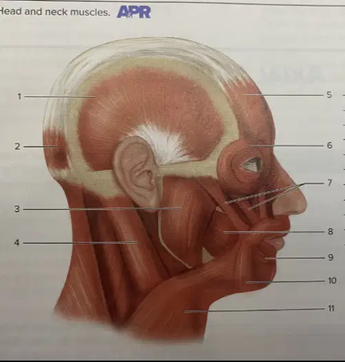
Name the structure identified with number 2.
Occipitalis
Frontalis
Temporalis
Sternocleidomastoid
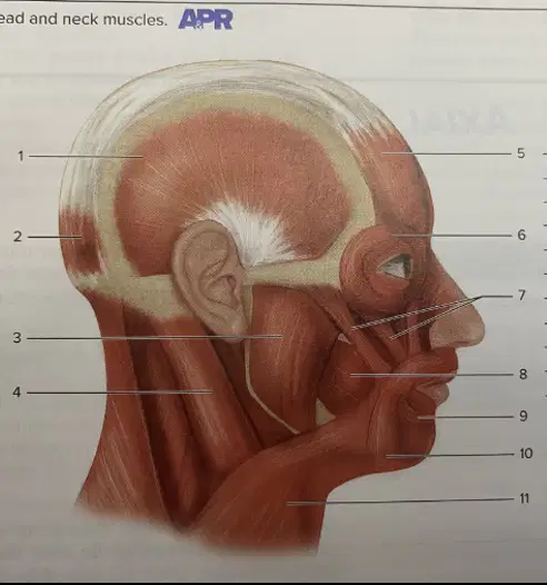
Name the structure identified with number 6.
Orbicularis oculi
Frontalis
Zygomaticus
Masseter
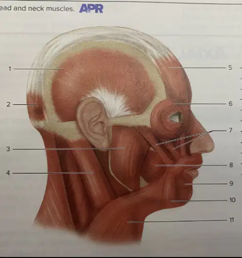
Name the structure identified with number 11.
Platysma
Sternocleidomastoid
Masseter
Temporalis
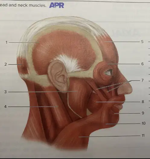
Name the structure identified with number 4.
Sternocleidomastoid
Trapezius
Platysma
Masseter
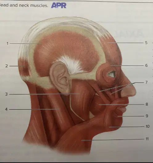
Name the structure identified with number 1.
Temporalis
Masseter
Sternocleidomastoid
Orbicularis oculi
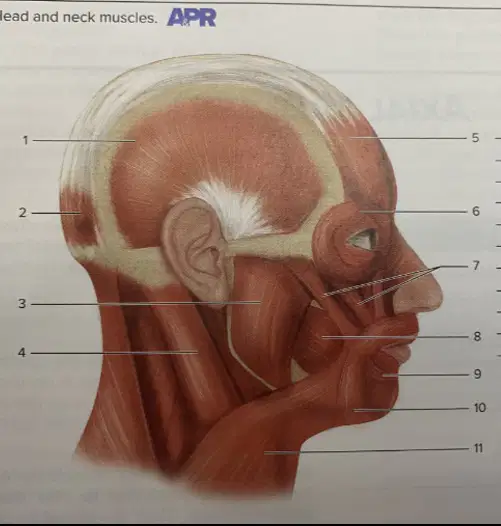
Name the structure identified with number 7.
Zygomaticus
Orbicularis oculi
Masseter
Buccinator
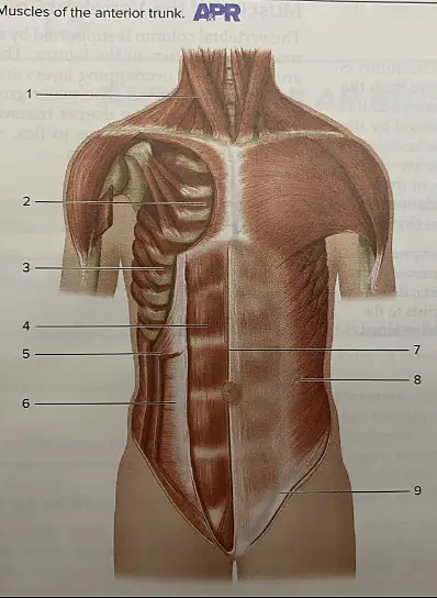
Name the structure identified with number 1.
Sternocleidomastoid
External intercostal
Internal oblique
Rectus abdominis
Name the structure identified with number 2.
Internal intercostal
External intercostal
Internal oblique
Transverse abdominis
Name the structure identified with number 3.
External intercostal
Internal intercostal
External oblique
Linea alba
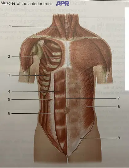
Name the structure identified with number 4.
Rectus abdominis
Transverse abdominis
Internal oblique
External oblique
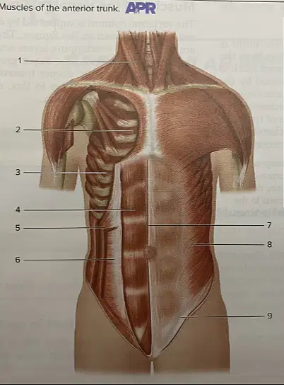
Name the structure identified with number 5.
Internal oblique
External intercostal
Linea alba
Inguinal ligament

Name the structure identified with number 6.
Transverse abdominis
External oblique
Rectus abdominis
Internal intercostal
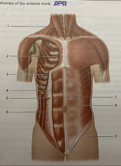
Name the structure identified with number 7.
Linea alba
Internal oblique
Transverse abdominis
External intercostal
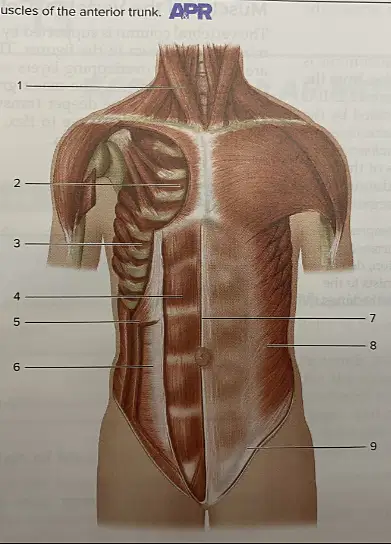
Name the structure identified with number 8.
External oblique
Internal oblique
Transverse abdominis
Rectus abdominis
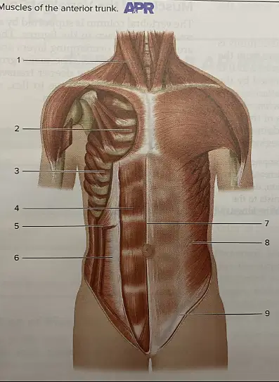
Name the structure identified with number 9.
Inguinal ligament
Sternocleidomastoid
Linea alba
External intercostal
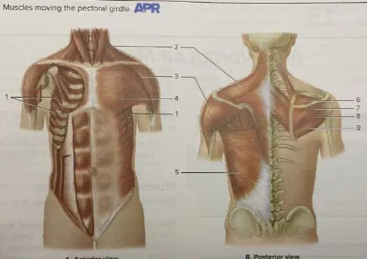
Name the structure identified with number 1.
Serratus anterior
Deltoid
Pectoralis major
Trapezius
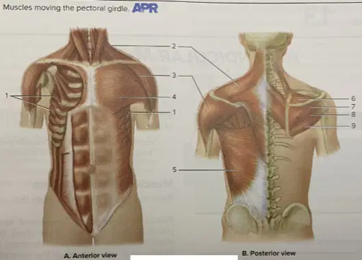
Name the structure identified with number 2.
Trapezius
Deltoid
Latissimus dorsi
Pectoralis major
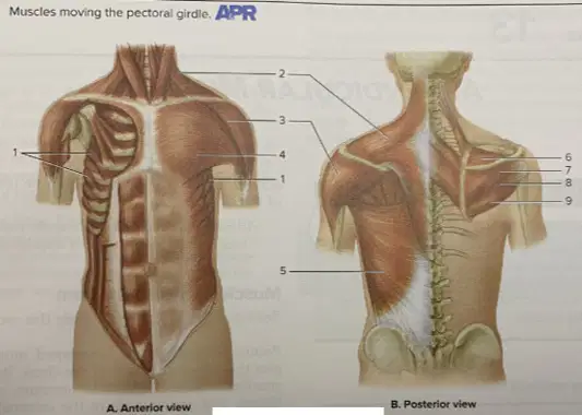
Name the structure identified with number 3.
Deltoid
Serratus anterior
Supraspinatus
Teres minor
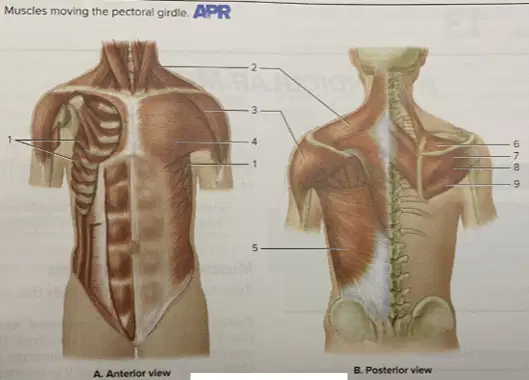
Name the structure identified with number 4.
Pectoralis major
Trapezius
Infraspinatus
Latissimus dorsi
Name the structure identified with number 5.
Latissimus dorsi
Teres major
Infraspinatus
Supraspinatus
Name the structure identified with number 6.
Supraspinatus
Infraspinatus
Teres minor
Deltoid
Name the structure identified with number 7.
Infraspinatus
Supraspinatus
Teres major
Latissimus dorsi

Name the structure identified with number 8.
Teres minor
Infraspinatus
Deltoid
Trapezius
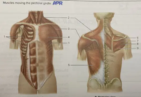
Name the structure identified with number 9.
Teres major
Teres minor
Supraspinatus
Pectoralis major
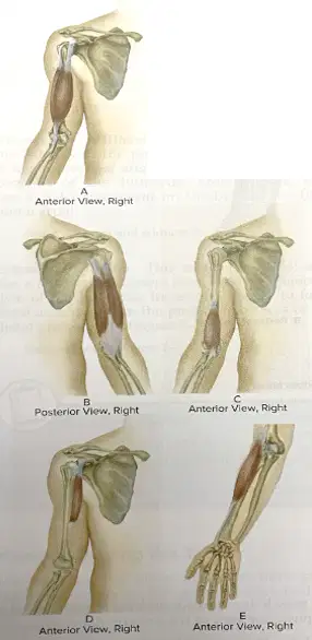
Name the structure identified with number A in the anterior view of the right upper limb.
Biceps brachii
Triceps brachii
Brachialis
Coracobrachialis
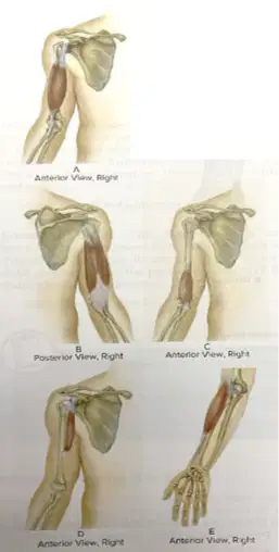
Figure showing posterior view of the right upper limb labeled B. Which muscle attached to the humerus is labeled B?
Brachioradialis
Biceps brachii
Triceps brachii
Coracobrachialis
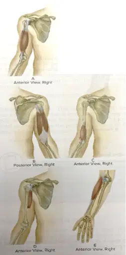
Figure showing anterior view of the right upper limb labeled C. Which muscle attached to the humerus is labeled C?
Brachialis
Coracobrachialis
Triceps brachii
Brachioradialis
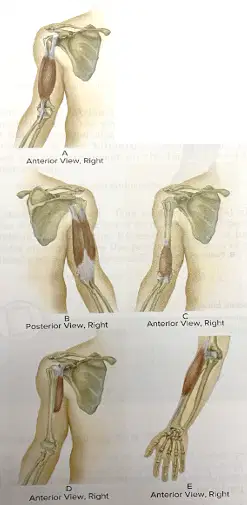
Figure showing anterior view of the right upper limb labeled D. Which muscle attached to the humerus is labeled D?
Coracobrachialis
Brachialis
Biceps brachii
Triceps brachii
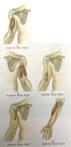
Figure showing anterior view of the right forearm labeled E. Which muscle attached to the humerus is labeled E?
Brachioradialis
Brachialis
Coracobrachialis
Triceps brachii
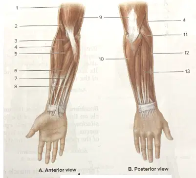
In the anterior view of the forearm diagram, which muscle is indicated by label 1 on the upper arm just above the elbow flexors?
Biceps brachii
Triceps brachii
Extensor carpi ulnaris
Extensor digitorum
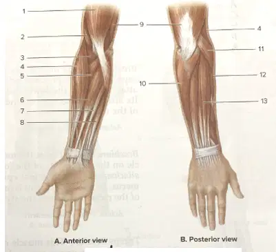
In the anterior view of the forearm diagram, which muscle is indicated by label 2 deep to the biceps near the anterior distal humerus?
Brachialis
Brachioradialis
Pronator teres
Palmaris longus
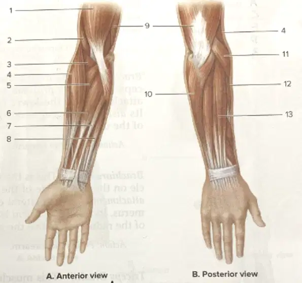
In the anterior view of the forearm diagram, which muscle is indicated by label 4 on the lateral (radial) side of the forearm?
Brachioradialis
Flexor carpi radialis
Palmaris longus
Flexor carpi ulnaris
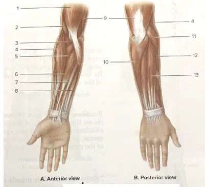
In the anterior view of the forearm diagram, which muscle is indicated by label 5 crossing obliquely from the medial epicondyle toward the radius?
Pronator teres
Palmaris longus
Flexor carpi radialis
Supinator
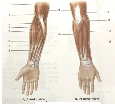
In the anterior view of the forearm diagram, which muscle is indicated by label 6 running along the anterior forearm toward the base of the second and third metacarpals?
Flexor carpi radialis
Palmaris longus
Flexor carpi ulnaris
Extensor carpi radialis longus
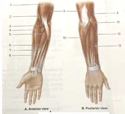
In the anterior view of the forearm diagram, which muscle is indicated by label 7 as a slender tendon in the midline of the wrist?
Palmaris longus
Flexor carpi radialis
Flexor carpi ulnaris
Brachioradialis
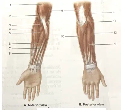
Name the structure identified with number 8.
Flexor carpi ulnaris
Palmaris longus
Flexor carpi radialis
Pronator teres
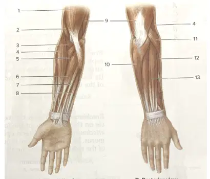
Name the structure identified with number 9.
Triceps brachii
Biceps brachii
Brachialis
Coracobrachialis
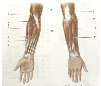
Name the structure identified with number 10.
Flexor carpi radialis
Extensor digitorum
Extensor carpi radialis longus
Extensor carpi radialis brevis
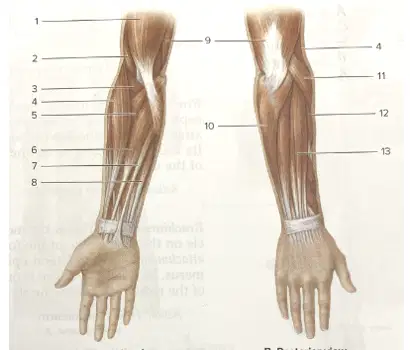
Name the structure identified with number 11.
Extensor digitorum
Extensor carpi ulnaris
Extensor carpi radialis longus
Extensor carpi radialis brevis
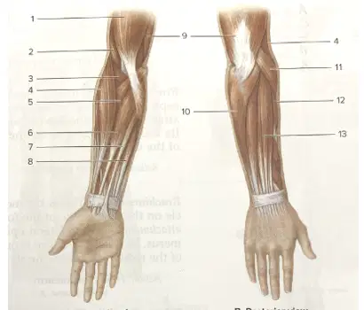
Name the structure identified with number 12.
Extensor carpi radialis longus
Extensor carpi radialis brevis
Extensor carpi ulnaris
Brachioradialis
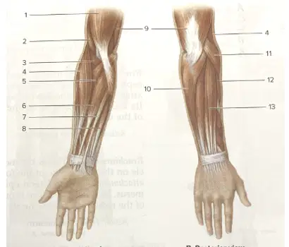
Name the structure identified with number 13 in the posterior view of the forearm diagram.
Extensor carpi radialis brevis
Extensor carpi radialis longus
Extensor digitorum
Flexor carpi radialis
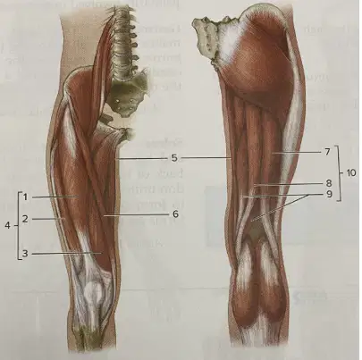
Name the structure identified with number 1.
Rectus femoris
Vastus lateralis
Vastus medialis
Sartorius
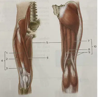
Name the structure identified with number 2.
Vastus lateralis
Vastus medialis
Rectus femoris
Gracilis
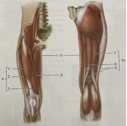
Name the structure identified with number 3.
Vastus medialis
Vastus lateralis
Sartorius
Rectus femoris
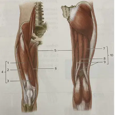
Name the structure identified with number 4.
Quadriceps femoris
Hamstrings
Adductor longus
Iliotibial tract
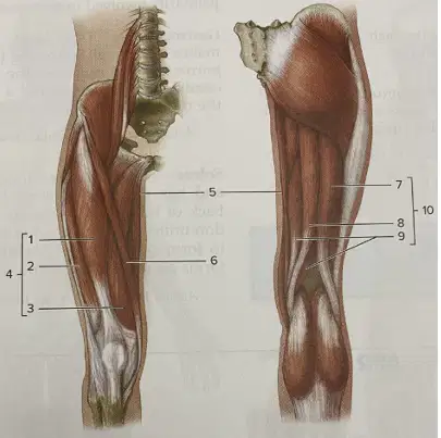
In the thigh anatomy diagram with labels, anterior view (A), identify the muscle labeled 5.
Gracilis
Sartorius
Rectus femoris
Pectineus
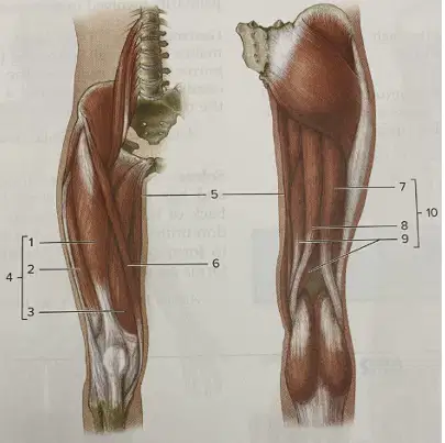
In the thigh anatomy diagram with labels, anterior view (A), identify the muscle labeled 6.
Sartorius
Gracilis
Vastus medialis
Adductor longus
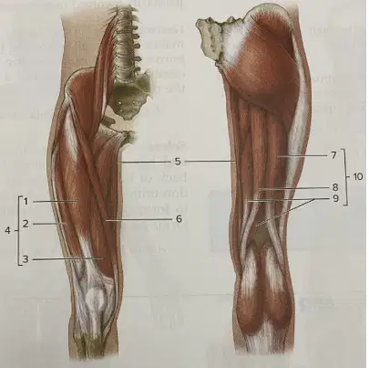
In the thigh anatomy diagram with labels, posterior view (B), identify the muscle labeled 7.
Biceps femoris
Semitendinosus
Semimembranosus
Gluteus maximus
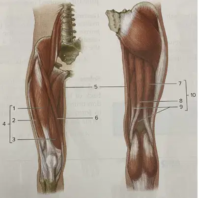
In the thigh anatomy diagram with labels, posterior view (B), identify the muscle labeled 8.
Semimembranosus
Semitendinosus
Biceps femoris
Adductor magnus
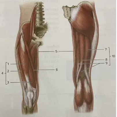
In the thigh anatomy diagram with labels, posterior view (B), identify the muscle labeled 9.
Semitendinosus
Semimembranosus
Biceps femoris
Gracilis
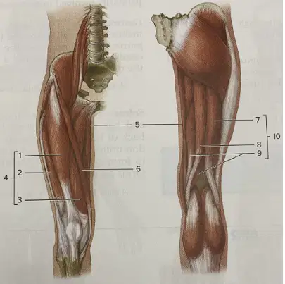
In the thigh anatomy diagram with labels, posterior view (B), identify the structure labeled 10.
Hamstrings
Quadriceps femoris
Iliotibial tract
Adductor group
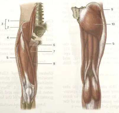
In the hip and proximal thigh anatomy diagram with labels, anterior view (A), identify the muscle labeled 1.
Psoas major
Iliacus
Iliopsoas
Tensor fasciae latae
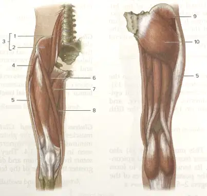
In the hip and proximal thigh anatomy diagram with labels, anterior view (A), identify the muscle labeled 2.
Iliacus
Psoas major
Pectineus
Adductor longus
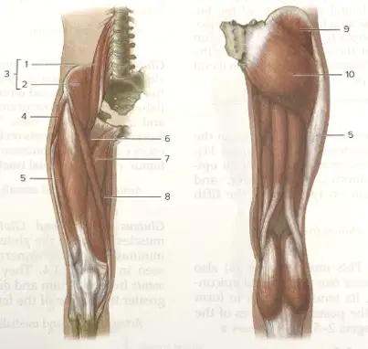
In the hip and proximal thigh anatomy diagram with labels, anterior view (A), identify the structure labeled 3.
Iliopsoas
Tensor fasciae latae
Iliotibial tract
Gluteus medius
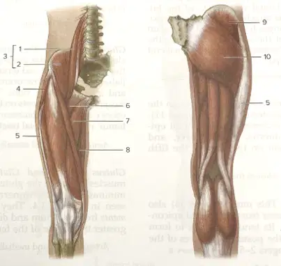
In the hip and proximal thigh anatomy diagram with labels, anterior view (A), identify the muscle labeled 4.
Tensor fasciae latae
Pectineus
Adductor longus
Gluteus medius
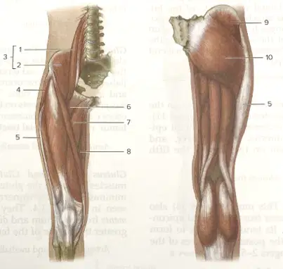
In the hip and proximal thigh anatomy diagram with labels, lateral aspect, identify the structure labeled 5.
Iliotibial tract
Gluteus maximus
Tensor fasciae latae
Psoas major
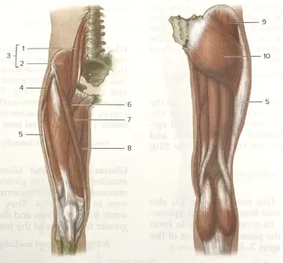
In the hip and proximal thigh anatomy diagram with labels, anterior view (A), identify the muscle labeled 6.
Pectineus
Adductor longus
Iliacus
Sartorius
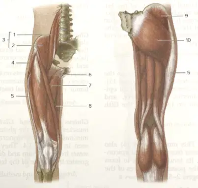
In the hip and proximal thigh anatomy diagram with labels, medial thigh, identify the muscle labeled 7.
Adductor longus
Adductor magnus
Pectineus
Gracilis
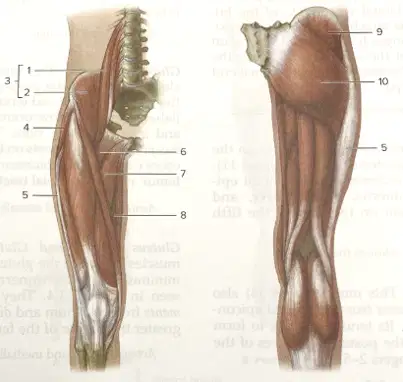
In the hip and proximal thigh anatomy diagram with labels, medial thigh, identify the muscle labeled 8.
Adductor magnus
Adductor longus
Gracilis
Gluteus medius
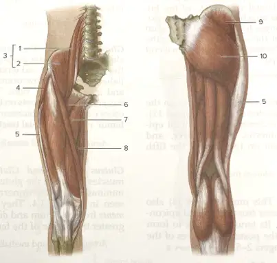
In the hip and gluteal region diagram with labels, posterior view (B), identify the muscle labeled 9.
Gluteus medius
Gluteus maximus
Tensor fasciae latae
Iliotibial tract
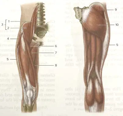
In the hip and gluteal region diagram with labels, posterior view (B), identify the muscle labeled 10.
Gluteus maximus
Gluteus medius
Adductor magnus
Iliotibial tract
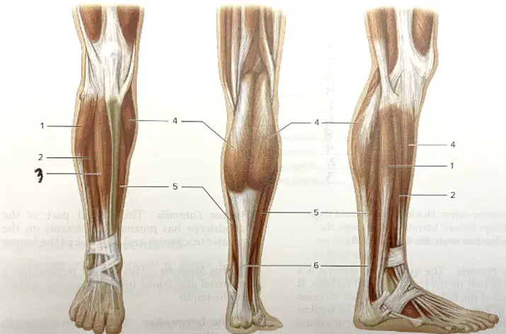
In the diagram of the lower leg showing three views (anterior, posterior, lateral) with numbered labels 1–6, identify the structure labeled 1.
Fibularis longus
Gastrocnemius
Soleus
Tibialis anterior
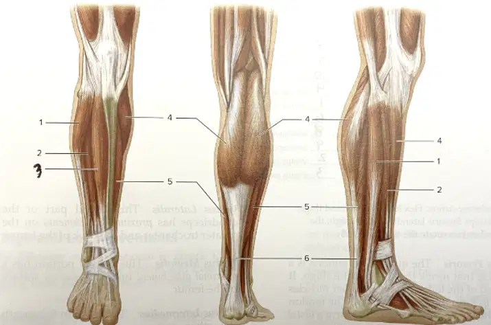
In the diagram of the lower leg showing three views (anterior, posterior, lateral) with numbered labels 1–6, identify the structure labeled 2.
Extensor digitorum longus
Tibialis anterior
Fibularis longus
Soleus
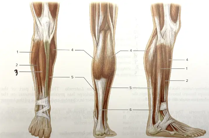
In the diagram of the lower leg showing three views (anterior, posterior, lateral) with numbered labels 1–6, identify the structure labeled 3.
Tibialis anterior
Extensor digitorum longus
Fibularis longus
Gastrocnemius
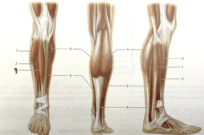
In the diagram of the lower leg showing three views (anterior, posterior, lateral) with numbered labels 1–6, identify the structure labeled 4.
Gastrocnemius
Soleus
Fibularis longus
Calcaneal tendon
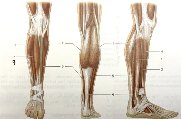
In the diagram of the lower leg showing three views (anterior, posterior, lateral) with numbered labels 1–6, identify the structure labeled 5.
Soleus
Gastrocnemius
Tibialis anterior
Calcaneal tendon
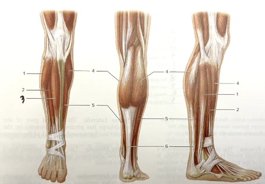
In the diagram of the lower leg showing three views (anterior, posterior, lateral) with numbered labels 1–6, identify the structure labeled 6.
Calcaneal tendon
Gastrocnemius
Soleus
Fibularis longus

