
 Worksheets
Patient Preparation and History
Total questions: 68
Worksheet time: 34mins
Which patient history detail most directly assists with menstrual cycle dating in pelvic sonography?
Date of last menstrual period
Gravidity and live birth count
Current hormone replacement plan
Prior pelvic surgical history
For a 28-year-old presenting with pelvic pain, which combination of history elements is MOST critical to guide a targeted pelvic ultrasound?
Last menstrual period timing
Current pelvic symptoms
Family history of ovarian cancer
Number of prior pregnancies and births
Recent Pap smear laboratory results
Gravidity and parity are obtained during preparation because they primarily inform which aspect of pelvic sonography interpretation?
Reproductive history and uterine changes
Medication adherence over months
Risk of immediate hemodynamic collapse
Likelihood of urinary tract anomalies
A patient had a hysterectomy two years ago. Which history element becomes most important to avoid misinterpretation of absent uterine structures?
Past pelvic surgeries
Family cancer risks
Recent biopsy results
Parity documentation
During intake for pelvic sonography, which findings should be documented to correlate with imaging and guide differential diagnosis?
Pelvic examination observations
Current symptoms description
Gravidity and parity counts
Family cancer history
Blood pressure thresholds
Family history is obtained before pelvic ultrasound primarily to assess which consideration?
Genetic risk for gynecologic cancers
Immediate need for IV hydration
Probability of bowel obstruction
Suitability for contrast agents
On ultrasound, how do the rectus abdominis muscles most typically appear in the anterior abdominal wall?
Hypoechoic bands with echogenic striations
Anechoic tubes without internal echoes
Uniformly echogenic slabs with shadowing
Heterogeneous masses with posterior enhancement
Which statement best describes the rectus abdominis muscles in relation to abdominal wall anatomy?
Paired parasagittal straps inserting on pubic rami
Single midline sheet attaching to iliac crests
Oblique paired bands inserting on xiphoid alone
Transverse sheet spanning from ribs to sacrum
What is the primary sonographic role of the rectus sheath in abdominal wall imaging?
Bright linear reflector separating muscle from fat and bowel
Low-level echo region enhancing bowel visualization
Anechoic plane indicating intraperitoneal free fluid
Diffuse echogenic band causing acoustic shadowing

In a transabdominal pelvic ultrasound at the level of the vagina and cervix, which structure is typically seen as hypoechoic, ovoid, and located at the posterolateral corners of the bladder?
Obturator internus muscle
Psoas major muscle
Iliacus muscle belly
Gluteus medius muscle
Which statement best describes the sonographic appearance and surrounding structures of the obturator internus muscle in the true pelvis?
Hypoechoic ovoid muscle surrounded by obturator fascia
Hyperechoic linear tendon deep to transversalis fascia
Isoechoic triangular muscle covered by thoracolumbar fascia
Heterogeneous round mass adjacent to inguinal ligament
On the provided ultrasound image, the obturator internus lies in which relative location with respect to the urinary bladder?
Posterolateral to the bladder
Anterosuperior to the bladder
Directly posterior to the bladder
Medial within the bladder lumen
Which group of structures are commonly identified within the true (lesser) pelvis in ultrasound at this level?
Urinary bladder
Reproductive organs
Levator ani muscles
Obturator internus muscles
Descending colon
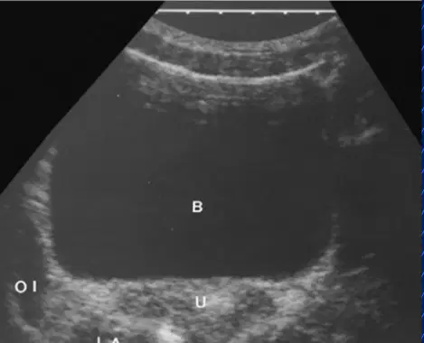
When scanning at the level of the cervix, which feature helps distinguish the obturator internus from adjacent pelvic floor muscles?
Ovoid hypoechoic shape abutting bladder corner
Bright echogenic linear fibers midline
Striated hyperechoic texture anteriorly
Cystic round structure with posterior enhancement
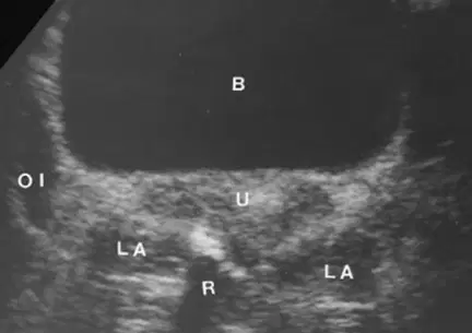
Which pairing correctly matches the labeled structures commonly seen together on the image at this level?
B with bladder
U with uterus
R with rectum
LA with levator ani
OI with obturator internus
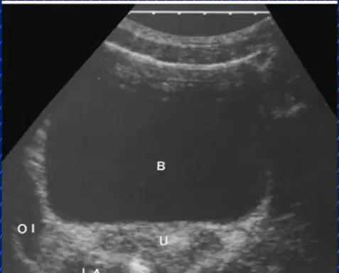
Which statement about pelvic anatomy visualization is most accurate for identifying obturator internus during sonography?
Seen at posterolateral bladder corners near cervix
Best seen anterior to pubic symphysis superiorly
Identified as echogenic fascia within perineum
Visualized within abdominal wall lateral to iliac crest
In pelvic ultrasound, which technique best visualizes the levator ani muscle as indicated by the arrows in the diagram?
Transverse plane with caudal angulation
Transverse plane with cephalic angulation
Sagittal plane with neutral angulation
Coronal plane with cranial angulation
When aiming to image the most inferior pelvic floor level, how should the transducer be oriented?
Transverse with slight caudal tilt
Transverse with slight cranial tilt
Sagittal with neutral alignment
Coronal with lateral sweeping
Why are the coccygeus and piriformis muscles not routinely distinguished on standard pelvic sonography?
They lie deep and blend with surrounding muscles
They move too rapidly to capture clearly
They are avascular and produce shadowing
They are superficial and overgain causes dropout
All of the following muscles are commonly considered part of the pelvic floor except:
Levator ani
Coccygeus
Puborectalis
Rectus abdominis
During a pelvis scan, you fail to visualize coccygeus clearly. Which adjustment is LEAST likely to help?
Increase cranial angulation to seek deeper posterior level
Use transverse plane with mild caudal tilt for levator ani
Recognize limitation that coccygeus is not routinely seen
Optimize depth and gain for posterior pelvis
In the ultrasound image labeled “Iliacus Muscle,” which relationship is correct about the iliacus at the superior two thirds of the iliac fossa?
It arises anterior to psoas major
It arises posterior to psoas major
It inserts on the greater trochanter
It is isolated from psoas major fascia
Which statement best describes the iliopsoas muscle as visualized in the pelvis on ultrasound?
It is the iliacus and psoas major combined
It is the psoas minor and iliacus only
It is the sartorius and iliacus complex
It is the tensor fasciae latae and psoas
Where does the iliopsoas tendon insert after coursing anterolaterally in the caudal direction?
Lesser trochanter of femur
Greater trochanter of femur
Anterior superior iliac spine
Pubic symphysis
On a transverse pelvic ultrasound, a hypoechoic band deep to the iliac fossa fascia is labeled with arrows. Which identification is most accurate?
Gluteus medius muscle belly
Iliacus muscle belly
Obturator internus muscle
Quadratus lumborum muscle
Select all accurate statements about the iliacus and iliopsoas complex.
Iliacus is contiguous with psoas major
The combined muscle continues caudally into the pelvis
The combined muscle courses posteromedially to the acetabulum
Insertion occurs on the lesser trochanter
A sonographer scanning the greater pelvis expects the iliopsoas to appear as:
A combined fusiform structure from iliacus and psoas major
A thin echogenic line superficial to abdominal wall
A round vascular structure lateral to femoral vein
An isolated psoas major without iliacus contribution
Which component is explicitly part of the iliopsoas muscle complex?
Psoas major
Gluteus minimus
Rectus femoris
Adductor magnus
Which arteries primarily supply blood to the uterus?
Uterine and vaginal arteries
Ovarian and renal arteries
Internal pudendal only
Common iliac branches
Which vessels provide arterial blood to the ovaries?
Ovarian arteries from the aorta
Branches of the uterine artery
Internal pudendal branches
Superior mesenteric branches
The ovarian arteries arise directly from which major vessel?
Abdominal aorta
External iliac artery
Inferior vena cava
Common hepatic artery
In color Doppler evaluation of pelvic vessels, what is the key benefit of color mapping?
Localizes the vessel for precise sample placement
Increases grayscale contrast resolution
Eliminates need for spectral analysis
Measures blood pressure noninvasively
2D imaging of pelvic vascularity is primarily used to do which of the following?
Provide anatomic context for Doppler assessment
Directly measure flow velocities in vessels
Calculate resistive and pulsatility indices
Quantify cardiac output in uterine arteries
Which statement best describes the S/D ratio in Doppler waveform analysis?
Peak systolic divided by end diastolic
End diastolic divided by mean velocity
Peak systolic minus end diastolic
Mean velocity divided by peak systolic
The Pourcelot resistive index (RI) is calculated using which formula?
(A − B) / A
A / B
(A − B) / mean
B / A
The pulsatility index (PI) is defined as which of the following?
(A − B) / mean velocity
A / mean velocity
(A + B) / mean velocity
B − A over mean velocity
When placing a spectral Doppler sample gate in a pelvic vessel, which practice optimizes accuracy?
Place gate exactly where color shows the lumen
Use the widest gate to include side walls
Avoid angle correction for small vessels
Place gate distal to the area of interest
A uterine artery waveform shows high PI and RI values. Which physiologic interpretation is most consistent?
High downstream resistance flow
Low downstream resistance flow
Arteriovenous shunting present
Measurement of venous waveform
Which labeled branch directly supplies the cervix in typical pelvic arterial anatomy?
Cervical branch of uterine artery
Tubal branch of uterine artery
Ovarian branch of uterine artery
Internal pudendal artery
In a nonpregnant uterus during the proliferative phase, which Doppler characteristic is most typical?
Low-resistance flow with RI near 0.4
High-resistance flow with RI around 0.88
Absent diastolic flow with RI undefined
Turbulent flow with variable RI values
On the day before ovulation, how does the uterine artery resistive index (RI) most commonly change from earlier proliferative values?
Increases markedly to above 1.0
Decreases slightly from earlier values
Remains exactly the same throughout
Reverses to negative diastolic values
Which statement best describes ovarian arterial flow during the follicular phase on Doppler?
Low-velocity and highly resistive waveform
High-velocity and low-resistance waveform
Turbulent high-velocity venous waveform
Monophasic continuous low-resistance waveform
Identify the vessel group often seen at the uterine periphery and its typical Doppler pattern.
Radial arteries with low-velocity, low-resistance
Arcuate vessels with high-velocity, high-resistance
Spiral arteries with absent diastolic flow
Uterine veins with triphasic arterial waveform

Which combination matches the physiologic state with its expected Doppler finding?
Nonpregnant uterine artery, resistive pattern predominant
Follicular-phase ovary, high resistance and low velocity
Arcuate uterine vessels, low-resistance diastolic augmentation
Pre-ovulatory day, slight RI decrease in uterine artery
A measured uterine artery RI of 0.82 in a nonpregnant patient most likely indicates which pattern?
Physiologic high-resistance arterial flow
Pathologic reversed diastolic flow state
Physiologic low-resistance trophoblastic flow
Venous triphasic spectral waveform
During the follicular phase, which spectral Doppler feature is most expected in the ovarian stroma?
Prominent diastolic forward flow
Minimal diastolic flow with sharp systolic peaks
To-and-fro flow with spectral broadening
Continuous high-velocity laminar flow
Which change would most strongly suggest transition from early proliferative toward periovulatory uterine hemodynamics?
RI increases from 0.88 to 1.05
RI decreases modestly from 0.88 to 0.80
PSV decreases while RI remains unchanged
EDV becomes negative indicating reversal
Where are arcuate vessels located and how do they typically appear on Doppler?
Within endometrium, low-resistance continuous flow
Periphery of uterus, high-velocity high-resistance spikes
Ovarian medulla, turbulent mixed venous signals
Broad ligament, biphasic low-velocity pattern
Which pairing is least consistent with normal physiology?
Nonpregnant uterine artery RI about 0.88
Follicular-phase ovary highly resistive flow
Arcuate vessels high-velocity high-resistance
Pre-ovulatory uterine RI increases substantially
Which uterine layer forms the middle muscular component typically evaluated on ultrasound?
Myometrium
Endometrium
Perimetrium
Serosa
Basalis
On sonography, the normal myometrium should most typically appear as
Homogeneous with smooth borders
Heterogeneous with irregular walls
Anechoic with indistinct margins
Cystic with lobulated contour
Highly echogenic with shadowing
Which uterine layer is generally not visualized sonographically as a distinct structure?
Outer serosa
Inner endometrium
Middle myometrium
Junctional zone
Subendometrium
When scanning the uterus, areas of altered echotexture within the myometrium should be
Noted and measured carefully
Ignored unless very large
Assumed physiological variants
Compressed to improve detail
Left for Doppler only

In a transvaginal sagittal image of the uterus, the echogenic stripe labeled within the cavity corresponds to the
Endometrial complex
Cervical stroma
Myometrial wall
Serosal surface
Arcuate vessels
Which statement best describes the endometrium relative to the surrounding myometrium on ultrasound?
Relatively echogenic stripe
Uniformly anechoic band
Markedly hyperechoic halo
Heterogeneous hypoechoic mass
Isoechoic indistinct layer
The hypoechoic layer that creates a subendometrial halo represents
Inner myometrium adjacent to endometrium
Endometrial functional layer only
Serosal peritoneal reflection
Cervical mucosa continuation
Arcuate vessel lumens
Which attributes typically describe the endometrium’s inner layer characteristics?
Thin and compact
Relatively hypovascular
Predominantly cystic appearance
Uniformly heterogeneous texture
Consistently thicker than myometrium
Arcuate vessels are best described as
Vessels separating outer and intermediate myometrium
Branches supplying the cervix exclusively
Portal venous collaterals within pelvis
Uterine veins coursing within endometrium
Ovarian arterial anastomoses at fundus
A normal uterine echotexture evaluation should prioritize identifying
Homogeneity of myometrium
Smooth-walled borders
Measurement of focal heterogeneity
Accurate layer identification
Routine depiction of serosa as a line
Which statement best describes the normal arcuate vessels seen at the uterine periphery in imaging?
Physiologic vessels not indicating pathology
Collateral veins suggesting venous obstruction
Ectopic arterial channels requiring embolization
Neoplastic neovessels suspicious for malignancy
Radial arteries in the uterus most directly arise from which vessels?
Arcuate arteries within the myometrium
Uterine veins near the cervix
Ovarian arteries in the adnexa
Spiral arteries of the endometrium
What is the principal direction of travel for uterine radial arteries after branching?
Centrally toward deeper uterine layers
Peripherally along the serosal surface
Laterally toward the fallopian tubes
Inferiorly toward the cervical canal
Which uterine layers receive the rich capillary network supplied by the radial arteries?
Myometrium deeper regions
Endometrium functional layer
Perimetrium serosal layer
Cervical epithelium
Before entering the endometrium, radial arteries give rise to which named vessels?
Straight and spiral endometrial arteries
Segmental and arcuate uterine arteries
Marginal and periuterine branches
Transverse and oblique collaterals
In the labeled uterine vascular diagram, which sequence best traces arterial flow from the uterine artery inward?
Uterine → arcuate → radial → straight/spiral
Uterine → spiral → arcuate → radial
Uterine → radial → arcuate → peripheral
Uterine → peripheral → arcuate → straight
Which finding on Power Doppler most likely represents normal physiology rather than pathology?
Prominent arcuate flow at the periphery
Absent flow throughout the endometrium
Tortuous neovessels crossing serosa
Chaotic flow replacing myometrium
Power Doppler depiction of myometrial versus endometrial vascularity typically shows which pattern?
Radial vessels coursing centrally
Capillary network within deeper layers
Exclusive flow only in periphery
No contribution from arcuate vessels
A trainee confuses peripheral arteries with radial arteries on a diagram. Which distinguishing feature best identifies radial arteries?
Central trajectory toward endometrium
Parallel course along uterine serosa
Origin directly from ovarian artery
Termination in venous sinusoids

