
 Worksheets
Femur (Distal End)
Total questions: 67
Worksheet time: 34mins
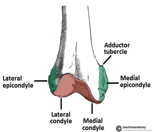
Refer to the diagram of the distal end of the femur. Which structure is labeled as the attachment point for muscles and ligaments?
Lateral condyle
Medial condyle
Lateral epicondyle
Adductor tubercle
Medial & Lateral Condyles articulate with the ______ to form the knee joint.
Tibia
Fibula
Femur
Patella
The medial epicondyle is an attachment point for muscles and ligaments.
True
False
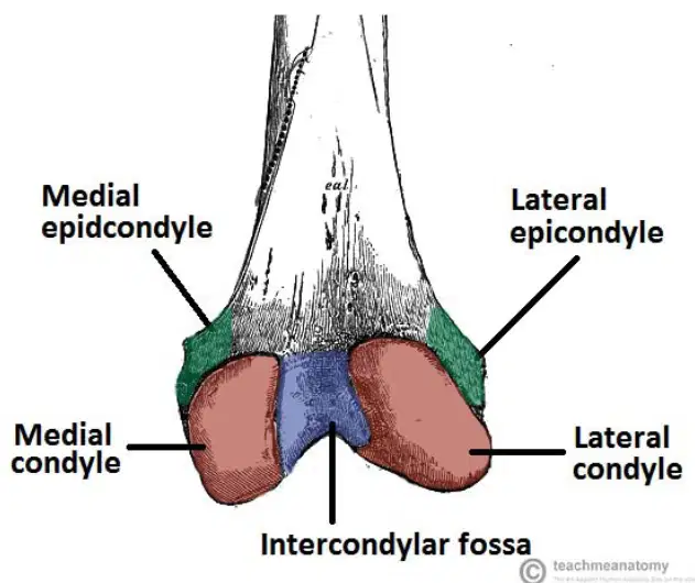
What is the intercondylar fossa of the femur?
A) A projection on the anterior aspect of femur
B) A depression on posterior aspect of femur where ligaments attach
C) A ridge on the lateral aspect of femur
D) A notch on the medial aspect of femur
Fill in the blank: The intercondylar fossa is a ______ on the posterior aspect of the femur where ligaments attach.
depression
ridge
notch
tuberosity
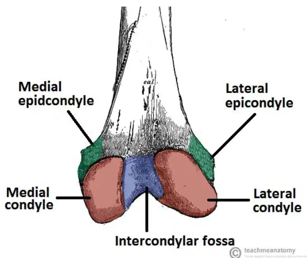
Match the following parts of the distal end of the femur to their correct locations as shown in the diagram:
Inner upper bump
Medial epicondyle
Outer upper bump
Lateral epicondyle
Inner lower bump
Medial condyle
Outer lower bump
Lateral condyle
Central depression
Intercondylar fossa
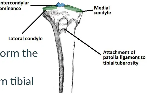
What is the function of the medial and lateral condyles of the tibia?
They articulate with the femur to form the knee joint.
They attach to the patella for movement.
They serve as the origin for the quadriceps muscle.
They provide attachment for the Achilles tendon.
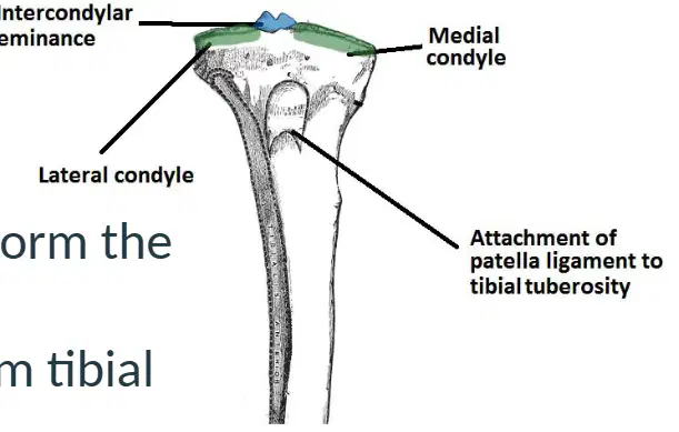
Which structure serves as the attachment point of the patellar tendon?
Tibial tuberosity
Medial malleolus
Femoral condyle
Fibular head
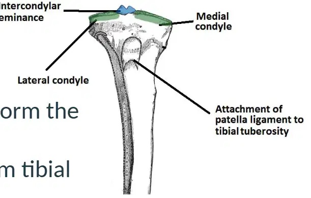
The medial and lateral condyles of the tibia widen at the proximal end to form the ________.
tibial plateau
intercondylar eminence
fibular head
patellar surface
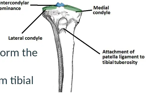
Match the following parts of the proximal tibia with their descriptions:
Medial condyle
Articulates with femur
Lateral condyle
Articulates with femur
Tibial tuberosity
Attachment point of patellar tendon
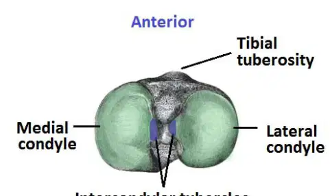
What is the area where ligaments and menisci attach on the proximal end of the tibia called?
Tibial tuberosity
Intercondylar eminence
Medial condyle
Lateral condyle
The intercondylar eminence contains _________.
intercondylar tubercles
medial malleolus
patellar ligament
fibular head
The intercondylar eminence articulates with the intercondylar fossa of the _________?
femur
tibia
fibula
patella
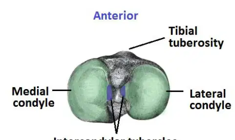
Match the following anatomical features of the proximal end of the tibia with their correct labels:
Anterior
1-A
Left side
2-B
Right side
3-C
Center between condyles
4-D
What is the largest sesamoid bone in the body?
Patella
Pisiform
Fabella
Hallux sesamoid
The patella articulates in the patellofemoral groove with which structures?
Tibial condyles
Medial & lateral condyles
Femoral shaft
Fibular head
Which of the following is a function of the patella?
Enhances leg extension by creating leverage for quadriceps tendon
Supports the ankle joint
Forms the hip joint
Protects the posterior aspect of the knee from trauma
The patella protects the anterior aspect of the knee from trauma.
True
False
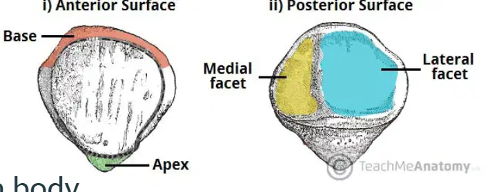
Label the following parts on the diagram of the patella:
Base, Apex, Medial facet, Lateral facet
Head, Neck, Shaft, Trochlea
Body, Greater trochanter, Lesser trochanter, Intercondylar fossa
Acromion, Coracoid process, Glenoid cavity, Spine
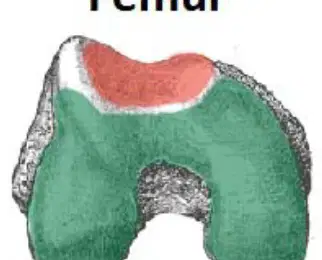
Which bone is shown on the left side of the image?
Tibia
Femur
Patella
Fibula
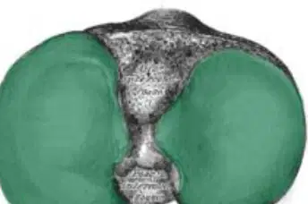
Refer to the diagram of articulating surfaces. The green color in the diagram represents which type of surface?
Patellofemoral surfaces
Tibiofemoral surfaces
Articular cartilage
Synovial membrane
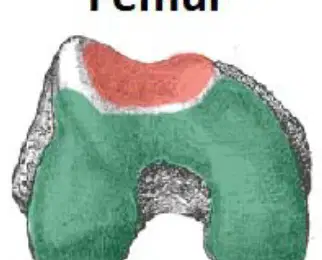
Refer to the diagram of articulating surfaces. Fill in the blank: The red color in the diagram represents __________ surfaces.
Patellofemoral
Tibiofemoral
Glenohumeral
Radiocarpal
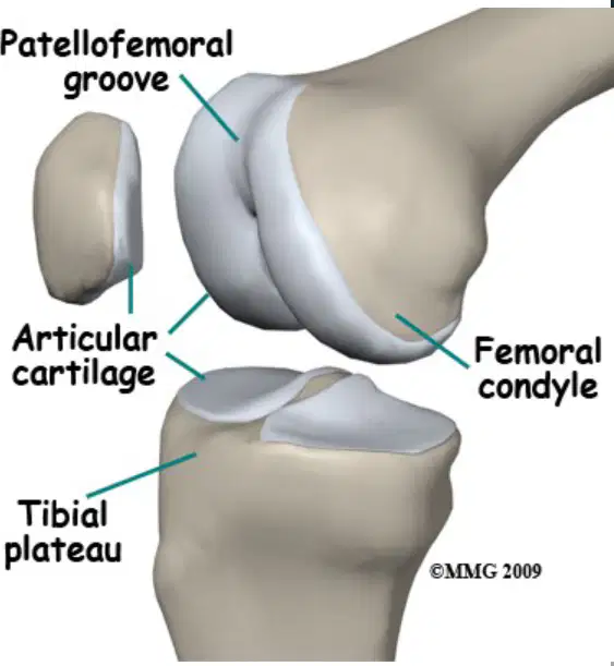
What does articulating cartilage cover in the knee joint?
It covers the condyles of femur & tibia as well as the posterior of patella.
It covers only the anterior surface of the femur.
It covers the ligaments of the knee joint.
It covers the menisci exclusively.
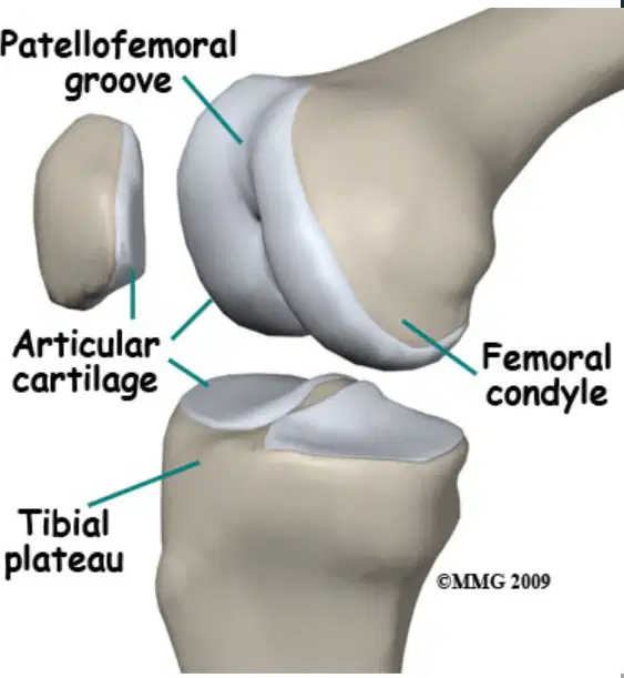
What is the function of the smooth, slippery surface of articulating cartilage?
It allows the bones to slide over each other without rubbing.
It helps in the production of red blood cells.
It stores calcium for the body.
It connects muscles to bones.
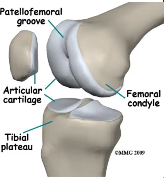
Which of the following is designed to minimize pressure and friction in the knee joint?
Muscle
Articulating cartilage
Ligament
Bone
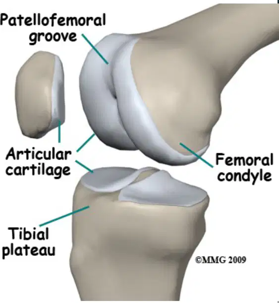
Match the following anatomical features with their labels in the diagram:
Upper groove near patella
Patellofemoral groove
Rounded end of femur
Femoral condyle
Top surface of tibia
Tibial plateau
What is one function of the meniscus? Fill in the blank: The meniscus helps to ______ the articulating surface of tibia to make the joint more stable.
deepen
flatten
widen
narrow
What is another function of the meniscus? Fill in the blank: The meniscus acts as ______ ______.
shock absorbers
muscle relaxants
nerve endings
blood vessels
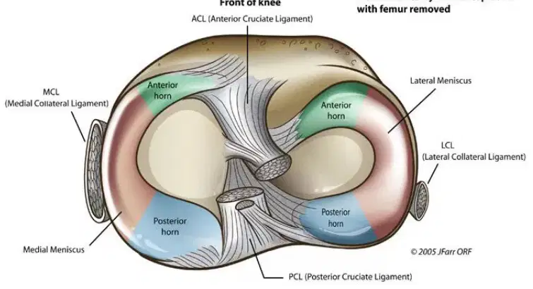
Which structure is labeled as 'Lateral Meniscus' in the diagram?
Medial Meniscus
Lateral Meniscus
Anterior horn
Posterior horn
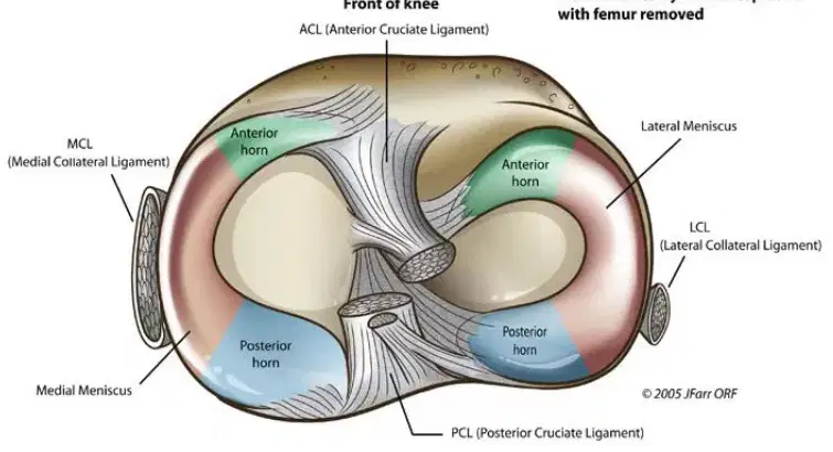
Match the following anatomical terms to their locations in the knee joint diagram:
Front of knee
A
Medial side
B
Lateral side
C
Posterior side
D
What type of structure is the meniscus?
Bone
Fibrocartilage
Muscle
Ligament
How many C shaped structures is the meniscus divided into?
1
2
3
4
The meniscus attaches to the ______ surface of the tibia.
intercondylar
anterolateral
posterior
medial
The medial meniscus also attaches to the medial (tibial) collateral ligament.
True
False
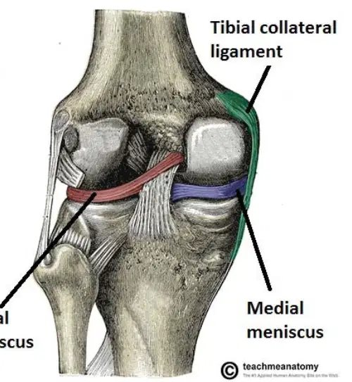
Identify the labeled structures in the diagram.
Tibial collateral ligament (green), Lateral meniscus (left, black label), Medial meniscus (right, black label)
Fibular collateral ligament (green), Lateral meniscus (right, black label), Medial meniscus (left, black label)
Anterior cruciate ligament (green), Lateral meniscus (left, black label), Medial meniscus (right, black label)
Patellar ligament (green), Lateral meniscus (left, black label), Medial meniscus (right, black label)
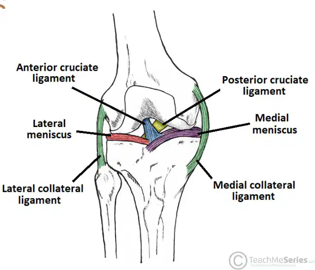
What do collateral ligaments connect?
Collateral ligaments connect the femur to the tibia.
Collateral ligaments connect the humerus to the radius.
Collateral ligaments connect the scapula to the clavicle.
Collateral ligaments connect the patella to the fibula.
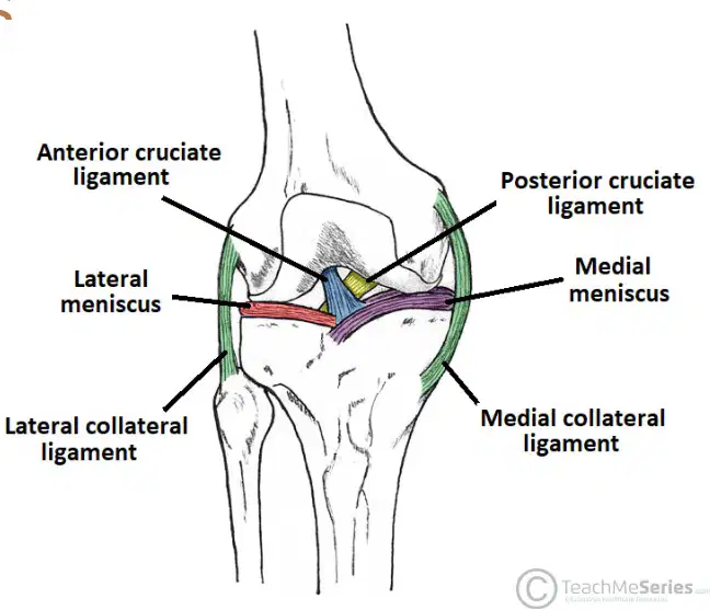
What is the function of collateral ligaments during knee movement?
Collateral ligaments stabilize the knee during flexion/extension.
Collateral ligaments produce synovial fluid for lubrication.
Collateral ligaments initiate muscle contraction in the thigh.
Collateral ligaments absorb shock during jumping.
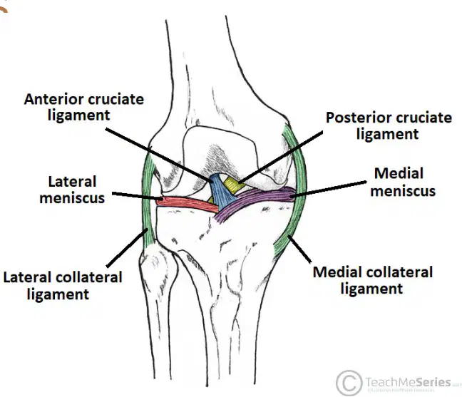
What movement do collateral ligaments prevent?
Collateral ligaments prevent medial/lateral movement.
Collateral ligaments prevent flexion/extension movement.
Collateral ligaments prevent rotational movement.
Collateral ligaments prevent anterior/posterior movement.
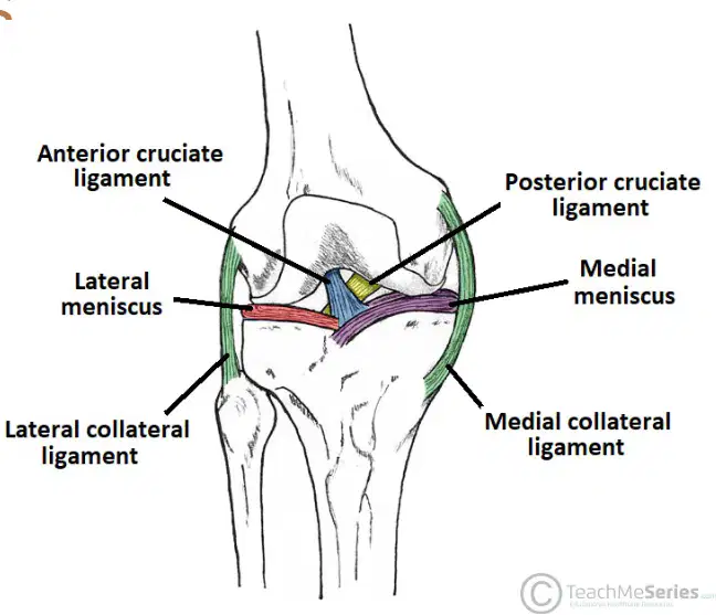
Match the following knee joint structures to their correct labels in the diagram:
Anterior cruciate ligament
Anterior cruciate ligament
Posterior cruciate ligament
Posterior cruciate ligament
Lateral meniscus
Lateral meniscus
Medial meniscus
Medial meniscus
Lateral collateral ligament
Lateral collateral ligament
What is another name for the Medial Collateral Ligament (MCL)?
Tibial Collateral Ligament
Fibular Collateral Ligament
Patellar Ligament
Anterior Cruciate Ligament
Where is the proximal attachment of the Medial Collateral Ligament (MCL)?
Medial epicondyle of femur
Lateral epicondyle of femur
Medial condyle of tibia
Head of fibula
Where is the distal attachment of the Medial Collateral Ligament (MCL)?
Medial condyle of tibia
Lateral condyle of femur
Head of fibula
Medial malleolus
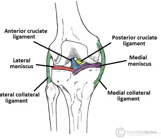
Which ligament is labeled in the diagram as connecting the medial side of the knee?
Lateral collateral ligament
Medial collateral ligament
Anterior cruciate ligament
Posterior cruciate ligament
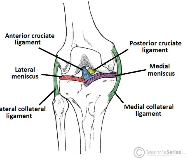
Match the following ligaments to their locations in the knee diagram:
Medial side
Medial collateral ligament
Lateral side
Lateral collateral ligament
Front center
Anterior cruciate ligament
Back center
Posterior cruciate ligament
What is another name for the Lateral Collateral Ligament (LCL)?
Tibial Collateral Ligament
Fibular Collateral Ligament
Medial Collateral Ligament
Cruciate Ligament
Which of the following best describes the Lateral Collateral Ligament (LCL)?
Thick, flat ligament
Thin, round ligament
Short, wide ligament
Long, narrow ligament
Where is the proximal attachment of the Lateral Collateral Ligament (LCL)?
Medial epicondyle of femur
Lateral epicondyle of femur
Head of tibia
Head of fibula
Where is the distal attachment of the Lateral Collateral Ligament (LCL)?
Head of tibia
Head of fibula
Lateral epicondyle of femur
Medial epicondyle of femur
Fill in the blank: The Lateral Collateral Ligament (LCL) is also known as the ________ Collateral Ligament.
Fibular
Tibial
Patellar
Femoral
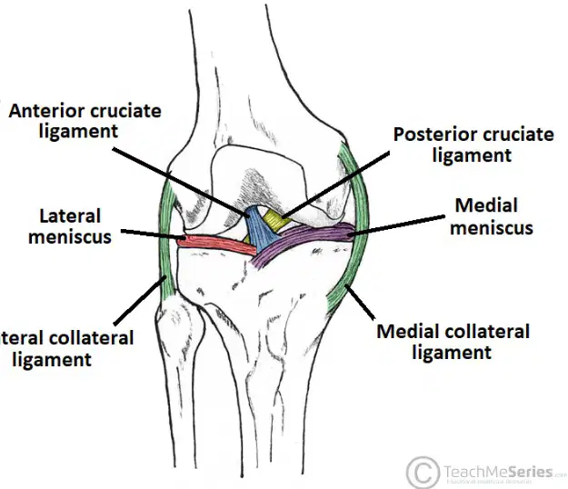
Identify the ligament labeled as 'Lateral collateral ligament' in the diagram.
Lateral Collateral Ligament (LCL)
Medial Collateral Ligament (MCL)
Anterior Cruciate Ligament (ACL)
Posterior Cruciate Ligament (PCL)
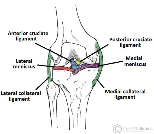
What do cruciate ligaments connect?
Femur to tibia
Femur to fibula
Tibia to patella
Femur to pelvis
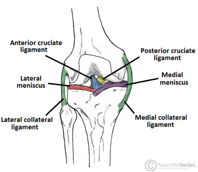
Which of the following is a function of cruciate ligaments?
Connect femur to tibia
Stabilize knee during medial/lateral rotation
Prevent excessive anterior/posterior movement
All of the above
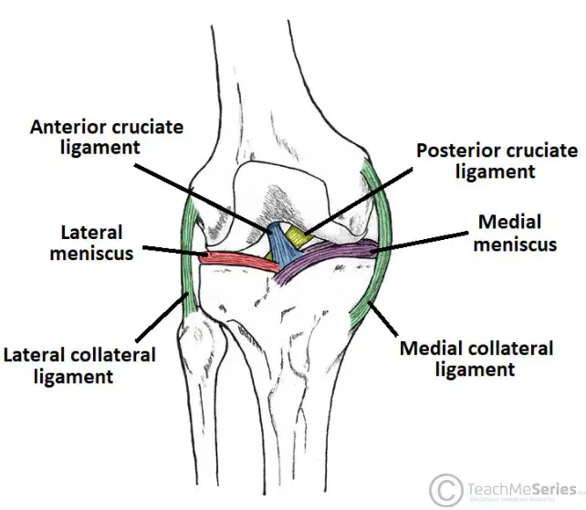
Cruciate ligaments help stabilize the knee during ______ rotation.
medial/lateral
anterior/posterior
superior/inferior
proximal/distal
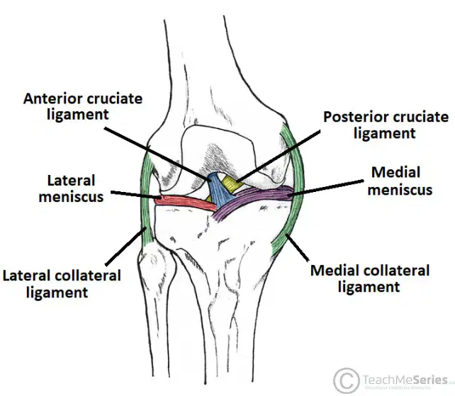
Match the following knee structures to their correct location in the diagram:
Anterior cruciate ligament
Anterior cruciate ligament
Posterior cruciate ligament
Posterior cruciate ligament
Lateral meniscus
Lateral meniscus
Medial meniscus
Medial meniscus
Lateral collateral ligament
Lateral collateral ligament
What is the function of the Anterior Cruciate Ligament (ACL)? Fill in the blank: The ACL prevents ________ dislocation of the knee.
anterior
posterior
lateral
medial
Where is the proximal attachment of the Anterior Cruciate Ligament (ACL)? Fill in the blank: The proximal attachment of the ACL is the ________ aspect of the intercondylar fossa.
posterior
anterior
medial
lateral
Where is the distal attachment of the Anterior Cruciate Ligament (ACL)? Fill in the blank: The distal attachment of the ACL is the ________ aspect of the intercondylar region of tibia.
anterior
posterior
medial
lateral
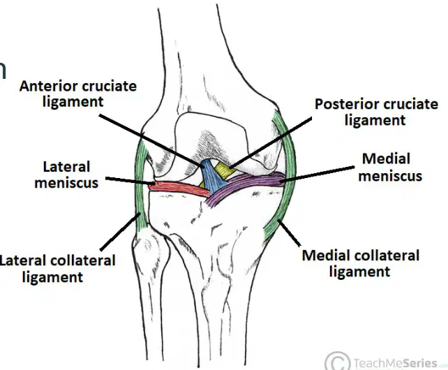
Refer to the diagram of the knee. Which ligament is labeled as the Anterior Cruciate Ligament?
Lateral collateral ligament
Posterior cruciate ligament
Anterior cruciate ligament
Medial collateral ligament
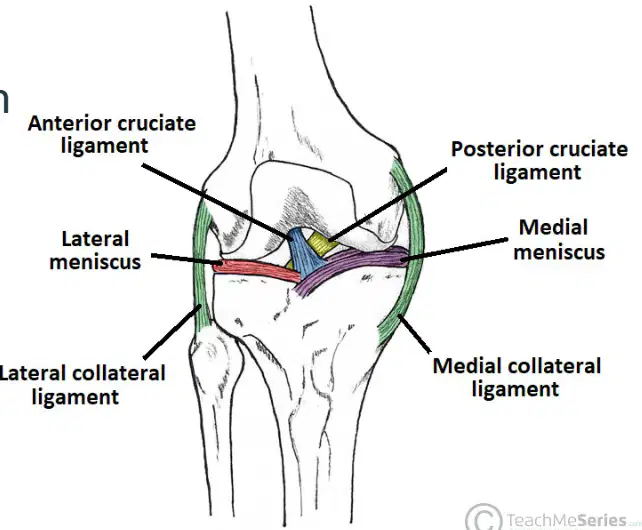
Match the following ligaments to their locations in the knee diagram:
Front center of knee
Anterior cruciate ligament
Back center of knee
Posterior cruciate ligament
Outer side of knee
Lateral collateral ligament
Inner side of knee
Medial collateral ligament
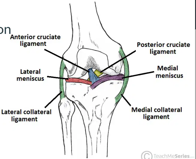
What is the main function of the Posterior Cruciate Ligament (PCL)? Fill in the blank: The PCL prevents _______ dislocation of the knee.
posterior
anterior
lateral
medial
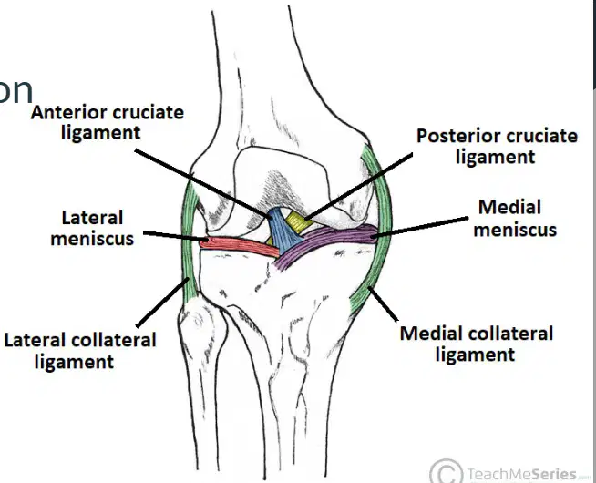
Where is the proximal attachment of the Posterior Cruciate Ligament (PCL)?
anterior
posterior
medial
lateral
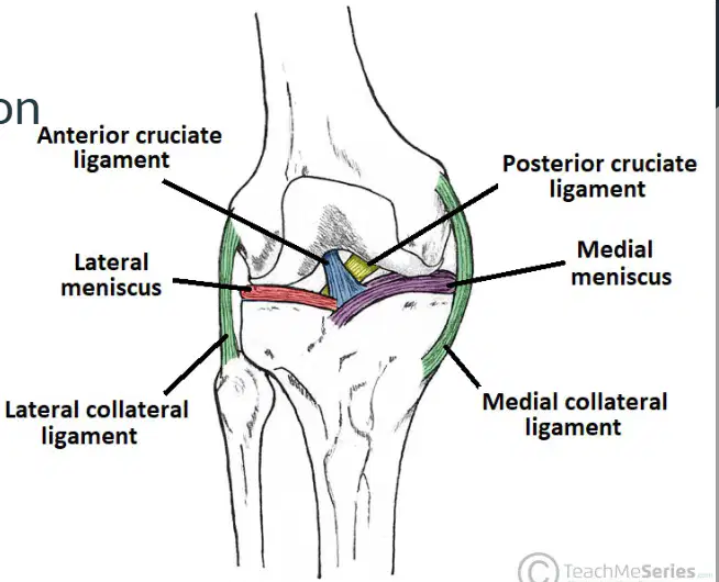
Where is the distal attachment of the Posterior Cruciate Ligament (PCL)? Fill in the blank: The distal attachment of the PCL is the _______ aspect of the intercondylar region of tibia.
posterior
anterior
medial
lateral
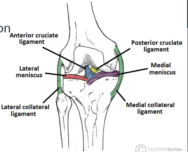
Which ligament is highlighted as the Posterior Cruciate Ligament (PCL) in the diagram?
A) Anterior cruciate ligament
B) Posterior cruciate ligament
C) Lateral collateral ligament
D) Medial collateral ligament
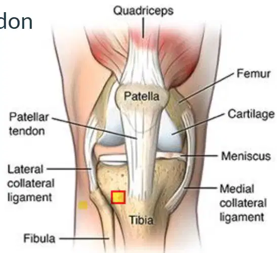
What is the patellar tendon a continuation of?
Quadriceps tendon
Hamstring tendon
Patella ligament
Achilles tendon
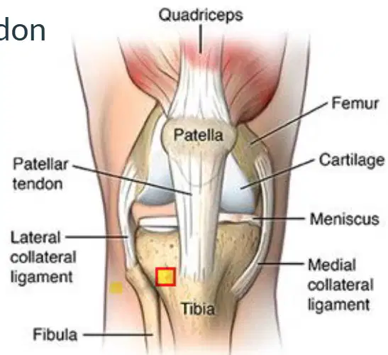
Where is the proximal attachment of the patellar tendon?
Apex of patella
Tibial tuberosity
Medial condyle of femur
Head of fibula
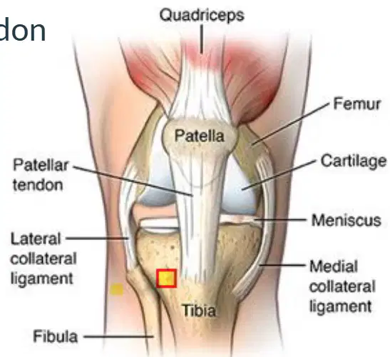
Where is the distal attachment of the patellar tendon?
Tibial tuberosity
Femoral condyle
Fibular head
Medial malleolus
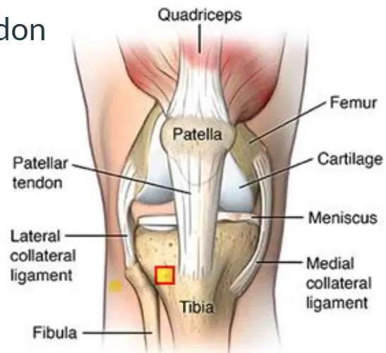
Which structure is labeled as the patellar tendon in the diagram?
The structure connecting the patella to the tibia, shown as 'Patellar tendon' in the diagram.
The structure connecting the femur to the patella, shown as 'Quadriceps tendon' in the diagram.
The structure running along the side of the knee, shown as 'Lateral collateral ligament' in the diagram.
The structure connecting the femur to the tibia, shown as 'Anterior cruciate ligament' in the diagram.

