
 Worksheets
Nervous System Test Part 2
Total questions: 103
Worksheet time: 54mins
All cells naturally have an equal charge on the inside versus the outside.
True
False
What do we call the difference in charge on the inside of a cell versus the outside?
Membrane potential
Osmotic pressure
Action threshold
Cell polarity
What causes the difference in charge on the inside versus the outside of a cell?
The unequal distribution of ions across the cell membrane
The equal distribution of water molecules across the cell membrane
The movement of proteins into the nucleus
The presence of DNA in the cytoplasm
Which two ions are involved in membrane potential?
Sodium (Na+) and Potassium (K+)
Calcium (Ca2+) and Chloride (Cl-)
Magnesium (Mg2+) and Iron (Fe2+)
Hydrogen (H+) and Phosphate (PO4^3-)
What is the charge inside of a cell naturally (positive or negative)? Why is it this way?
Negative; because there are more negatively charged ions and proteins inside the cell than outside
Positive; because there are more positively charged ions inside the cell than outside
Neutral; because the number of positive and negative ions are equal inside the cell
Negative; because there are more positively charged ions inside the cell than outside
What is a concentration gradient?
A difference in the concentration of a substance across a space or membrane
A type of chemical reaction
A measure of temperature change
A form of energy transfer
What does a concentration gradient cause?
Movement of substances from high concentration to low concentration (diffusion)
Production of energy in mitochondria
Formation of new cells
Increase in temperature
If there are 6 K+ ions on the outside of a cell and 2 K+ ions on the inside of a cell, is there a concentration gradient?
Yes
No
Only if there are more ions inside
Only if there are equal ions on both sides
What must happen for a cell i to reach equilibrium?
K+ ions must move from outside to inside until the concentrations are equal
Na+ ions must move from inside to outside until the concentrations are equal
K+ ions must move from inside to outside until the concentrations are equal
No ions need to move; the cell is already at equilibrium
If there are 3 Na+ ions on the inside of a cell and 1 Na+ ion on the outside of a cell, is there a concentration gradient?
Yes, there is a concentration gradient.
No, there is no concentration gradient.
There is an electrical gradient but not a concentration gradient.
There are equal concentrations of Na+ ions inside and outside the cell.
What do voltage-gated channels require to open?
A change in membrane potential (voltage).
The presence of neurotransmitters.
Binding of hormones to the channel.
An increase in temperature.
What are the two types of voltage-gated channels?
Sodium (Na+) channels and potassium (K+) channels.
Calcium (Ca2+) channels and chloride (Cl-) channels.
Magnesium (Mg2+) channels and iron (Fe2+) channels.
Hydrogen (H+) channels and zinc (Zn2+) channels.
What do sodium-potassium pumps require to open?
ATP (energy).
Oxygen.
Glucose.
Calcium ions.
What happens to sodium and potassium when the sodium-potassium pumps open?
Sodium is pumped out of the cell and potassium is pumped into the cell.
Sodium is pumped into the cell and potassium is pumped out of the cell.
Both sodium and potassium are pumped out of the cell.
Both sodium and potassium are pumped into the cell.
Does an action potential travel quickly or slowly?
Quickly.
Slowly.
At a constant rate regardless of conditions.
It does not travel at all.
Before an action potential, what is the charge inside of a neuron?
Negative.
Positive.
Neutral.
Alternating.
During depolarization, what happens to the charge inside of the neuron?
It becomes positive.
It becomes more negative.
It stays the same.
It fluctuates randomly.
Does the action potential travel down the axon all at once or little by little?
Little by little.
All at once.
It does not travel.
It travels randomly.
The action potential passes from one neuron to the next.
True
False
Which are the 4 stages of action potential in order?
Resting potential, depolarization, repolarization, Refractory Period
Depolarization, resting potential, Refractory Period, repolarization
Refractory Period, repolarization, depolarization, resting potential
Repolarization, Refractroy Period, resting potential, depolarization
When a neuron is not sending any message, what stage is the neuron in?
Resting potential
Action potential
Depolarization
Repolarization
During resting potential, what is the charge inside the axon?
Negative
Positive
Neutral
Alternating
During resting potential, what is the charge outside the axon?
Positive
Negative
Neutral
No charge
During resting potential, which pumps/channels are open?
Potassium leak channels and sodium-potassium pump
Voltage-gated sodium channels and calcium channels
Ligand-gated chloride channels and sodium-potassium pump
Voltage-gated potassium channels and calcium channels
Describe the concentration of K+ ions during resting potential.
Higher inside the axon than outside
Higher outside the axon than inside
Equal inside and outside the axon
No K+ ions present during resting potential
During resting potential is there a concentration gradient of K+ ions?
Yes
No
Only during action potential
Only outside the cell
Describe the concentration of Na+ ions during resting potential.
Higher outside the axon than inside
Higher inside the axon than outside
Equal inside and outside the axon
Absent inside the axon
During resting potential is there a concentration gradient of Na+ ions?
Yes
No
Only during action potential
Only in muscle cells
Which stage of action potential comes when an impulse arrives at the axon?
Depolarization
Repolarization
Hyperpolarization
Resting potential
The impulse causes which of the following during depolarization?
Opening of sodium channels
Release of neurotransmitters
Closure of potassium channels
Increase in ATP production
When the Na+ channels open during depolarization, sodium moves into the cell.
Sodium moves into the cell.
Sodium moves out of the cell.
Sodium remains outside the cell.
Sodium is converted to potassium.
What does this rush in of Na+ cause?
It causes depolarization of the neuron membrane.
It causes hyperpolarization of the neuron membrane.
It causes the release of neurotransmitters.
It causes the neuron to become more negative.
Once Na+ rushes in and is at equilibrium (ex. 4 inside, 4 outside) is there a concentration gradient of Na+ anymore?
Yes, there is still a concentration gradient.
No, there is no concentration gradient anymore.
The concentration gradient increases.
The concentration gradient fluctuates.
If there is no concentration gradient, what happens to the sodium voltage-gated channels?
They do not open because there is no driving force for sodium movement.
They open and allow sodium to flow freely.
They close permanently and cannot be activated.
They become leaky and allow other ions to pass through.
After depolarization, which stage of action potential comes next?
Repolarization
Hyperpolarization
Resting potential
Threshold potential
At the beginning of repolarization, what happens?
Potassium channels open and potassium ions leave the cell
Sodium channels open and sodium ions enter the cell
Calcium channels open and calcium ions enter the cell
Chloride channels open and chloride ions enter the cell
When the K+ channels open, what happens to potassium?
Potassium moves out of the cell.
Potassium moves into the cell.
Potassium remains stationary.
Potassium binds to sodium channels.
What does this rush out of K+ cause?
It causes repolarization of the membrane.
It causes depolarization of the membrane.
It causes hyperpolarization of the membrane.
It causes the release of neurotransmitters.
Once K+ rushes out and is at equilibrium (ex. 4 inside, 4 outside) is there a concentration gradient of K+ anymore?
No, there is no concentration gradient of K+ anymore.
Yes, there is still a concentration gradient of K+.
The concentration gradient increases.
The concentration gradient fluctuates constantly.
If there is no concentration gradient, what happens to the potassium voltage-gated channels?
They open but no net movement of potassium occurs.
They close permanently.
They allow potassium to move freely across the membrane.
They become inactive and degrade.
After repolarization, is the axon back at resting potential? Why or why not?
Yes, because the membrane potential has returned to its resting state.
No, because the axon remains depolarized.
No, because the axon is hyperpolarized after repolarization.
Yes, because the action potential is still ongoing.
Which channel/pump needs to open to move ions even when they are at equilibrium?
Leak channel
Voltage-gated channel
Ligand-gated channel
ATPase pump
The sodium-potassium pumps require ______ to open.
ATP
Glucose
Oxygen
Calcium ions
The phase of action potential that occurs after repolarization is called:
Refractory Period
depolarization
threshold phase
resting potential
When the sodium-potassium pumps open during the refractory period, what happens?
Sodium ions are pumped out and potassium ions are pumped in, restoring the resting potential.
Sodium ions rush into the cell, causing depolarization.
Potassium ions rush into the cell, causing hyperpolarization.
The cell becomes more positive inside than outside.
The sodium-potassium pumps accomplish which of the following, and how do they help restore resting potential?
They actively transport sodium out of the cell and potassium into the cell, helping to restore the resting potential by maintaining the correct ion gradient.
They passively allow sodium and potassium to move across the membrane, restoring resting potential by diffusion.
They only transport potassium out of the cell, which restores the resting potential.
They only transport sodium into the cell, which restores the resting potential.
Why is it important to reset the axon to resting potential?
It is important to reset the axon to resting potential to ensure that the neuron can fire another action potential and maintain proper signaling. Resetting restores the original charge difference across the membrane, allowing the neuron to respond to new stimuli.
It is important to reset the axon to resting potential so that the neuron can permanently stop sending signals.
Resetting the axon to resting potential allows the neuron to absorb more nutrients from the surrounding tissue.
Resetting the axon to resting potential causes the neuron to become inactive and unable to respond to any stimuli.
Which of the following best describes the stages of an action potential, including the charge inside and outside the cell, the status of pumps and channels, and the movement of ions?
Depolarization: inside becomes positive, Na+ channels open, Na+ enters; Repolarization: inside becomes negative, K+ channels open, K+ exits; Hyperpolarization: inside more negative, K+ channels remain open, K+ continues to exit; Resting: inside negative, Na+/K+ pump active, ions at equilibrium.
Depolarization: inside becomes negative, K+ channels open, K+ enters; Repolarization: inside becomes positive, Na+ channels open, Na+ exits; Hyperpolarization: inside more positive, Na+ channels remain open, Na+ continues to enter; Resting: inside positive, Na+/K+ pump inactive, ions at equilibrium.
Depolarization: inside becomes positive, K+ channels open, K+ enters; Repolarization: inside becomes negative, Na+ channels open, Na+ exits; Hyperpolarization: inside more negative, Na+ channels remain open, Na+ continues to enter; Resting: inside negative, Na+/K+ pump inactive, ions at equilibrium.
Depolarization: inside becomes negative, Na+ channels open, Na+ exits; Repolarization: inside becomes positive, K+ channels open, K+ enters; Hyperpolarization: inside more positive, K+ channels remain open, K+ continues to enter; Resting: inside positive, Na+/K+ pump active, ions at equilibrium.
Allows 'muscle memory': ________
Cerebellum
Medulla Oblongata
Thalamus
Hypothalamus
Also known as the 'interbrain': ________
Diencephalon
Cerebellum
Medulla Oblongata
Cerebrum
Attaches the spinal cord to the cerebrum. : ________
Brain stem
Cerebellum
Thalamus
Cerebrum
Broken into 2 hemispheres. ________
Cerebrum
Cerebellum
Medulla
Thalamus
Connects the cerebrum to the brainstem. ________
Diencephalon
Cerebellum
Medulla Oblongata
Corpus Callosum
Connects the left and right hemisphere of the cerebrum. ________
Corpus callosum
Cerebellum
Medulla oblongata
Thalamus
Contains the midbrain, pons, and medulla oblongata. ________
Brain stem
Cerebellum
Cerebrum
Thalamus
Contains the thalamus, hypothalamus, and pituitary gland. Structure of the Brain: ________
Diencephalon
Cerebellum
Medulla Oblongata
Cerebrum
Controls other hormone releasing glands. ________
Pituitary gland
Cerebellum
Medulla oblongata
Amygdala
Divided into 4 lobes. ________
Cerebrum
Cerebellum
Medulla
Thalamus
Helps with muscle coordination. ____
Cerebellum
Medulla Oblongata
Thalamus
Hypothalamus
Largest part of the brain. ________
Cerebrum
Cerebellum
Medulla
Pons
Major part of the brain that cannot be seen from the outside. ________
Diencephalon
Cerebellum
Cerebrum
Brainstem
Master endocrine gland. Structure of the Brain: ________
Pituitary gland
Cerebellum
Medulla oblongata
Thalamus
Means 'little brain' in Latin.
Cerebellum
Medulla
Thalamus
Cortex
Meets basic needs (breathing, circulation, and digestion).
Brainstem
Cerebellum
Cerebrum
Thalamus
Outer layer of the brain.
Cerebral cortex.
Medulla oblongata.
Thalamus.
Cerebellum.
Outer layer of the cerebrum.
Cerebral cortex
Medulla oblongata
Thalamus
Cerebellum
Peaks in the surface of the cerebrum.
Gyri
Sulci
Lobes
Cortex
Site of integration (understanding sensory input and dictating motor output).
Cerebral cortex
Cerebellum
Medulla oblongata
Spinal cord
Valleys in the surface of the cerebrum.
Sulci
Gyri
Lobes
Cortex
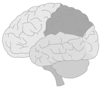
Which of the following correctly labels the cerebrum with the terms frontal lobe, parietal lobe, occipital lobe, temporal lobe, and places Broca’s area, the sensory cortex, the motor cortex, the amygdala, the hippocampus, and Wernicke’s area in their proper lobes?
Frontal lobe: Broca’s area, motor cortex; Parietal lobe: sensory cortex; Temporal lobe: Wernicke’s area, amygdala, hippocampus; Occipital lobe: none
Frontal lobe: sensory cortex, amygdala; Parietal lobe: Broca’s area; Temporal lobe: motor cortex, hippocampus; Occipital lobe: Wernicke’s area
Frontal lobe: hippocampus, sensory cortex; Parietal lobe: amygdala; Temporal lobe: Broca’s area, motor cortex; Occipital lobe: Wernicke’s area
Frontal lobe: Wernicke’s area, amygdala; Parietal lobe: motor cortex; Temporal lobe: sensory cortex, Broca’s area; Occipital lobe: hippocampus
Lobe of the brain that allows you to develop memories.
Temporal lobe
Frontal lobe
Occipital lobe
Parietal lobe
Complete the chart: Lobe of the brain that allows you to know what is around you (spatial awareness).
Parietal lobe
Frontal lobe
Temporal lobe
Occipital lobe
Lobe of the brain that contains Broca’s area.
Frontal lobe.
Parietal lobe.
Occipital lobe.
Temporal lobe.
Lobe of the brain that contains the amygdala.
Temporal lobe.
Frontal lobe.
Occipital lobe.
Parietal lobe.
Lobe of the brain that contains the hippocampus.
Temporal lobe
Frontal lobe
Occipital lobe
Parietal lobe
Lobe of the brain that contains the motor cortex.
Frontal lobe.
Parietal lobe.
Occipital lobe.
Temporal lobe.
Lobe of the brain that contains the sensory cortex.
Parietal lobe.
Frontal lobe.
Occipital lobe.
Temporal lobe.
Lobe of the brain that contains Wernicke’s area.
Temporal lobe
Frontal lobe
Occipital lobe
Parietal lobe
Lobe of the brain that interprets (understands) sensory information.
Parietal lobe
Frontal lobe
Occipital lobe
Temporal lobe
Lobe of the brain that receives and interprets auditory information.
Temporal lobe
Frontal lobe
Occipital lobe
Parietal lobe
Lobe of the brain that receives and interprets smell information.
Temporal lobe
Occipital lobe
Parietal lobe
Frontal lobe
Lobe of the brain that receives and interprets visual information.
Occipital lobe
Frontal lobe
Temporal lobe
Parietal lobe
Lobe of the cerebrum responsible for “executive function” – planning, organizing.
Frontal lobe
Parietal lobe
Temporal lobe
Occipital lobe
Lobe of the cerebrum that determines your personality.
Frontal lobe
Parietal lobe
Occipital lobe
Temporal lobe
Lobe of the cerebrum that helps with decision making.
Frontal lobe
Occipital lobe
Parietal lobe
Temporal lobe
Lobe of the cerebrum that helps you to control your emotions. (Fill in the blank)
Frontal lobe
Parietal lobe
Occipital lobe
Temporal lobe
Part of the brain that allows you to move your mouth/tongue right to form speech. (Fill in the blank)
Broca's area (Frontal lobe)
Wernicke's area (Temporal lobe)
Cerebellum
Occipital lobe
Part of the brain that allows you to understand speech. (Fill in the blank)
Wernicke's area (Temporal lobe)
Broca's area (Frontal lobe)
Cerebellum
Occipital lobe
Part of the brain that changes your short-term memories into long-term memories.
Hippocampus
Cerebellum
Medulla
Amygdala
Part of the brain that receives most sensory information (ex. Touch, hot, cold). (Fill in the blank)
Thalamus
Cerebellum
Medulla
Amygdala
Part of the brain that starts all voluntary movements.
Motor cortex (Frontal lobe)
Occipital lobe
Cerebellum
Temporal lobe
Part of the brain that triggers fear and anxiety.
Amygdala
Cerebellum
Hippocampus
Medulla
“Bridge” between the cerebellum and motor cortex/sensory cortex: __________
Pons
Medulla oblongata
Thalamus
Midbrain
Allows the cerebellum to better coordinate movements: __________
Pons
Thalamus
Medulla oblongata
Hypothalamus
Below the thalamus: __________
Hypothalamus
Medulla oblongata
Pons
Cerebellum
Controls how our blood vessels dilate and constrict: __________
Medulla oblongata
Thalamus
Pons
Hypothalamus
Which ion is primarily responsible for depolarizing the neuron membrane during an action potential?
Sodium (Na+)
Potassium (K+)
Calcium (Ca2+)
Chloride (Cl-)
What happens to the membrane potential when potassium ions exit the neuron during repolarization?
The membrane potential becomes more negative.
The membrane potential becomes more positive.
The membrane potential remains unchanged.
The membrane potential fluctuates randomly.
Which structure helps maintain the concentration gradients of Na+ and K+ across the neuron membrane?
Ligand-gated channel
Sodium-potassium pump
Chloride pump
Calcium channel
Label the parts of the brain listed:
Cerebellum
Brainstem
Frontal lobe
Temporal lobe
Parietal lobe
Occipital lobe


Frontal Lobe

Parietal lobe

Temporal lobe

Occipital lobe
Drag the appropriate labels from below onto the diagram of the brain.

Match the parts of the brain with the labels in the diagram.
Region labeled A
Cerebrum
Region labeled B
Brain stem
Region labeled C
Cerebellum
Region labeled D
Corpus callosum
Region labeled E
Diencephalon/ Thalaums

