
Font size
 Worksheets
Bone Practical Exam – Identification
Total questions: 88
Worksheet time: 44mins
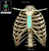
In the anterior view of the thoracic cage image, which structure is highlighted in the center of the sternum?
Body of sternum
Manubrium
Xiphoid process
True ribs
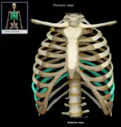
In the anterior view of the thoracic cage image, which ribs are highlighted and indirectly attached to the sternum via costal cartilage?
False ribs
True ribs
Floating ribs
Sternum
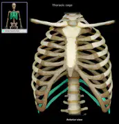
In the anterior view of the thoracic cage image, which ribs are highlighted and do not attach to the sternum?
Floating ribs
True ribs
False ribs
Body of sternum
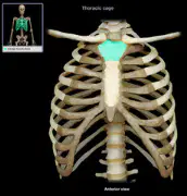
In the anterior view of the thoracic cage image, which superior portion of the sternum is highlighted?
Manubrium
Body of sternum
Xiphoid process
Floating ribs
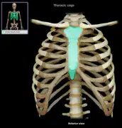
In the anterior view of the thoracic cage image, which bone of the anterior thorax is highlighted as a whole?
Sternum
Clavicle
Scapula
Ribs
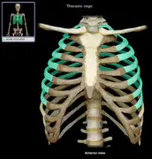
In the anterior view of the thoracic cage image, which ribs are highlighted and directly attach to the sternum by their own costal cartilage?
True ribs
False ribs
Floating ribs
Manubrium
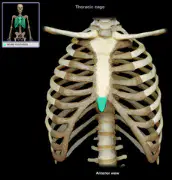
In the anterior view of the thoracic cage image, which small inferior tip of the sternum is highlighted?
Xiphoid process
Manubrium
Body of sternum
Costal cartilage
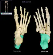
In the foot skeleton images, which heel bone is highlighted?
Calcaneus
Talus
Navicular
Cuboid
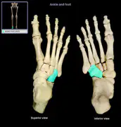
In the foot skeleton images, which lateral tarsal bone anterior to the calcaneus is highlighted?
Cuboid
Navicular
Medial cuneiform
Talus
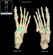
In the foot skeleton images, which middle cuneiform bone is highlighted between the medial and lateral cuneiforms?
Intermediate cuneiform
Medial cuneiform
Lateral cuneiform
Navicular
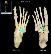
In the foot skeleton images, which cuneiform bone on the lateral side is highlighted?
Lateral cuneiform
Medial cuneiform
Intermediate cuneiform
Cuboid
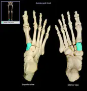
In the foot skeleton images, which cuneiform bone on the medial side is highlighted?
Medial cuneiform
Intermediate cuneiform
Lateral cuneiform
Navicular
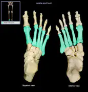
In the foot skeleton images, which long bones of the forefoot are highlighted?
Metatarsals
Phalanges
Tarsals
Sesamoid bones
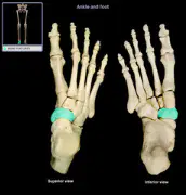
In the foot skeleton images, which boat-shaped tarsal bone proximal to the cuneiforms is highlighted?
Navicular
Cuboid
Talus
Calcaneus
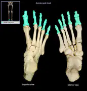
In the foot skeleton images, which bones of the toes are highlighted?
Phalanges
Metatarsals
Tarsals
Sesamoid bones
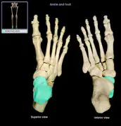
In the foot skeleton images, which superior tarsal bone that articulates with the tibia is highlighted?
Talus
Calcaneus
Navicular
Cuboid
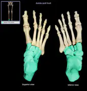
In the foot skeleton images, which collective group of proximal foot bones is indicated by the highlight?
Tarsals
Metatarsals
Phalanges
Sesamoid bones
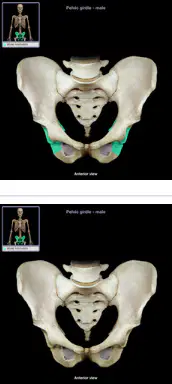
Identify the entire bony structure formed by the two hip bones and the sacrum as shown.
Pelvic girdle
Thoracic cage
Pectoral girdle
Vertebral column
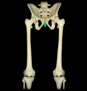
Identify the small terminal bone at the base of the sacrum highlighted in the pelvis image (posterior view).
Coccyx
Sacrum
Pubis
Ischium
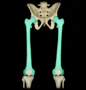
Identify the long bone of the thigh highlighted bilaterally in the lower limb image (anterior view).
Femur
Tibia
Fibula
Humerus
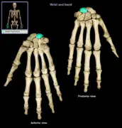
Identify the central carpal bone highlighted in the wrist image.
Capitate
Hamate
Lunate
Scaphoid
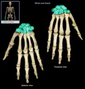
Identify the group of eight small bones forming the proximal part of the hand highlighted in the image.
Carpals
Metacarpals
Phalanges
Tarsals
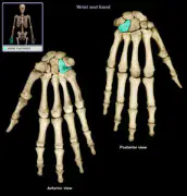
Identify the carpal bone with a hook-like projection highlighted in the wrist image.
Hamate
Capitate
Pisiform
Trapezoid
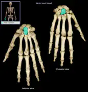
Identify the crescent-shaped carpal bone highlighted in the wrist image.
Lunate
Scaphoid
Triquetral
Capitate
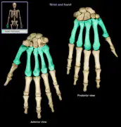
Identify the long bones of the palm highlighted in the hand image.
Metacarpals
Carpals
Phalanges
Tarsals
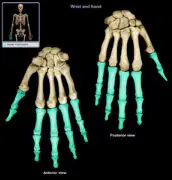
Identify the finger bones highlighted in the hand image.
Phalanges
Metacarpals
Carpals
Tarsals
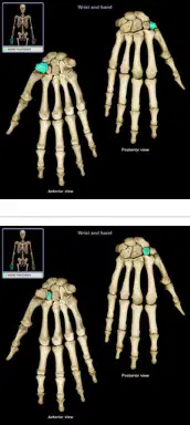
Identify the pea-shaped carpal bone highlighted on the medial side of the wrist.
Pisiform
Triquetral
Hamate
Scaphoid
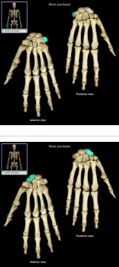
Identify the boat-shaped carpal bone highlighted near the base of the thumb.
Scaphoid
Lunate
Trapezium
Capitate
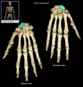
Identify the carpal bone that articulates with the first metacarpal and is highlighted near the thumb base.
Trapezium
Trapezoid
Capitate
Hamate
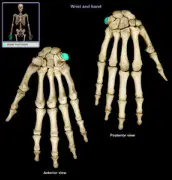
Identify the wedge-shaped carpal bone highlighted adjacent to the trapezium.
Trapezoid
Capitate
Lunate
Hamate

Identify the triangular carpal bone highlighted on the medial side of the wrist.
Triquetral
Pisiform
Hamate
Lunate
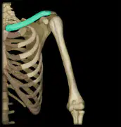
Identify the slender S-shaped bone of the shoulder girdle highlighted above the ribcage.
Clavicle
Scapula
Humerus
Sternum
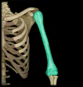
Identify the long bone of the upper arm highlighted.
Humerus
Radius
Ulna
Clavicle
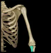
Identify the highlighted bone in the skeleton image.
Radius
Ulna
Humerus
Fibula
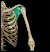
Identify the highlighted bone in the skeleton image.
Scapula
Clavicle
Sternum
Pelvic girdle
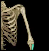
Identify the highlighted bone in the skeleton image.
Ulna
Radius
Humerus
Tibia
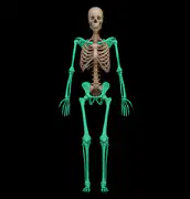
Identify the highlighted division of the skeleton in the image.
Appendicular skeleton
Axial skeleton
Thoracic cage
Vertebral column
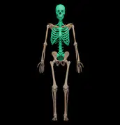
Identify the highlighted division of the skeleton in the image.
Axial skeleton
Appendicular skeleton
Upper limb
Lower limb
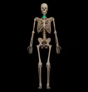
Identify the highlighted region of the vertebral column in the image.
Cervical vertebrae
Thoracic vertebrae
Lumbar vertebrae
Sacral vertebrae
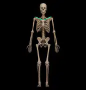
Identify the highlighted bone in the skeleton image.
Clavicle
Scapula
Sternum
Humerus
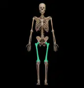
Identify the highlighted bone in the skeleton image.
Femur
Fibula
Tibia
Humerus
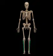
Identify the highlighted bone in the skeleton image.
Fibula
Tibia
Femur
Radius
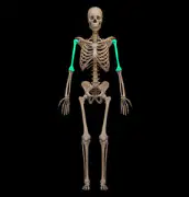
Identify the highlighted bone in the skeleton image.
Humerus
Radius
Ulna
Clavicle
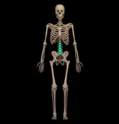
Identify the highlighted region of the vertebral column in the image.
Lumbar vertebrae
Thoracic vertebrae
Cervical vertebrae
Sacral vertebrae
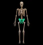
Identify the highlighted structure in the skeleton image.
Pelvic girdle
Sternum
Scapula
Sacrum
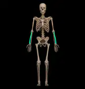
Identify the highlighted bone in the skeleton image.
Radius
Ulna
Humerus
Tibia
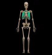
Identify the highlighted bones in the skeleton image.
Ribs
Sternum
Clavicles
Scapulae
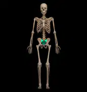
Identify the highlighted bone in the skeleton image.
Sacrum
Coccyx
Lumbar vertebrae
Pelvic girdle
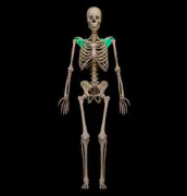
Identify the highlighted bone in the skeleton image.
Scapula
Clavicle
Sternum
Humerus
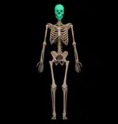
Identify the highlighted structure in the skeleton image.
Skull
Sternum
Mandible
Cervical vertebrae
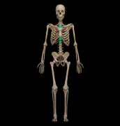
Identify the highlighted bone in the skeleton image.
Sternum
Ribs
Clavicle
Scapula
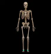
Identify the highlighted bones in the skeleton image.
Tarsals
Metatarsals
Phalanges of toes
Carpals
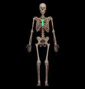
Identify the highlighted region of the vertebral column in the image.
Thoracic vertebrae
Cervical vertebrae
Lumbar vertebrae
Sacral vertebrae
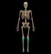
Identify the highlighted bone in the image.
Tibia
Fibula
Radius
Humerus
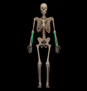
Identify the highlighted bone in the image.
Ulna
Radius
Tibia
Fibula
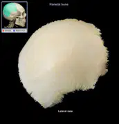
Identify the highlighted bone in the image.
Parietal
Occipital
Temporal
Frontal
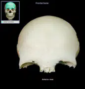
Identify the highlighted bone in the image.
Frontal
Parietal
Occipital
Temporal
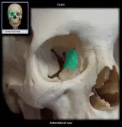
Identify the highlighted bone in the image.
Ethmoid
Sphenoid
Lacrimal
Vomer

Identify the highlighted bone in the image.
Frontal
Parietal
Occipital
Temporal
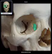
Identify the highlighted bone in the image.
Lacrimal
Ethmoid
Zygomatic
Nasal
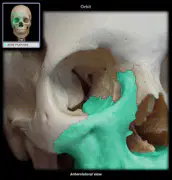
Identify the highlighted bone in the image.
Maxilla
Mandible
Zygomatic
Palatine
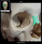
Identify the highlighted bone in the image.
Nasal
Lacrimal
Vomer
Zygomatic
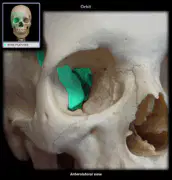
Identify the highlighted bone in the image.
Sphenoid
Ethmoid
Temporal
Occipital
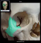
Identify the highlighted bone in the image.
Zygomatic
Maxilla
Temporal
Mandible
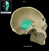
Identify the highlighted bone in the image.
Occipital
Parietal
Temporal
Frontal
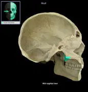
Identify the highlighted bone in the image.
Palatine
Maxilla
Vomer
Zygomatic
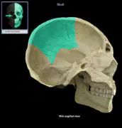
Identify the highlighted bone in the image.
Parietal
Occipital
Temporal
Frontal
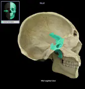
Identify the highlighted bone in the image.
Sphenoid
Ethmoid
Temporal
Occipital
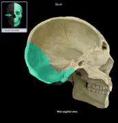
Identify the highlighted bone in the image.
Temporal
Parietal
Frontal
Sphenoid
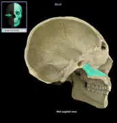
Identify the highlighted bone in the image.
Vomer
Nasal
Palatine
Ethmoid
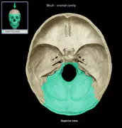
Identify the highlighted bone in the image.
Occipital
Parietal
Temporal
Frontal
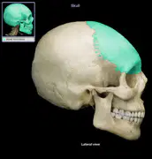
Identify the highlighted bone in the image.
Frontal
Parietal
Occipital
Temporal
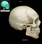
Identify the highlighted bone on the lateral view of the skull shown in the image.
Lacrimal
Nasal
Zygomatic
Maxilla
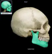
Identify the highlighted bone on the lateral view of the skull shown in the image.
Mandible
Maxilla
Temporal
Zygomatic
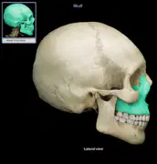
Identify the highlighted bone on the lateral view of the skull shown in the image.
Maxilla
Mandible
Vomer
Zygomatic
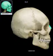
Identify the highlighted bone on the lateral view of the skull shown in the image.
Nasal
Lacrimal
Ethmoid
Frontal
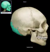
Identify the highlighted bone on the lateral view of the skull shown in the image.
Occipital
Parietal
Temporal
Sphenoid
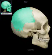
Identify the highlighted bone on the lateral view of the skull shown in the image.
Parietal
Occipital
Frontal
Temporal
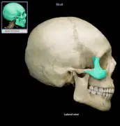
Identify the highlighted bone on the lateral view of the skull shown in the image.
Sphenoid
Ethmoid
Frontal
Parietal
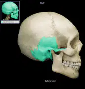
Identify the highlighted bone on the lateral view of the skull shown in the image.
Temporal
Parietal
Zygomatic
Occipital
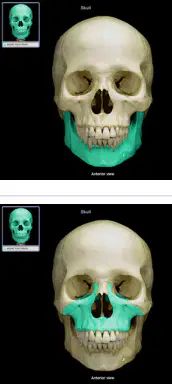
Identify the highlighted bone on the anterior view of the skull shown in the image.
Ethmoid
Sphenoid
Vomer
Lacrimal
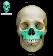
Identify the highlighted bone on the anterior view of the skull shown in the image.
Lacrimal
Nasal
Zygomatic
Maxilla
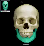
Identify the highlighted bone on the anterior view of the skull shown in the image.
Mandible
Maxilla
Vomer
Zygomatic
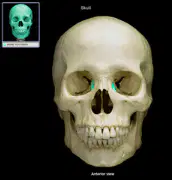
Identify the highlighted bone on the anterior view of the skull shown in the image.
Maxilla
Mandible
Vomer
Zygomatic
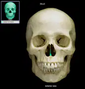
Identify the highlighted bone on the anterior view of the skull shown in the image.
Sphenoid
Ethmoid
Frontal
Parietal
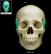
Identify the highlighted bone on the anterior view of the skull shown in the image.
Temporal
Zygomatic
Parietal
Occipital
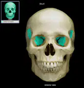
Identify the highlighted bone on the anterior view of the skull shown in the image.
Vomer
Maxilla
Nasal
Palatine
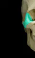
Identify the highlighted bone on the anterior view of the skull shown in the image.
Zygomatic
Maxilla
Temporal
Mandible

