
 Worksheets
Lab Practical Report 2 (HW)
Total questions: 77
Worksheet time: 48mins

Name the highlighted muscle.
orbicularis oculi
orbicularis oris
pectoralis
quadriceps femoris

Name the highlighted muscle.
external oblique
internal oblique
rectus abdominus
diaphragm

Name the highlighted muscle.
orbicularis oculi
orbicularis oris
frontalis
sternocleidomastoid

Name the highlighted muscle.
gluteus minimus
gluteus maximus
hamstring
triceps brachii

Name the highlighted muscle.
quadriceps
hamstrings
gastrocnemius
peroneus longus

Name the highlighted muscle.
deltoids
pectoralis
triceps
trapezius

Name the highlighted muscle.
external oblique
internal oblique
rectus abdominus
pectoralis

Name the highlighted muscle.
triceps brachii
biceps brachii
flexors to fingers
deltoids

Name the highlighted muscle.
masseter
frontalis
temporalis
orbicularis oculi

Name the highlighted muscle.
frontalis
temporalis
masseter
orbicularis oris

Name the highlighted muscle.
Temporalis
Frontalis
Masseter
Trapezius

Name the highlighted muscle.
Pectoralis
Frontalis
Sternoicleidomastoid
Trapezius

Name the highlighted muscle.
Trapezius
Latissimus dorsi
Gluteus maximus
Triceps brachii

Name the highlighted muscle.
biceps brachii
brachioradialis
triceps brachii
finger extensors

What muscle is the arrow pointing to?
Pectoralis (Pecs)
Biceps
Triceps
Lats

Name the highlighted muscle.
deltoids
rectus abdominis
external oblique
triceps

Name the highlighted muscle.
Brachioradialis
Trapezius
Biceps
Triceps

Name the highlighted muscle.
Hamstrings
Quadriceps
Gastrocnemius
Tibialis Anterior

Name the muscle highlighted in the diagram.
Hamstring
Quadriceps
Gastrocnemius
Sartorius
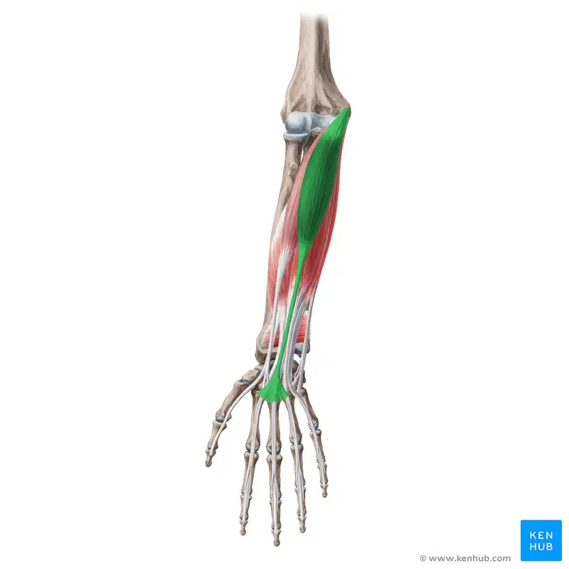
Which muscle is this?
(look at the markings to help you determine)
Extensor digitorum
Palmaris Longus
Flexor carpi ulnaris
Flexor carpi radialis
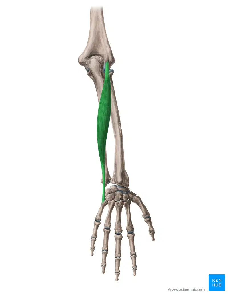
Identify the muscle.
Flexor carpi ulnaris
Flexor carpi radialis
Extensor carpi ulnaris
Brachioradialis

Identify the endocrine gland/part shown by letter B
Pineal
Hypothalamus
Pituitary
Parathyroid
Identify the endocrine gland/part shown by letter A
Pineal
Hypothalamus
Pituitary
Parathyroid
Identify the endocrine gland/part shown by letter C
Pineal
Hypothalamus
Pituitary
Parathyroid

Identify the endocrine gland/part shown by letter D
Thyroid
Adrenal
Thymus
Parathyroid
Identify the endocrine gland/part shown by letter C
Thyroid
Adrenal
Thymus
Parathyroid
Identify the endocrine gland/part shown by letter B
Thyroid
Adrenal
Thymus
Parathyroid
Identify the endocrine gland/part shown by letter A
Thyroid
Adrenal
Thymus
Parathyroid

Identify the endocrine gland/part shown by letter A
Thyroid
Adrenal
Thymus
Parathyroid
Identify the endocrine gland/part shown by letter B
Ovaries
Adrenal
Testes
Pancreas
Identify the endocrine gland/part shown by letter C
Ovaries
Adrenal
Testes
Pancreas
Identify the endocrine gland/part shown by letter D
Ovaries
Adrenal
Testes
Pancreas

Identify #2
Choroid
Cornea
Pupil
Sclera

Identify #4.
cornea
Iris
Pupil
Tapetum Lucidum
Identify #5.
Ciliary Body
Iris
Pupil
Tapetum Lucidum
Lens

Identify #3.
Choroid
Ciliary Body
Optic Nerve
Tapetum Lucidum
Sclera

Identify #9 the reflective colorful part at the back of the eye.
Ciliary Body
Optic Nerve
Tapetum Lucidum
Vitreous Humor

The white part of the eye is the:
Retina
Sclera
Iris
Tapetum

The thin layer of cells at the back of the eye that resembles skin is the:
Cornea
Optic nerve
Sclera
Retina

What jelly-like structure is the lens sitting in?
Sclera
Aqueous Humor
Retina
Vitreous Humor
Posterior Chamber
Which part of the eye surrounds the pupil and can be different colors?
Iris
Pupil
Cornea
Retina

What is number 4?
lens
cornea
pupil
retina

What is number 1?
Lens
cornea
ciliary bodies
retina

What is this?
internal auditory canal (eustachian tube)
external auditory canal
pinna
cochlea

What is this?
tympanic membrane
external auditory canal
ossicles
cochlea

What is this?
tympanic membrane
external auditory canal
semicircular canal
vestibulocochlear nerve

What is this?
tympanic membrane
external auditory canal
semicircular canal
vestibulocochlear nerve

What is this?
internal auditory canal (eustachian tube)
external auditory canal
pinna
ossicles

What is this?
pinna (auricle)
external auditory canal
ossicles
cochlea

What is this? (hard covering)
bony labyrinth
membranous labyrinth
ossicles
cochlea
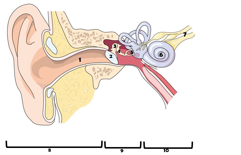
Name the part of the ear labeled as 6 in the diagram.
anvil
ear canal
stirrup
cochlea

What organ of the inner ear is NOT involved in hearing, but in equilibrium/bakance? (half loops)
Ear drum
Cochlea
Semicircular canals

What part is #10?
Incus/Anvil
Malleus/Hammer
Cochlea/Snail Shell
Stapes/Stirrup
What part is #12?
Incus/Anvil
Malleus/Hammer
Cochlea/Snail Shell
Stapes/Stirrup
What part is #13?
Incus/Anvil
Malleus/Hammer
Cochlea/Snail Shell
Stapes/Stirrup

What is labeled A?
Corpus callosum
Cerebrum
Cerebellum
Midbrain
Pons
What is labeled B?
Corpus callosum
Cerebrum
Cerebellum
Midbrain
Pons
What is labeled C?
Corpus callosum
Cerebrum
Cerebellum
Midbrain
Pons
What is labeled D?
Medulla oblongata
Spinal cord
Cerebellum
Midbrain
Pons
What is labeled E?
Medulla oblongata
Spinal cord
Cerebellum
Midbrain
Pons
What is labeled F?
Medulla oblongata
Spinal cord
Cerebellum
Midbrain
Pons
What is labeled G?
Medulla oblongata
Spinal cord
Cerebellum
Midbrain
Pons

which lobe is pictured?
Cerebellum
Temporal
Parietal
Occipital
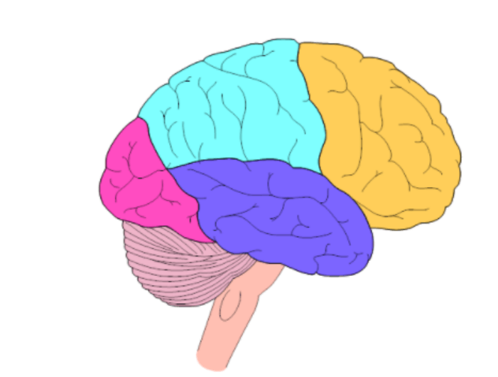
What lobe is bright pink in this picture?
Frontal
Parietal
Temporal
Occipital
What lobe is orange in this picture?
Frontal
Parietal
Temporal
Occipital
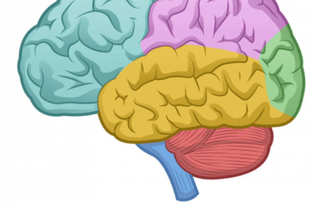
What structure is blue in this picture?
Cerebrum
Cerebellum
Brain Stem
The brain is covered in a membrane called:
choroid plexus.
meninges.
ventricles.
cerebral aqueduct.

Identify structure A (tree of life)
(a)

Identify structure B
(a)

Identify structure H
(a)

identify structure E
(a)

identify structure G
(a)

identify structure F
(a)

identify structure A
(a)

Identify the Thalamus.
B
C
E
F

What is 10? (whole part)
(a)

