
NEW
Font size
 Worksheets
Nerve Tissue
Total questions: 23
Worksheet time: 12mins
Progenitor cells for neurons and glial cells of peripheral nerves are directly derived from what embryonic structure(s)?
Neural folds
Neural crest
Neural tube
Neural plate
Somites
What are the most numerous cells of the central nervous system?
Neurons of the gray matter
Oligodendrocytes of the white matter
Astrocytes
Ependymal cells
Microglial cells
Flattened epithelial-like fibroblasts in what connective tissue layer immediately surround the grouped axons of a large peripheral nerve?
Perineurium
Schwann sheath
Epineurium
Endoneurium
Dura mater
The central nervous system is surrounded by layers of support tissue collectively called the meninges. Which part of the meninges directly contacts cells of the neural tissue?
Dura mater
Subarachnoid space
Arachnoid mater
Ependymal layer
Pia mater
Which of the following best describes a “peripheral nerve”?
An axon and cell body of a single neuron
A structure with nerve processes of many neurons, including both afferent and efferent fibers
A structure with many processes originating from a single neuron
A structure with unmyelinated fibers typically surrounding a central area of myelinated fibers
A structure with many axons, each of which is surrounded by multiple layers from Schwann cells
You read in a pathology report that a patient has peripheral neuropathy with partial demyelination in the larger nerves of the lower limbs. You know immediately that this involves what cells?
Satellite cells
Bipolar neurons
Oligodendrocytes
Schwann cells
Astrocytes
Accumulations or plaques of proteins between CNS neurons is one classic pathologic feature of Alzheimer’s disease. These proteins stimulate activity of what local cell related to the monocyte lineage with an immunosurveillance role in the brain?
Astrocytes
Satellite cells
Microglia
Schwann cells
Oligodendrocytes
Which part of a synapse is most likely to show the presence of secretory vesicles in a transmission electron micrograph?
Schwann cell
Terminal bouton
Primary synaptic cleft
Secondary synaptic cleft
Postsynaptic cell
Dense irregular connective tissue comprises what layer in large peripheral nerves?
Perineurium
Dura mater
Epineurium
Endoneurium
Arachnoid layer
In many peripheral nerves there are long neurons whose axons end with junctions at muscle fibers. Where are the perikarya of these neurons located?
In gray matter of the spinal cord
In white matter of the spinal cord
In ganglia
Just outside the muscle the axons innervate
In the cerebellum
The ventricles of the brain are filled with cerebrospinal fluid and lined by what cells?
Astrocytes
Cells of the pia mater
Satellite cells
Ependymal cells
Purkinje cells
Nodes of Ranvier represent the junctions between successive Schwann cells along an axon. Where are these nodes found in nervous tissue?
White matter of the CNS
Myelinated axons of peripheral nerves
All peripheral nerves
Both white and gray matter of the CNS
In ganglia
What property or structure common to almost all animal cells is utilized by neurons in their specialized conduction of a nerve impulse?
Microtubules
Electron transport system
Microfilaments
Contractility
Excitability
Remodeling of synaptic connections in the central nervous system occurs as needed throughout one’s life. This represents primarily which of the following?
Formation of new neurons
Changes in axonal targeting
Progenitor cell proliferation
Neural plasticity
Neural stem cell proliferation
In the central nervous system the microenvironments of perikarya, synapses, and capillaries are all surrounded by and regulated by what processes from what glial cell?
Ependymal cells
Schwann cells
Astrocytes
Satellite cells
Microglia
What is the term for aggregations of neuronal cell bodies lying in peripheral sites outside the central nervous system?
Terminal boutons
Perikaryons
Ganglia
Nuclei
Perineurium
Neuronally associated satellite cells are found in what region or structures of the nervous system?
Ganglia of the peripheral nervous system
White matter
Gray matter
Unmyelinated axons
Cortex of the cerebellum
Where in a typical synapse are receptors for the neurotransmitters located?
In the presynaptic membrane
In the postsynaptic membrane
In the terminal bouton
White matterIn the synaptic cleft
In the membranes of surrounding satellite cells
The functional significance of the material generally referred to as “Nissl substance” is reflected in its neuronal location. What is its location?
In the perikaryon
Outside the neurons, surrounding perikaryons and axons
In the axon and dendrites
In the axon only
In the neuron’s nucleus
Patients with cerebral edema (swelling) may have cognitive deficits and reduced function within the blood-brain barrier. What are the two most important cell types in forming this CNS “barrier”?
Capillary endothelial cells and oligodendrocytes
Astrocytes and ependymal cells
Astrocytes and capillary endothelial cells
Microglial cells and capillary endothelial cells
Schwann cells and capillary endothelial cells
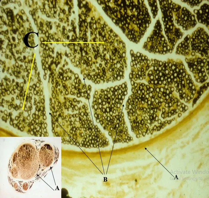
Identify the pointed structure labeled as B
Epineurium
Perineurium
Endoneurium
Exoneurium
Which type of glial cell is NOT derived from the neuroepithelial stem cell?
Satellite cells
Microglial cell
Ependymal cells
Oligodendrocytes
Schwann cells
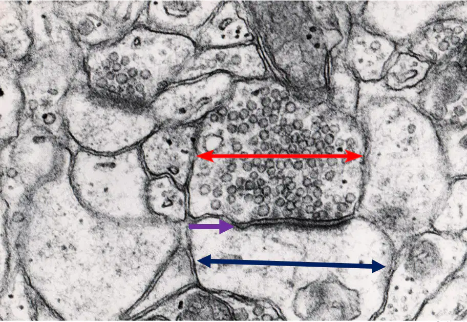
An impulse in the red arrow region would cause an _______, releasing neurotransmitter molecules onto the violet arrow region via exocytosis. These molecules would bind to the receptors in the blue arrow region, which then results to an _______
influx of potassium,
efflux of sodium
influx of calcium, efflux of sodium
efflux of sodium, efflux of calcium
efflux of calcium, influx of sodium
Influx of calcium, influx of sodium

