
 Worksheets
Ch. 16: The Nervous System Practice Review #1
Total questions: 115
Worksheet time: 2hrs 4mins

Match the following letters to part/location.
A
Dendrites
C
Axon
D
Schwann Cell
E
Nodes of Ranvier
F
Axon Terminals
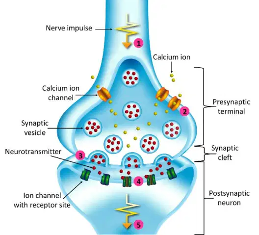
What substance is released at axon terminals propagating a nerve impulse?
Neurotransmitter
Calcium ion
Sodium
Potassium
Sends impulses away from the CNS
Efferent Nerves
Afferent Nerves
Autonomic Nervous System
Somatic Nervous System
Label the parts of the Neuron
Axon Terminal
Nucleus
Dendrites
Axon
Myelin Sheath
Cell Body

Which of the following is the nervous system?


The basic unit of structure and function in the nervous system is a nerve cell. Nerve cells are also called-
Osteocytes
Dendrites
Neurons
Spinal Cords
What is the function of a nerve?
To transport needed food around the body
To control body movement for reflexes.
To carry messages (impulses) to and form the Central Nervous System
All of the above
The Central Nervous system includes what two organs?
Brain and Nerves
Brain and Spinal Cord
Spinal cord and Nerves
Nerves and Neurons



____________ ---> ____________ ---> _______________
Response, Stimulus, Interpret
Interpret, Stimulus, Response
Stimulus, Interpret, Response
Response, Interpret, Stimulus
Select the INVOLUNTARY action.


Select the VOLUNTARY action.


This part of the neuron receives chemical messages from the neurotransmitters of other neurons. It is labeled A in the diagram.
Dendrite
Axon
Axon Terminal
Cell Body
What is the gap between two neurons called?
Impulse
Synapse
CNS
Inter neuron
Which statement best describes a reflex arc?
Slow response that you have to think about
Rapid response to protect body from harm

Where would the nerve impulse start in this reflex arc?
A
C
D
E

_____ neurons carry messages away from the central nervous system and out to muscles or glands. The RED neuron in diagram.
sensory
motor
inter
central

What type of neuron is A?
Sensory
Inter
Motor
What type of neuron is C?
Sensory
Inter
Motor

Name an effector in this diagram.
A
B
D
E
F

Select two examples of effectors.
Neuron
Gland
Muscle
Receptor
Which statement best describes a reflex?
Slow response that you have to think about
Rapid response to protect body from harm
_____ neurons carry messages away from the central nervous system and out to muscles or glands. The RED neuron in diagram.
sensory
motor
inter
central
Which type of neuron causes a muscle to contract in a reflex arc?
Motor Neuron
Sensory Neuron
Interneuron
Effector
Where would the nerve impulse start in this reflex arc?
A
C
D
E
What type of neuron is A?
Sensory
Inter
Motor
Which is the correct order of neurons in a reflex arc?
Sensory neuron, motor neuron, relay neuron
Relay neuron, sensory neuron, motor neuron
Sensory neuron, relay neuron, motor neuron
What is the final component of a reflex arc?
Effector
Receptor
Afferent Pathway

Which number represents the stimulus?
1
2
3
4
Which number represents the effector?
4
5
6
7
Which number represents the sensory neuron?
3
4
5
7
Which numbers represents the neurons in a reflex arc?
3
4
5
7
In a reflex arc, the sensory neuron leads to the ___________ and the motor neuron leads to the __________
Spinal cord, CNS
CNS, muscle
Spinal cord, muscle
Muscle, CNS
Reflex actions are important because
They provide a quick, involuntary response
They prevent injury
They are essential to the survival of organisms
All of the above
Muscles that you control are known as....
Involuntary
Fast Twitch
Voluntary
Slow Twitch
Muscles that you DO NOT control are known as ...
Involuntary
Fast Twitch
Voluntary
Slow Twitch
Which type of human body tissue can contract, or shorten?
epithelial tissue
muscle tissue
connective tissue
nerve tissue
How do pairs of skeletal muscles work together?
One muscle in the pair pulls on a bone, while the second muscle pulls on the first muscle.
While one muscle in the pair contracts, the other returns to its original length.
Both muscles extend at the same time.
One contracts and the other stretches.
Straightening a joint or increasing the angle between two bones
Flexion
Extension
Hyperextension
Abduction
Bending a joint or decreasing the angle between two bones
Flexion
Extension
Hyperextension
Adduction

Which muscle is identified here?
Bicep
Tricep
Latissimus Dorsi
Trapezius

What muscle is this that is highlighted?
Hamstrings
Gastrocnemius
Quadriceps
Biceps
Which of these two muscles are in an antagonistic pair?
Hamstrings
Abdominals
Triceps
Quadriceps

What type of movement is happening at the elbow joint?
Extension
Circumduction
Rotation
Flexion
Which of these connects muscles to bone?
Cartilage
Ligaments
Tendons
Which part of the ear is responsible for collecting sound waves from the environment?
Cochlea
Pinna
Eustachian tube
Semicircular canals
Which structure in the inner ear is primarily responsible for hearing?
Semicircular canals
Cochlea
Tympanic membrane
Auditory canal
Which part of the ear is responsible for collecting sound waves from the environment?
Cochlea
Pinna
Eustachian tube
Semicircular canals
What is the main function of the ossicles in the middle ear?
To produce earwax
To transmit and amplify sound vibrations
To maintain balance
To detect smell
Which structure in the inner ear is primarily responsible for hearing?
Semicircular canals
Cochlea
Tympanic membrane
Auditory canal
What is the function of the semicircular canals in the ear?
To collect sound waves
To help maintain balance and equilibrium
To transmit sound to the brain
To produce earwax
Which part of the ear separates the outer ear from the middle ear?
Cochlea
Tympanic membrane (eardrum)
Auditory nerve
Oval window
During the hearing process, which sequence correctly describes the path of sound from the outer ear to the brain?
Pinna → Cochlea → Ossicles → Brain
Pinna → Ear canal → Eardrum → Ossicles → Cochlea → Auditory nerve → Brain
Cochlea → Ossicles → Pinna → Brain
Eardrum → Pinna → Cochlea → Brain
Which component of the middle ear connects to the throat and helps equalize air pressure?
Cochlea
Eustachian tube
Pinna
Semicircular canals
What is the main function of the auditory nerve?
To produce earwax
To transmit sound signals from the ear to the brain
To maintain balance
To amplify sound
Which part of the ear contains fluid that moves in response to sound vibrations and helps with hearing?
Pinna
Cochlea
Eustachian tube
Ossicles
If a person has trouble maintaining balance, which part of the ear is most likely affected?
Pinna
Semicircular canals
Eardrum
Ossicles

function of iris
adjusting the light
allow light to enter
allow eye hydration
changing the size of pupil

function of cornea
refract light onto lens
reflect light onto lens
reflect light onto retina
refract light onto retina
function of iris
adjusting the light
allow light to enter
allow eye hydration
changing the size of pupil
function of sclera
protect the eyeball
connecting to brain nerve
give eye a square shape
shaped the eyeball
function of retina
projector similarity
allow eye to see
act as a screen of the image
internal busy cell
How does light affect the pupil?
The pupil gets larger as light decreases
The pupil gets larger as light increases
The pupil's color depends on the amount of light
The pupil isn't affected by light

Which structure is represented by the letter "B"?
Cornea
Retina
Pupil
Iris
Which structure is represented by the letter "A"?
Cornea
Sclera
Lens
Iris
Which structure is represented by the letter "C"?
Optic Nerve
Pupil
Lens
Iris
Which structure is represented by the letter "D"?
Optic Nerve
Sclera
Cornea
Blind Spot
Which structure is represented by the letter "E"?
Sclera
Lens
Optic Nerve
Retina
Which structure converts light waves to electrical signals that the brain uses to understand what it is seeing?
Lens
Sclera
Retina
Iris
Which structure carries the electrical signals from retina to the brain?
Optic Nerve
Sclera
Blind Spot
Pupil
Is a transparent, jelly-like mass located behind the lens. It acts as a ‘suspension’ for the lens so that the delicate lens is not damaged. It helps to maintain the shape of the posterior chamber of the eyeball.
Anterior Humour
Posterior Humour
Aqueous Humour
Vitreous humour


Which part of the eye is made up of rods and cones?
Lens
Retina
Iris
Sclera

Name the type of the vision defect (look in your textbook pg. 93)

Which image shows normal vision? (look in your textbook pg. 93)
A
B
C
D

Which image shows a person with nearsightedness? (look in your textbook pg. 93)
A
B
C
D
Label the Ear
Match the word to its definition.
The fleshy outer part of the ear
Pinna
Tiny, string-like projections that move due to sound vibrations
Hair Cells
A small, tightly stretched membrane
Ear Drum
Full of gel-like material. Indicates if you are right side up or upside down. (Balance)
Semicircular Canals
Vibrates against Oval Window, carrying sound
Stirrup
The (a) in the cochlea transmit sound information via the (b) to the (c) lobe of the brain.

What are the bones within the middle ear that help in amplifying the vibrations from sound waves (hearing)?
Cochlea, ear drum, and Eustachian tube
Incus, stapes, and malleus
Hair cells, nerve fibers, and brain
Ear canal, outer ear, and lobule
Identify the semi circular canals in the diagram.
Identify the semi ear drum (tympanic membrane) in the diagram.
Match the following
the chamber between the eardrum and cochlea containing three tiny bones that concentrate the vibrations of the eardrum on the cochlear oval window
middle ear
a coiled, bony, fluid-filled tube in the inner ear through which sound waves trigger nerve impulses
cochlea
this part of the ear contains the cochlea, semicircular canals, and vestibular sacs
inner ear
a device for converting sounds into electrical signals and stimulating the auditory nerve through electrodes threaded into the cochlea.
cochlear implant
Identify the hammer (malleus) in the diagram.

