
 Worksheets
Exam 1 A&P Review
Total questions: 129
Worksheet time: 1hrs 13mins
What is a nutritional deficiency syndrome caused by thiamine deficiency, usually associated with alcoholism?
Metabolic disorder
Meningionoma
astyrocytoma
The genu, rostrum, and splenium make up the:
posterior commisure
anterior commisure
arcuate faciculus
Where is the parietal lobe located in the brain?
between the occipital lobe and central sulcus
laterally in the cerebral hemispheres
anterior part of the brain
posterior part of the brain
Where is the temporal lobe located in the brain?
anterior part of the brain
posterior part of the brain
laterally in the cerebral hemispheres
between the occipital lobe and central sulcus
Where is the frontal lobe located in the brain?
posterior part of the brain
anterior part of the brain
laterally in the cerebral hemispheres
between the occipital lobe and central sulcus
Where is the occipital lobe located in the brain?
laterally in the cerebral hemispheres
between the occipital lobe and central sulcus
anterior part of the brain
posterior part of the brain
CVAs are the leading cause of disability in the U.S.
True
False
Most of the fourth ventricle is:
located in the brain stem
in the cerebellar cortex
in the lateral ventricles
within the frontal lobe
The right and left hemispheres are connected by large bundles of axons called:
(a)
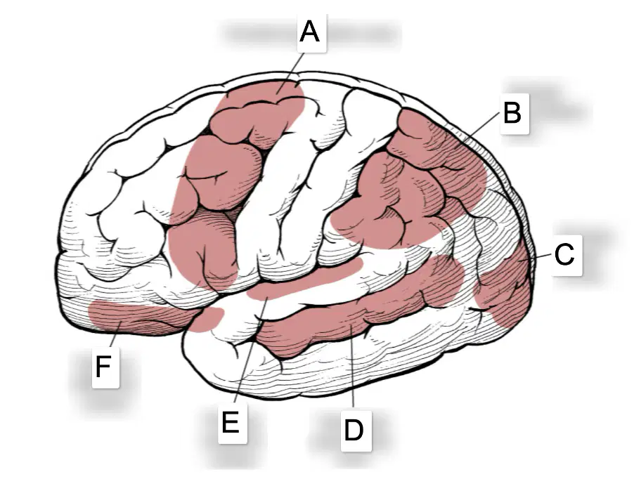
In the labeled lateral brain diagram, what area is labeled A?
Frontal association area
Parietal association area
Primary auditory cortex
Primary visual cortex
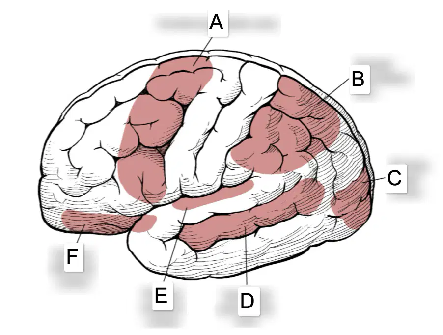
In the labeled lateral brain diagram, what area is labeled B?
Primary olfactory cortex
Temporal association area
Parietal association area
Primary visual cortex
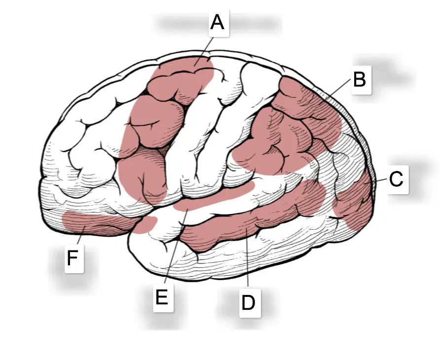
In the labeled lateral brain diagram, what area is labeled C?
Primary visual cortex
Frontal association area
Primary auditory cortex
Temporal association area
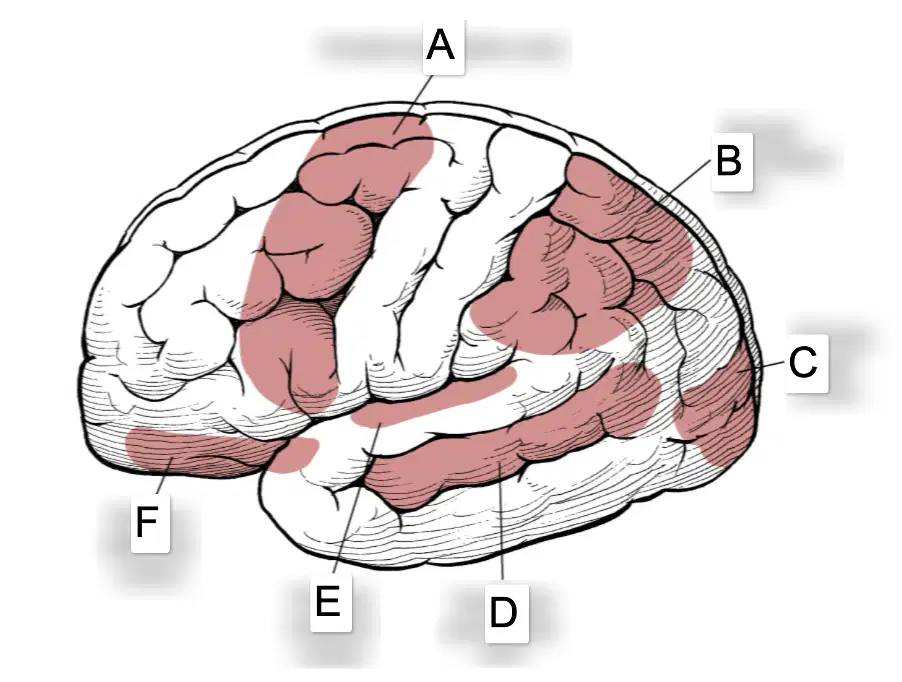
In the labeled lateral brain diagram, what area is labeled D?
Temporal association area
Primary olfactory cortex
Parietal association area
Primary visual cortex
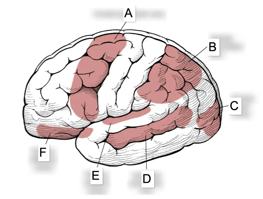
In the labeled lateral brain diagram, what area is labeled E?
Primary auditory cortex
Frontal association area
Primary visual cortex
Parietal association area

In the labeled lateral brain diagram, what area is at label F?
Primary olfactory cortex
Primary visual cortex
Parietal association area
Temporal association area
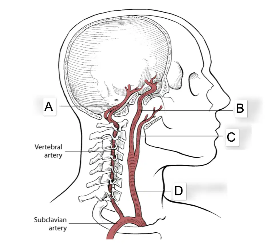
In the labeled sagittal head and neck artery diagram, what artery is at label A?
Basilar artery
Internal carotid artery
External carotid artery
Common carotid artery
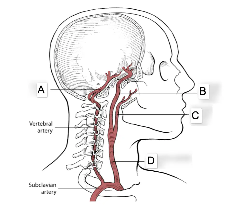
In the labeled sagittal head and neck artery diagram, what artery is at label B?
External carotid artery
Basilar artery
Internal carotid artery
Common carotid artery
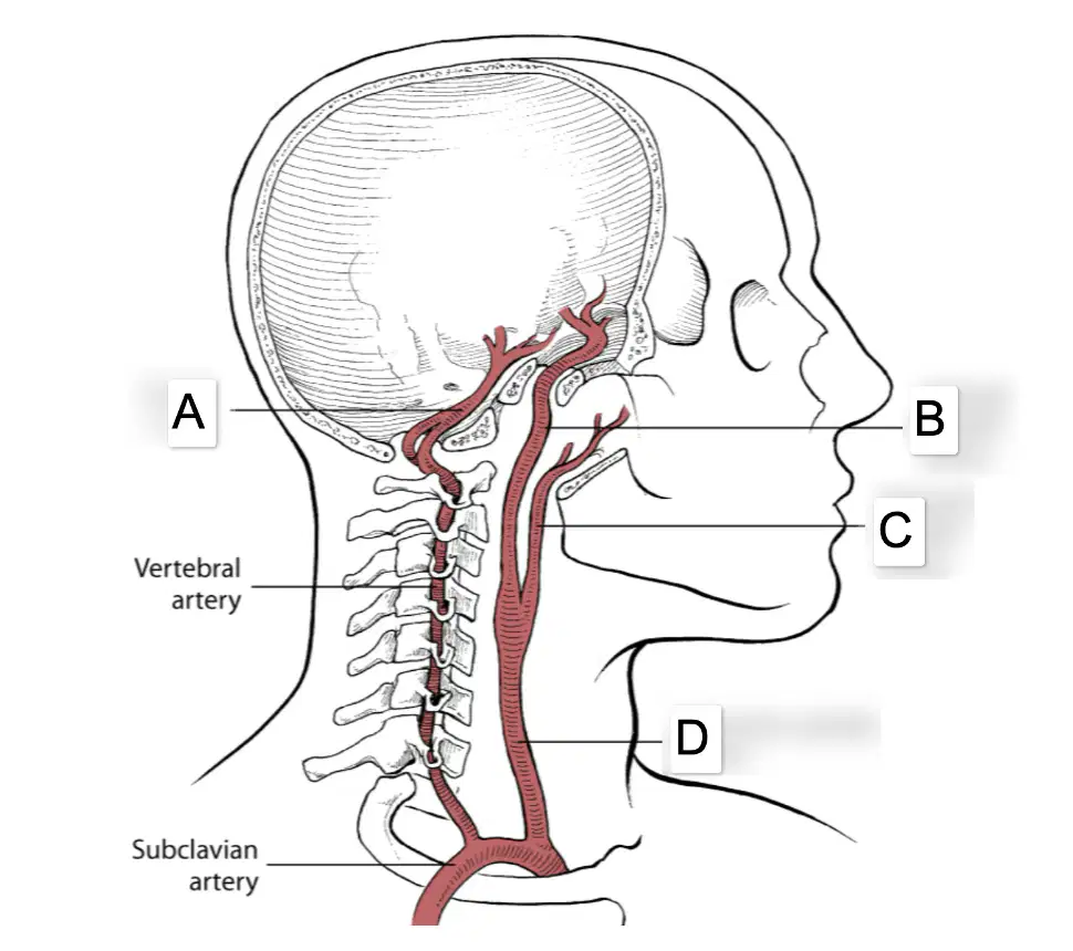
In the labeled sagittal head and neck artery diagram, what artery is at label C?
Internal carotid artery
External carotid artery
Basilar artery
Common carotid artery

In the labeled sagittal head and neck artery diagram, what artery is at label D?
Basilar artery
Common carotid artery
Internal carotid artery
External carotid artery
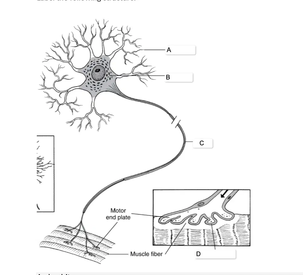
In the labeled neuron and neuromuscular junction diagram, what structure is at label A?
dendrite
axon
cell body
synaptic cleft

In the labeled neuron and neuromuscular junction diagram, what structure is at label B?
synaptic cleft
axon
cell body
dendrite
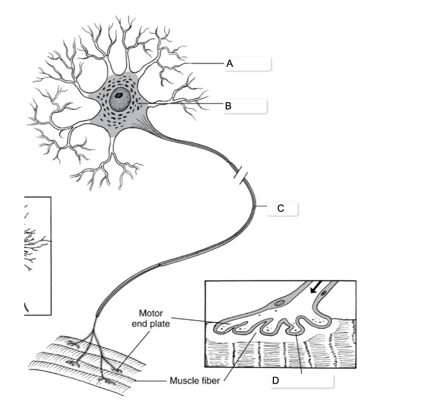
In the labeled neuron and neuromuscular junction diagram, what structure is at label C?
axon
cell body
dendrite
synaptic cleft
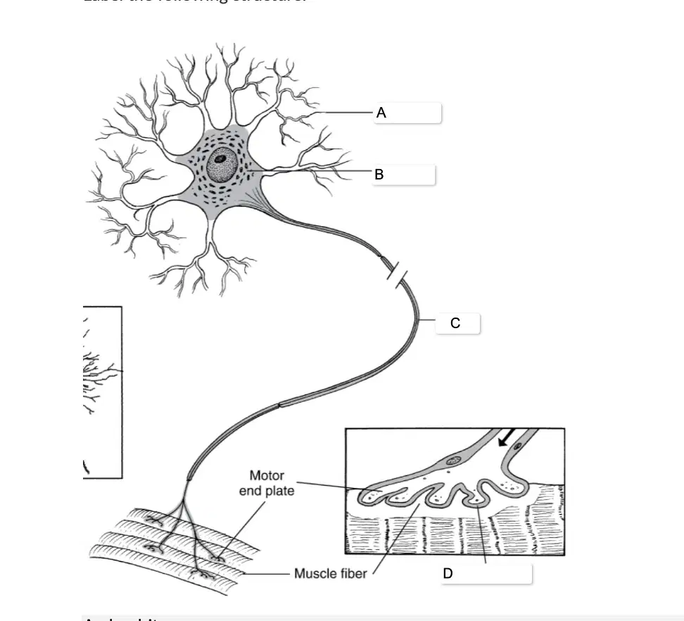
In the labeled neuron and neuromuscular junction diagram, what structure is at label D?
cell body
synaptic cleft
axon
dendrite
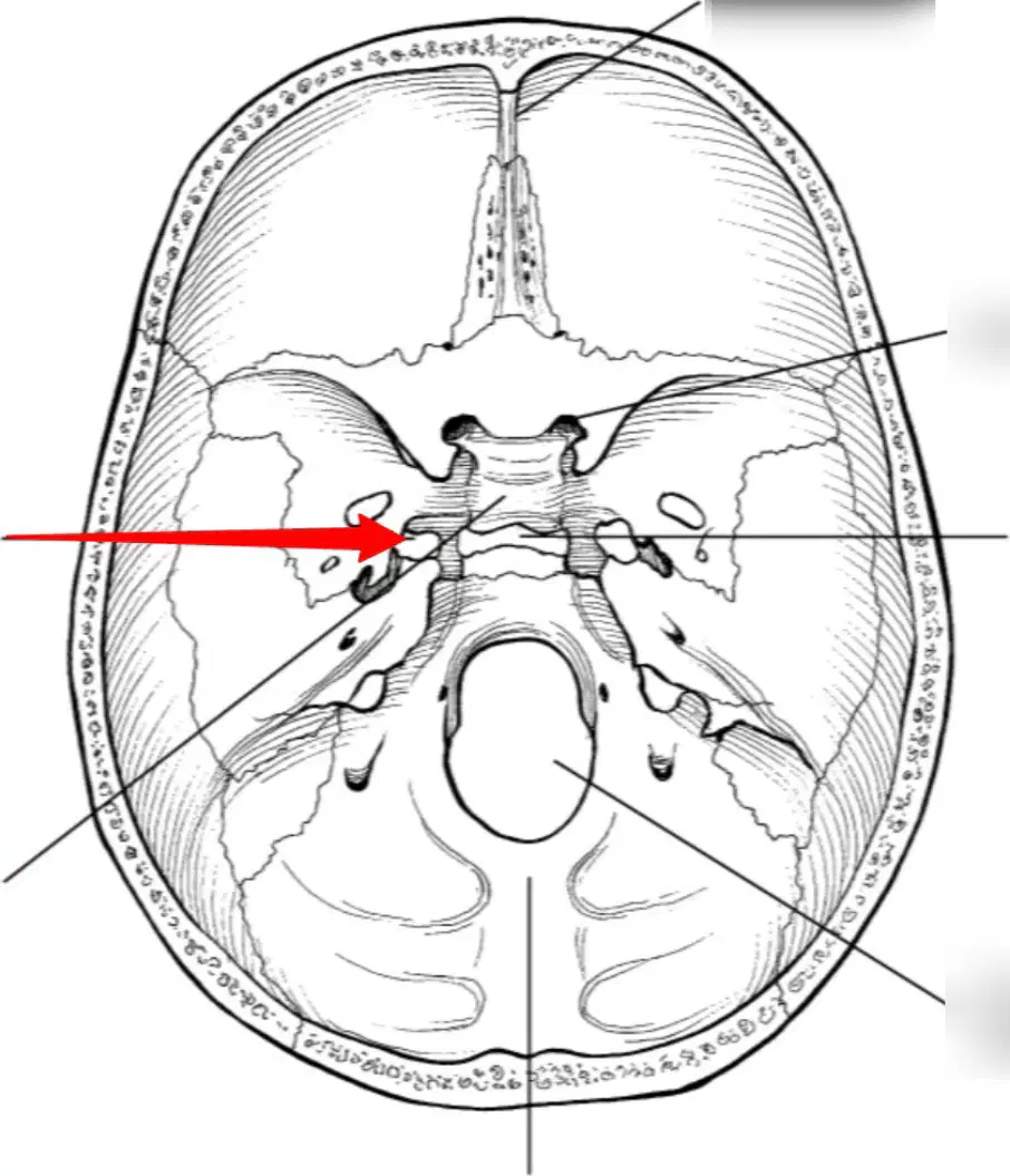
In the skull diagram with an arrow, what structure is the arrow pointing to?
carotid canal
foramen magnum
jugular foramen
optic canal
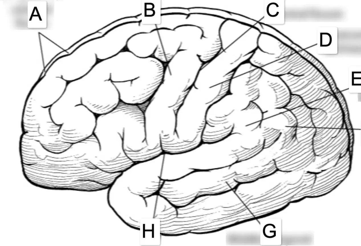
Match the following with the correct label on the lateral view brain diagram. Select the label that identifies the Longitudinal cerebral fissure.
A
C
H
F
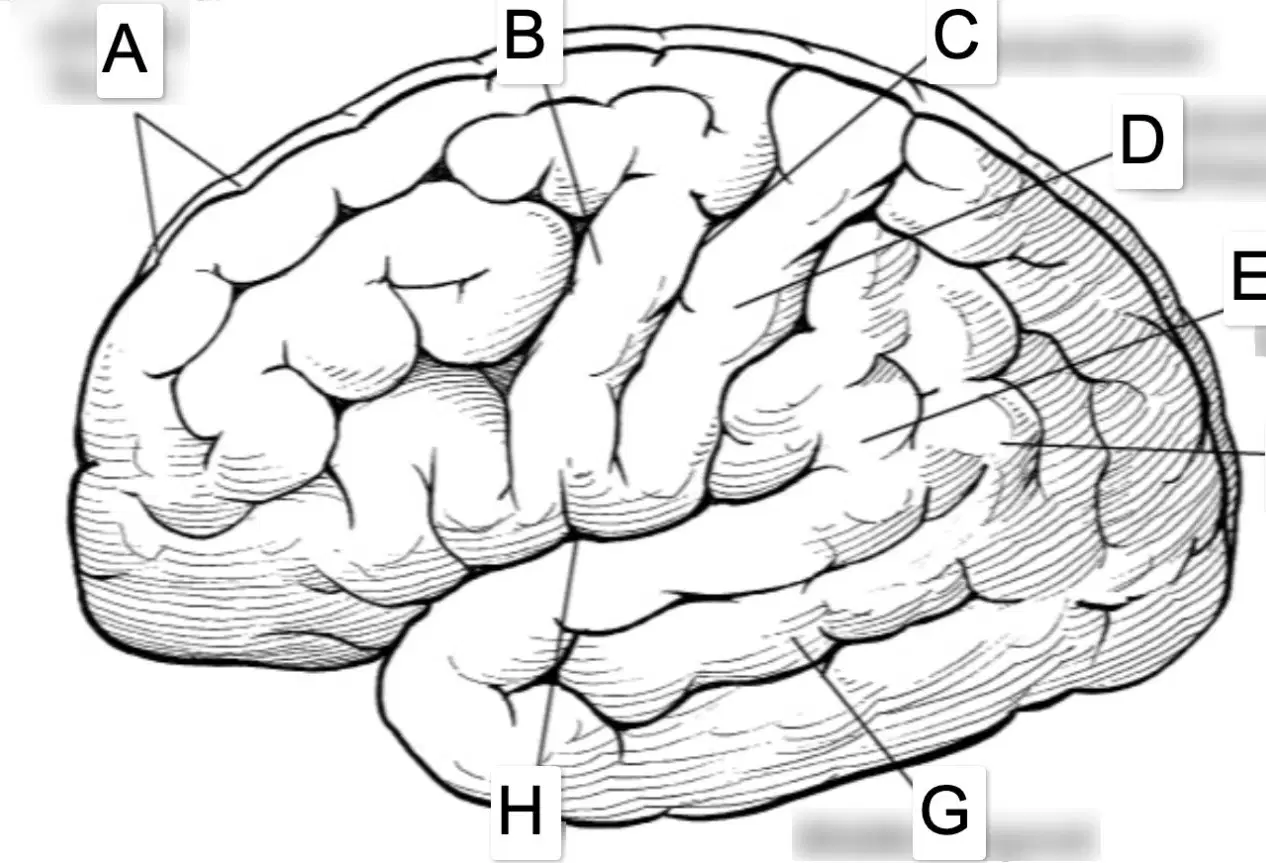
Match the following with the correct label on the lateral view brain diagram. Select the label that identifies the Precentral gyrus (primary motor cortex).
B
D
E
G

Match the following with the correct label on the lateral view brain diagram. Select the label that identifies the Central fissure.
C
A
H
B
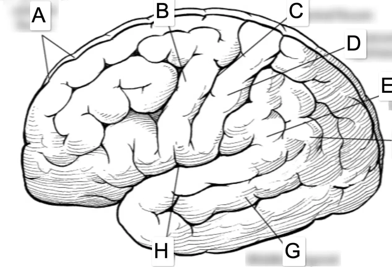
Match the following with the correct label on the lateral view brain diagram. Select the label that identifies the Postcentral gyrus (primary somatosensory cortex).
D
E
F
B

Match the following with the correct label on the lateral view brain diagram. Select the label that identifies the Supramarginal gyrus.
E
F
G
H
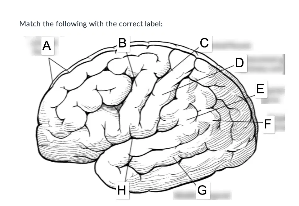
Match the following with the correct label on the lateral view brain diagram. Select the label that identifies the Angular gyrus.
F
E
C
D

Match the following with the correct label on the lateral view brain diagram. Select the label that identifies the Middle temporal gyrus.
G
H
A
C
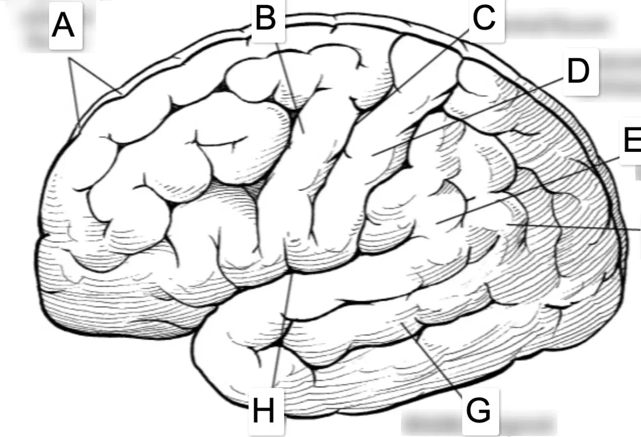
Match the following with the correct label on the lateral view brain diagram. Select the label that identifies the Lateral cerebral fissure (Sylvian fissure).
H
A
B
G
__________ strokes are rarer than __________ strokes but progress more quickly and are usually more severe in symptoms.
hemorrhagic; ischemic
ischemic; hemorrhagic
ischemic; ischemic
hemorrhagic; hemorrhagic
Most strokes can be categorized as hemorrhagic.
True
False
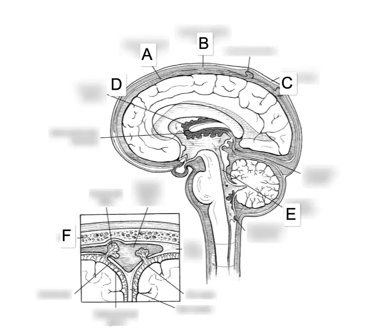
On the sagittal brain and meninges diagram, which label indicates the Subarachnoid space?
A
B
C
D

On the sagittal brain and meninges diagram, which label indicates the Superior sagittal sinus?
A
B
C
D
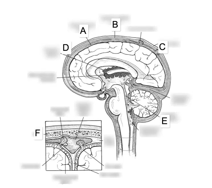
On the sagittal brain and meninges diagram, which label indicates the Dura mater?
A
C
D
E
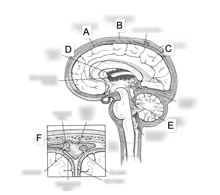
On the sagittal brain and meninges diagram, which label indicates the Choroid plexus?
B
D
E
F
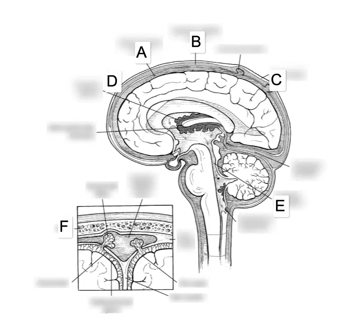
On the sagittal brain and meninges diagram, which label indicates the Cerebral aqueduct?
C
E
F
A
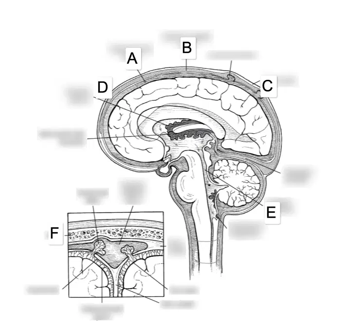
Which layer is the skull
B
D
F
E

Label the Mandible
A
C
B
D
(a) is a shock absorber, circulates nutrients, and removes waste from the brain.
There is considerable variability across brains in the location, shape, and prominence of the landmarks, sometimes making them difficult to identify.
True
False
Classify the following cranial nerve as either sensory, motor, or both: CN V
Sensory
Motor
Both
Multiple sclerosis is a disease that destroys (a)
Which cranial nerve moves the stylopharyngeus muscle to elevate the pharynx during speech and swallowing?
CN V (trigeminal)
CN IX (glossopharyngeal)
CN X (vagus)
CN XII (hypoglossal)
Error in distance judgment that can be noted during speech production is called
Ataxia
Apraxia
Dysmetria
Dysarthria
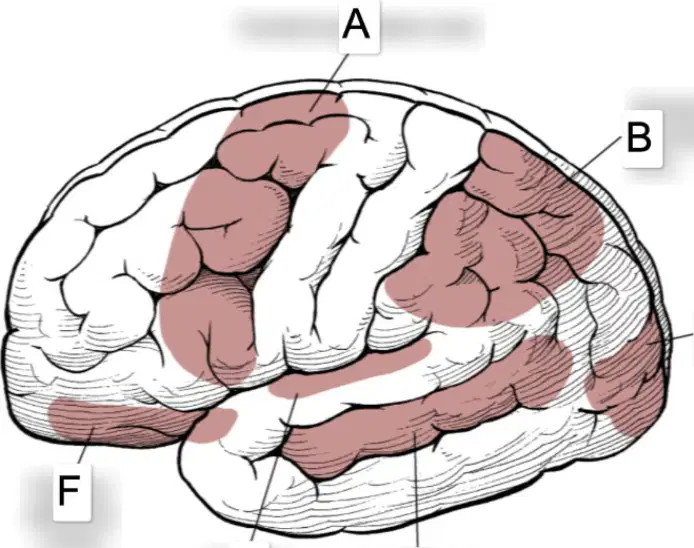
What hemisphere is pictured here?
Left hemisphere
Right hemisphere
Classify the following as sensory, motor, or both: CN XII
Sensory
Motor
Both
The precentral gyrus is located in the
Frontal lobe
Parietal lobe
Temporal lobe
Occipital lobe
Breathing, heartbeat, glands, and blood pressure are all regulated by the
Somatic nervous system
Autonomic nervous system
Central nervous system
Enteric nervous system
Damage to which of the following arteries typically causes visual field cuts? Select all that apply.
Anterior cerebral artery (ACA)
Middle cerebral artery (MCA)
Posterior cerebral artery (PCA)
Posterior inferior cerebellar artery (PICA)
Occlusions ______ the circle of Willis may not cause as much damage as occlusions ______ the circle of Willis.
below; above
above; below
within; outside
outside; within
When referring to a (a) stroke, one might use terms like thrombus and embolus.
Approximately what percentage of available oxygen within the body does the brain use?
10%
20%
30%
40%
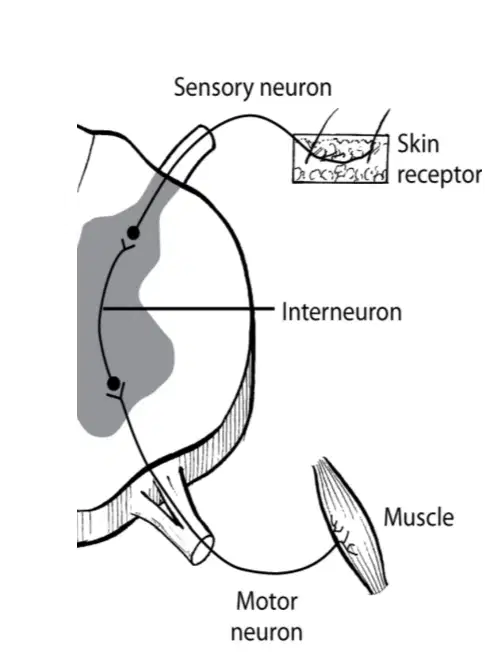
When stimulation of a sensory nerve is transmitted to a motor nerve via an interneuron, making possible rapid responses to stimuli without participation of higher centers in the nervous system, this is called a
Action potential
Reflex arc
Saltatory conduction
Summation
When using the terms afferent and efferent, we are referring to the
types of synapses
functions of neurons
cell types in neuroglia
brain lobes
Afferent and efferent are terms used to refer to nerves, not neurons. Sensory and motor are terms used to describe neurons.
True
False
The chemical process involved in the transfer of nerve impulses from one neuron to another occurs when a chemical neurotransmitter is released across the
axon hillock
dendritic spine
synaptic cleft
myelin sheath

Which of the following allows messages to go between hemispheres of the brain?
diencephalon
corpus collosum
cerebrospinal fluid
meninges


The green line in the picture represents which of the following structures?
Central Sulcus
Lateral Fissure
Longitudinal Fissure
Sylvian Fissure


The ORANGE part of the brain is called?
Frontal lobe
partial lobe
temporal lobe
occipital lobe

IDENTIFY THE PART
DENDRITE
AXON
AXON TERMINAL
SCHWANN CELL
NODE OF RANVIER

In this image, What is the PURPLE part of the brain called.
Frontal lobe
occipital lobe
partial lobe
temporal lobe

Label the sensory cortex
B
E
J
I
Which lobe of the brain is primarily responsible for processing visual information?
Frontal lobe
Parietal lobe
Occipital lobe
Temporal lobe
Which structure connects the left and right cerebral hemispheres, allowing communication between them?
Thalamus
Corpus callosum
Medulla oblongata
Pons


The brain stem/medulla is labeled as #...
1
2
3


What part of the brain is pictured in purple?
cerebellum
brain stem
temporal lobe

Label the frontal bone
C
B
E
A

Label the Occipital bone
B
A
C
E

Label the temporal bone
A
B
C
D

Which color represents the parietal lobe?
Blue
Pink
Yellow
Green
Olfactory
CN IV
CN II
CN I
cn XII
Olfactory
CN IV
CN II
CN I
cn XII
Olfactory
CN IV
CN II
CN I
cn XII
Trigeminal
CN V
CN X
CN III
CN IV
OPTIC
CN II
CN X
CN III
CN IV
OCULOMOTOR
CN IV
CN III
CN I
cn XII
TROCHLEAR
CN IV
CN III
CN I
cn XII
ABDUCENS
CN VI
CN X
CN III
CN IV
FACIAL
CN VI
CN VII
CN III
CN IV
VESTIBULOCOCHLEAR
CN VI
CN VIII
CN III
CN IV
GLOSSOPHARYNGEAL
CN V
CN VIII
CN IX
CN X
VAGUS
CN V
CN VIII
CN IX
CN X
ACCESSORY
CN V
CN VIII
CN XI
CN X
HYPOGLOSSAL
CN XII
CN VIII
CN XI
CN X

Controls most of the movement of the eye
Abducens
Trochlear
Oculomotor
Hypoglossal

VII MOTOR AND SENSORY NERVE TO 5 PARTS
OF THE FACE - FACIAL EXPRESSION
abducens
facial
trochlear
accessory

sense of smell
olfactory
optic
oculomotor
trigimenal;

hearing and balance
trigeminal
olfactory
hypoglossal
vestibulocochlear

Motor function of the movement of the eye
trochlear
trigeminal
glossopharyngeal
optic

SENSORY AND MOTOR NERVES TO PART OF THE FACE - CHEWING
Trochlear
Trigeminal
Abducens
Facial

Movement of the eyes - abducts the eyes
Trigeminal
Abducens
oculomoter
facial

Tasting and swallowing
vestibulochelear
vagus
hypoglossal
glossopharyngeal

vision
ociulomotor
trigeminal
optic
abducen

MOTOR MOVEMENT OF NECK AND SWALLOWING
accessory
hypoglossal
facial
vagus

CN XII MOTOR MOVEMENT OF THE TONGUE
TALKING - SPEECH
trochlear
vestibulocochlear
hypoglossal
vagus

PARASYMPATHETIC CONTROL OF INTERNAL ORGANS
trochlear
vestibulocochlear
hypoglossal
vagus
Which body section cuts the body into left and right portions?
Coronal
Transverse
Superior
Sagittal
Which body section cuts the body into left and right portions?
Coronal
Transverse
Superior
Sagittal
Which body section cuts the body into top and bottom portions?
Sagittal
Coronal
Transverse
Superior
____________ means "toward the back".
Superior
Inferior
Anterior
Posterior
____________ means "toward midline" or the middle of the body.
Medial
Lateral
Proximal
Distal
Which of the following terms means "opposite sided"?
ipsilateral
contralateral
Medial
Lateral
Which of the following terms means "bringing structures together"?
ipsilateral
contralateral
adduct
abduct
The part of the nervous system that includes the brain and spinal cord is the:
central nervous system
peripheral nervous system
autonomic nervous system
somatic nervous system
Which structures make up the brainstem?
Midbrain
pons
medulla
all of the above
The ______________ acts as a relay station for sensory fibers.
hypothalamus
basal ganglia
thalamus
corpus callosum
Which part of the nervous system includes cranial and spinal nerves?
Central nervous system
Peripheral nervous system
autonomic nervous system
somatic nervous system
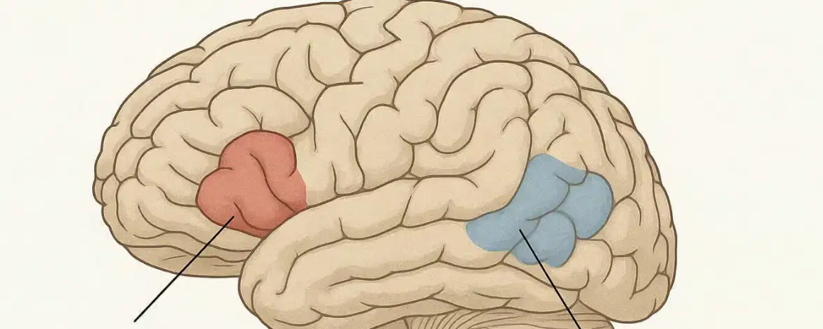
LABEL The blue area
Motor
Visual
Broca's
Wernicke's

Label the red area
Visual
Broca's
Wernicke's
Motor

Label the Carotid Canal
A
B
C
D

Label the Sella Turcica
A
C
B
E

Label the internal occipital crest
A
E
C
G

Label the Crista Gailli
A
B
E
F

Label the optic foramen
G
F
E
D

Label the Posterior Clinoid process
G
F
E
D

Label the Foramen Magnum
F
B
E
D

This type of stroke is characterised by a rupture.
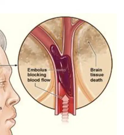
This type of stroke is characterised by a blockage.

