
Microscopes
Presentation
•
Biology
•
9th - 12th Grade
•
Easy
+1
Standards-aligned

Amy Kirkwood
Used 2+ times
FREE Resource
25 Slides • 102 Questions
1
2

3

4
5

6

7
8
9
10
11

12
Multiple Choice
13
Dropdown
14

15

16
Multiple Choice
17
Match
18
Multiple Choice
19
20
21

22
Multiple Choice
23
24
Multiple Choice
25
26
Drag and Drop
27

28

29

30
Multiple Choice

Which image shows the correct way to carry a microscope?
31
32
Multiple Choice
33
Multiple Choice
34
Multiple Choice
35
Multiple Choice
36
37
Multiple Choice
38
39
Multiple Choice
40
Multiple Choice
41
Multiple Choice
42
Multiple Choice
43

44
Hotspot
45
Hotspot
46
Hotspot
47
Hotspot
48
Hotspot
49
Hotspot
50
Hotspot
51
Multiple Choice
52
Multiple Choice
53
Multiple Choice
54
Reorder
55
Fill in the Blanks

56
Fill in the Blanks
57
Fill in the Blanks
58
Drag and Drop

59
Drag and Drop

60
Drag and Drop

61
Drag and Drop

62
Multiple Choice

How do you adjust the amount of light shining through your microscope?
63
Drag and Drop

64
Drag and Drop

You can hit the slide and
65
Match
66
Multiple Choice

What happened to your field of view as you moved from low to high power?
67
Multiple Choice
68
Multiple Choice
69
Multiple Choice
70
Multiple Choice
71
Match
72
Multiple Select

Read the attached passage:
Why does a specimen placed under the microscope have to be thin?
Check all that apply.
73
Labelling
74
Labelling
75
Multiple Choice

Part B
76
Multiple Choice

Part A
77
Labelling
78
Drag and Drop
79
Multiple Choice
80
Multiple Choice
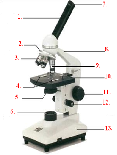
What is Part 12
81
Multiple Choice

What is Part 13
82
Multiple Choice

What is Part 7
83
Multiple Choice
84
Multiple Choice
85
Multiple Choice
86
Multiple Choice

The largest field of view shown is under the total magnification _____X.
87
Multiple Choice

If you wanted to center the image what direction would you move your slide in?
88
Multiple Choice
89
Multiple Choice
90
Multiple Choice
91
Multiple Choice
92
Multiple Choice
93
Multiple Choice

If you look at a letter such as "e" under the microscope, then how does it look when you are viewing the image?
94
Multiple Choice

When making a wet mount, place the coverslip at a 45-degree angle to reduce air bubbles.
95
Multiple Choice

This picture shows that the microscope:
96
Multiple Choice
97
Multiple Choice
98
Multiple Choice

Which laboratory procedure is represented in the diagram below?
99
Multiple Choice

Which laboratory technique is shown in the diagram?
100
Multiple Choice
101
Multiple Choice
102
Multiple Choice
103
Multiple Choice
104
Multiple Choice
105
Multiple Choice
106
Multiple Choice
107
Multiple Choice
108
Multiple Choice

What does a microscope do?
109
Multiple Choice
110
Multiple Choice

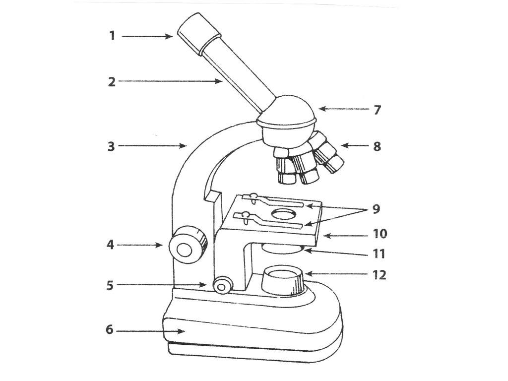
Which part is #8?
111
Multiple Choice

What part is # 9?
112
Multiple Choice

What part is #1?
113
Multiple Choice

What part is AB?
114
Multiple Choice

What part is E?
115
Multiple Choice

Which part is D?
116
Multiple Choice

Which part is C?
117
Multiple Choice

Which part is B?
118
Multiple Choice

Which part is A?
119
Multiple Choice

120
Multiple Choice

121
Multiple Choice

122
Multiple Choice

123
Multiple Choice
124
Multiple Choice

125
Multiple Choice
126
Multiple Choice

127
Multiple Choice
Show answer
Auto Play
Slide 1 / 127
SLIDE
