
 Worksheets
EKG CMA
Total questions: 108
Worksheet time: 2hrs 36mins
An atrial pacemaker site that changes from location to location has
P waves that continually change in appearance
One pacemaker site from which the impulse arise
Inverted P’ waves
A chaotic baseline preceding each QRS complex
Which of the following has more P waves than QRS complexes?
A dysrhythmia where the pacemaker changes from site to site with each beat
Early beats that arise from the atria
AV heart block
Dysrhythmias that arise from the AV junction
The ___________ represents the end of ventricular depolarization and beginning of ventricular repolarization.
T wave
U wave
QT interval
ST segment
The ___________ represents ventricular repolarization.
T wave
U wave
QT interval
ST segment
1. Prominent U waves are seen in:
Hypokalemia
myocardial infarction.
digitalis toxicity.
All of these
The point at which the QRS complex meets the ST segment is called the:
QT junction.
ST interval.
J point.
RS point.
Impulses that arise from the ventricles produce QRS complexes that:
look the same as the QRS complexes which are produced by impulses originating from the SA node.
are called R prime or R' waves.
are typically notched and biphasic.
appear wide and bizarre-looking.
The PR intervals are not measurable in:
wandering atrial pacemaker.
atrial flutter
2nd - degree AV heart block, Type I.
premature atrial complexes.
The PR interval denotes depolarization of the heart from the
Bundle of His through the ventricles
SA node through the atria, AV node, and His-Purkinje system
AV node through the bundle of His
SA node through the atria and AV node
ST segment depression suggests:
Myocardial ischemia
Myocardial injury/infarction
Pericarditis
A healthy heart
The QT interval:
represents the time needed for ventricular repolarization.
is prolonged when ventricular repolarization is accelerated.
extends from the beginning of the P wave to the end of the T wave.
is usually between 0.36 and 0.44 seconds in duration.
Blood enters the heart from the _______?
Superior and inferior vena cava
Bicuspid and Tricuspid
aorta
pulmonary artery
From the pulmonary valve blood enters the _______?
aortic artery
pulmonary valve
pulmonary artery
aortic valve
After the blood returns from the lungs, it is now____?
deoxygenated
oxygenated
purple
Once blood passes through the mitral/ bicuspid valve, it enters the________?
Right atrium
Left atrium
Right ventricle
Left ventricle
Blood is forced from the left ventricle up through the_______?
pulmonary valve
bicuspid valve
tricuspid valve
aortic valve
Normal heart rate for an adult is?
20 to 100/min
40 to 120/min
60 to 100/min
20 to 60/min
The normal hear rhythm is called?
Sinus brady
ventricle fibrillation
Normal sinus rhythm
Junctional Rhytms
deoxygenated blood coming from____?
body to lungs
head to body
lungs to body
lungs to head
oxygenated blood returning from ___?
Lungs to body
Body to lungs
head to body
lungs to head
The SA Node is known as?
Pacemaker
gatekeeper
ventricle
cardiac muscle
The AV Node is known as?
pacemaker
gatekeeper
ventricle
cardiac muscle
The SA Node generates impulses at?
60-100 times per minute
60-40 times per minute
40-20 time per minute
20-100 times per minutes
AV Node can fire at a rate of?
60-100 times per minute
40-60 times per minute
40-20 times per minute
60-20 times per minute
which is the correct order?
SA Node, AV Node, Bundle of His, Lt and Rt Bundle Branches, Purkinje Fibers
AV Node, SA Node, Lt and Rt Bundle Branches, Bundle of His, Purkinje Fibers
Bundle of His, Lt and Rt Bundle Branches, Bundles of His, Purkinje Fibers, AV Node, SA Node
Purkinje Fibers, Bundle of His, AV Node, Lt and Rt Bundle Branches
Define Depolarization
Contraction
Relaxation
Define repolarization
Contraction
Relaxation
The QRS complex is normally __________ seconds in duration
0.06 to 0.10
0.04 to 0.10
0.08 to 0.12
0.10 to 0.12
P waves contract the __________
ventricles
atria
AV junction
SA node
PR interval is the same at _________ seconds
0.06 to 0.10
0.10 to 0.12
0.12 to 0.20
0.20 to 0.25
What is cardiac output?
The amount of O2 carries through the veins per minute
Heart rate speeding up
The amount of blood pump through the heart per minute
The amount blood pump through the lungs per minute
Rhythm that is slow or fast is considered
Tachycardia
Bradycardia
Irregular
Abnormal
Four the 4 methods of the determining the pulse on an EKG tracing
adding, subtracting, multiplying, dividing
6 seconds X 10, 300 150 100 75 60 50, 1500, rate calculators
seconds, minutes, hours, days
6 seconds X 10, 300 200 150 100 75 60 50, 1500, rate rulers
Adult systolic blood pressure
> 70
70 + 2* age
90 to 120 mm Hg
60 to 90 mm Hg
Adult Diastolic blood pressure
60 to 90 mm Hg
70 + 2* age
90 to 120 mm Hg
N/A
In a lifetime, how much blood will your heart pump?
1.5 million
A thousand
2.4 million
10,000
Why is the SA node known as the pacemaker of the heart?
Because when impulses come too quickly, it slows or allows only select impulses through.
Initiates and maintains the rhythm of the heart and maintains the pace of the heart beat.
Because it splits into two and impulses travel much faster.
Because they conduct impulses rapidly through the muscles to assist in it's contractions
Can tell us if the impulse that initiated arose from the SA node and traveled through the atria and AV junction at a normal fashion:
QRS Complexes
PR Intervals
T waves
P waves
P waves, QRS complexes, and T waves are:
Depolarization
A type of measurement
Repolarization
The path of the electrical activity through the heart
They are so small that they sometimes are not seen on the ECG
ST segments
T wave
U waves
Q wave
The advantages of the 300, 150 method is all of the following except
Quick
Fairly accurate
Can be used only when rhythm is regular
More precise
How do you determine the 1500 method
Count the number of small squares between two consecutive R waves
Find a R wave located on a bold line and then find the next consecutive R wave. If it is on a bold line then the heart rate is the value of that line
Identify two successive 3-second marks. Count the number of QRS complexes in that 6 second section. Multiply by 10
Position the start mark on an R wave, then find the next consecutive R wave. Where the next R wave lines up is the approximate heart rate.
Identify the 4 chambers of the heart
Tricuspid valve, Bicuspid valve, Right atrium, and Left ventricle
Right atrium, Right ventricle, Left atrium, and Left ventricle
Left ventricle, Right atrium, Right ventricle, tricuspid valve
Left ventricle, Left atrium, bicuspid valve
Which of the following isn't apart of the nine main elements of the ECG tracing
T waves
QRS complexes
1500 method
PR intervals
P waves can tell us..
If the heart will beat in a consecutive, rhythmic fashion
If the impulse that initiated arose from the SA node and traveled through the atria and AV junction at a normal fashion
That there's sometimes the presence of a U wave
About movement of the electrical impulse through the ventricles, causing their contraction.
What has more oxygen than the veins?
Arteries
Heart
Lungs
Organs
The heart pumps around how many liters of blood?
3-4
4-8
5-6
8-9
What to not forget to hand to patient before leaving the room
Sticker
EKG machine
Call light
Gloves
Where is V1 located
Nipple line
Axillary line
5th intercostal space
4th intercostal space
Where is V6 located
Between V3 and V4
Axillary line
4th intercostal space
Nipple line
What do you do if the EKG comes out messed up
Leave the patient to figure it out themselves
Get a doctor or whose in charge to help you
Ignore it
Let the patient leave
Where is V4 located
4th intercostal space
inbetween V2 and V3
Axillary line
Nipple line
Where is V2 located
Across from V1
Right below V1
In between V3 and V4
Axillary line
Where is V3 located
Nipple line
In between V2 and V4
Below V1
Axillary line
Where is V5 located
Below V2
Nipple line
Beside V3
In between V4 and V6
What do you do when you first walk in the room
Knock on the door
Wash your hands
Introduce yourself
Provide privacy
Explain procedure


A patient complaining about left sided arm pain, nausea, and diaphoresis should be evaluated for:
Congestive Heart Failure (CHF)
Diabetes
Myocardial Infarction (MI)
Chronic Obstructive Pulmonary Disease (COPD)
Which would be on a chemistry panel?
Liver functions, kidney function, and electrolytes
Cholesterol, tryiglycerides, LDL, HDL
WBC, RBC, HGB, HCT
PT,PTT, INR
What is the measurement of the standardization mark?
10mm high and 5mm across
5mm high and 10mm across
5mm high and 5mm across
None of the above
How would you correct artifact from somatic (muscle) tremor?
Turn their cell phone off
Check the leads
Give the patient a blanket
Push the "AC Interference" button on the machine
The lead on 5th intercostal space, mid-axillary is:
V4
V6
V1
V5
The lead that is on the right side at the 4th intercostal space/sternal border is:
V2
V1
V3
V4
Who would be a candidate for a right sided EKG on?
A patient with Dextrocardia
A patient with Situs Inversus
A patient that needs nuclear testing
A patient with a L. lower extremity amputation
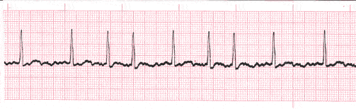

The nurse is reviewing a patient’s electrocardiogram. Which wave should the nurse recognize as being the first positive wave in a normal cardiac cycle?
P wave
Q wave
R wave
S wave
The nurse is assessing the client’s electrocardiography (ECG). What does the P wave on the ECG tracing represent?
Contraction of the atria
Contraction of the ventricles
Depolarization of the atria
Depolarization of the ventricles
The nurse is reviewing cardiac anatomy with a patient scheduled for pacemaker insertion. Where should the nurse identify the location of the sinus node for the patient?
Left atrium
Right atrium
Left ventricle
Right ventricle
A patient has a normal heartbeat of 72 beats per minute. What area of the conduction system should the nurse consider as functioning normally in this patient?
Bundle of His
Sinoatrial node
Purkinje’s fibers
Atrioventricular node
The nurse is determining the functioning of a patient’s sinoatrial node. How many times per minute should the nurse expect the node to normally fire?
20 to 40
25 to 55
40 to 85
60 to 100
The nurse determines that a patient’s electrocardiogram is demonstrating normal sinus rhythm. What did the nurse observe to come to this conclusion? (Select all that apply.)
Regular rhythm
P wave before each QRS complex
Wide QRS intervals
Heart rate 88 beats per minute
The nurse is analyzing a client’s electrocardiogram tracing. Which of the following complexes is not normally seen on an electrocardiogram tracing?
P wave
QRS complex
T wave
U wave

