
Font size
 Worksheets
Axial Skeleton + Processes
Total questions: 100
Worksheet time: 16hrs 21mins

Identify bone "F"
Temporal
Parietal
Sphenoid
Zygomatic

Identify Bone "H"
Zygomatic
Maxilla
Sphenoid
Lacrimal

identify Bone "B"
Maxilla
Mandible
Zygomatic
Sphenoid


frontal
sphenoid
zygomatic
nasal

maxilla
mandible
zygomatic
parietal


What bone is pictured here
Zygomatic
Lacrimal
Nasal bone
Ethmoid

The purple bone(s) is (are) called the
palatine bone(s)
lacrimal bone(s)
septum bone(s)
vomer bone(s)


Which bone is #6 pointing to?
Lacrimal
Inferior nasal concha
Zygomatic
Mandible

Identify cranial bone "B"
Lacrimal
Ethmoid
Parietal
Sphenoid

Identify cranial bone "A"
Ethmoid
Sphenoid
Lacrimal
Temporal

Identify Bone "D"
Sphenoid
Ethmoid
Lacrimal
Nasal

Identify Bone "B"
Lacrimal
Ethmoid
Sphenoid
Nasal

Identify Bone "C"
Lacrimal
Ethmoid
Sphenoid
Nasal

Identify Bone "E"
Vomer
Nasal
Maxilla
Inferior Nasal Conchae

Identify Bone "D"
Ethmoid
Vomer
Inferior Nasal Conchae
Maxilla

Bone #9 is...
Palatine Bone
Mandible
Parietal Bone
Zygomatic Bone

What bone is pictured here?
Palatine Bone
Volmer Bone
Ethmoid Bone
Sphenoid Bone
Identify bone "E"
Temporal
Parietal
Sphenoid
Zygomatic
Identify bone "D"
Temporal
Parietal
Sphenoid
Ethmoid
Which bone is #1 pointing to?
Lacrimal
Inferior nasal concha
Zygomatic
Palatine
Which bone is #3 pointing to?
Lacrimal
Inferior nasal concha
Vomer
Palatine


What are the structures in the picture?
foramen magnum
occipital condyles
external auditory meatus
zygomatic process

What is the structure in the picture?
foramen magnum
occipital condyles
external auditory meatus
zygomatic process

What is the structure on this picture?
infraorbital foramen
palatine process
zygomatic arch
mental foramen

The depressions on the temporal bones (within the red ovals) are called...
mandibular notch
occipital crest
mandibular fossae
zygomatic arch

What is the structure in this picture?
infraorbital foramen
zygomatic arch
horizontal plate
mental foramen

What is the structure in the picture?
foramen magnum
occipital condyles
external auditory meatus
zygomatic process
sella turcica

What is the structure in the picture?
foramen magnum
occipital condyles
styloid process
zygomatic process
mastoid process

What is the structure in this picture?
infraorbital foramen
palatine process
zygomatic arch
horizontal plate
mental foramen

What type of suture is this?
coronal
squamosal
lamboidal
sagittal

What is the structure in this picture?
infraorbital foramen
palatine process
zygomatic arch
horizontal plate
mental foramen

What type of suture is this?
coronal
squamosal
lamboidal
sagittal

What type of suture is this?
coronal
squamosal
lambdoidal
sagittal

What type of suture is this?
coronal
squamosal
lambdoidal
sagittal

What feature is this?
Zygomatic arch
Occipital condyles
Foramen magnum
Vomer

What feature is this?
External acoustic meatus
Temporomandibular joint (TMJ)
Zygomatic arch
Mastoid process

What processes is #2
Mastoid Process
Maxillary Process
Zygomatic Process
Mandibular Process

What process is letter A
Styloid
External Acoustic Meatus
Mastoid Notch
Mandibular Fossa
What process is letter C
Styloid
External Acoustic Meatus
Zygomatic Process
Mandibular Fossa

What process is letter C
Lesser Wing of Sphenoid
Greater Wing of Sphenoid


What feature is number 7?
foramen magnum
external occipital protuberance/EOP
occipital condyles
styloid process
What process is letter F
Styloid
External Acoustic Meatus
Mastoid Notch
Mandibular Fossa
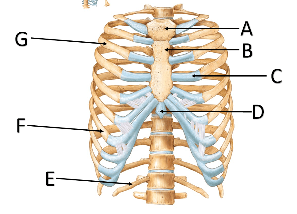


What is # 8 pointing to?
Intervertebral discs
intra-thoracic discs
Lumbar
sacrum
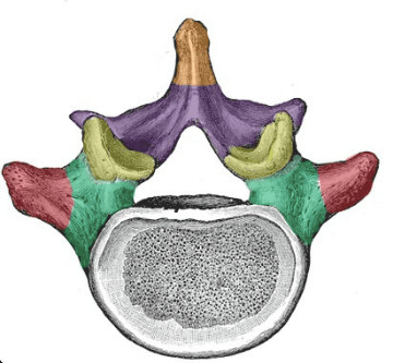
The yellow represents
transverse process
spinous process
superior articulating process
centrum (body)


What is number 1? Bony projection posteriorly.
Transverse Process
Spinous Process
Body
Pedicle
What is number 3? Bony projection laterally from the vertebra.
Demifacet
Superior Articular Facet
Transverse Process
Lamina
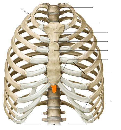
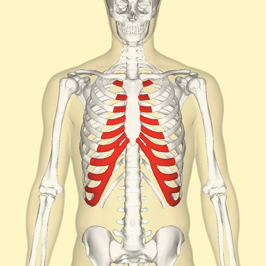

Label 3 is
the body
the vertebral foramen
a transverse process
for muscle attachment
Label 7 is
the transverse foramen
the vertebral foramen
a transverse process
for muscle attachment

This is the second cervical vertebrae (C2); it allows the head to rotate.
Atlas
Axis

What is structure 22?
true ribs
false ribs
floating ribs
costal cartilage
What is structure 21?
true ribs
false ribs
floating ribs
costal cartilage
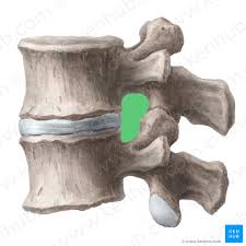
The green portion is? Where the spinal nerve roots exit the spinal cord/column.
Vertebral Foramen
Intra-spinous foramen
Intervertebral Foramen
Intra-vertebral Foramen

What is Letter F?
Sacral Promontory
Transverse Ridges
Apex
Median Sacral Crest
What is Letter A?
Sacral Promontory
Transverse Ridges
Apex
Median Sacral Crest
What is Letter B?
Sacral Promontory
Transverse Ridges
Apex
Median Sacral Crest
What is Letter E?
Sacral Promontory
Transverse Ridges
Apex
Median Sacral Crest

What is the proper name of your "tail bone?"
Pelvis
Pubis
Sacrum
Coccyx

What type of vertebrae is this?
Lumbar
Thoracic
Cervical
Sacrum

What type of vertebrae is this?
Lumbar
Thoracic
Cervical
Sacrum

What type of vertebrae is this?
Lumbar
Thoracic
Cervical
Sacrum

What type of vertebrae is this?
Lumbar
Thoracic
Cervical
Sacrum

What process is this pointing to?
Zygomatic process of temporal bone
Zygomatic Arch
Temporal process of zygomatic bone
Zygomatic Process

What process is this pointing to?
Zygomatic process of temporal bone
Zygomatic Arch
Temporal process of zygomatic bone
Zygomatic Process

What is the black line indicating
Mandibular notch
Zygomatic notch
Palatine Plate
Mandibular fossa

What is the name of the hole being indicated by the arrow?
superior orbital fissure
Inferior orbital fissure
Optic foramen
Lacrimal forament

What is the bone indicated by the arrow?
Sphenoid
Vomer
Perpendicular plate of the ethmoid
Sella Turcica
What is the bone indicated by the highlighted area?
Sphenoid
Vomer
Perpendicular plate of the ethmoid
Sella Turcica
What is the name of the back surface of the eye?
Orbital Surface
Orbital Sulcus
Optic Foramen
Orbital Foramen

