
 Worksheets
Nervous System III (Senses) Review
Total questions: 100
Worksheet time: 52mins

What is structure number 1?
Iris
Cornea
Lens
Pupil
What is structure number 2?
Iris
Cornea
Lens
Pupil
What is structure number 3?
Iris
Cornea
Lens
Pupil
What is structure number 4?
Iris
Cornea
Lens
Pupil
What is structure number 5?
Iris
Cornea
Aqueous humour
Ciliary muscle
What is structure number 6?
Lens
Suspensory ligaments
Aqueous humour
Ciliary muscle
What is structure number 7?
Lens
Suspensory ligaments
Aqueous humour
Ciliary muscle
What is structure number 8?
Lens
Suspensory ligaments
Aqueous humour
Ciliary body
What is structure number 9?
Sclera
choroid
Fovea
Aqueous humour
What is structure number 10?
Sclera
choroid
Fovea
Aqueous humour
What is structure number 11?
Sclera
choroid
Fovea
Retina
What is structure number 12?
Sclera
arteries and veins
Fovea
Retina
What is structure number 13?
Sclera
arteries and veins
Fovea
Vitreous humour
What is structure number 14?
Sclera
arteries and veins
Fovea
Vitreous humour
What is structure number 15?
Optic nerve
arteries and veins
Blind spot
Vitreous humour
Controls the shape of the lens.
Ciliary Body
Extrinsic Eye Muscles
Suspensory Ligaments
Optic Nerve
Iris
Produces aqueous humor
Ciliary Body
Extrinsic Eye Muscles
Suspensory Ligaments
Optic Nerve
Iris
Move the entire eyeball around
Ciliary Body
Extrinsic Eye Muscles
Suspensory Ligaments
Optic Nerve
Iris
Assist in holding the lens in an upright position so its shaped can be controlled
Ciliary Body
Extrinsic Eye Muscles
Suspensory Ligaments
Optic Nerve
Iris
Controls the size of the pupil
Ciliary Body
Extrinsic Eye Muscles
Suspensory Ligaments
Optic Nerve
Iris
Lets light into the anterior surface of the eye
Cornea
Vitreous Humor
Sclera
Aqueous Humor
Choroid
Balance pressure places on the eye by the extrinsic eye muscles
Cornea
Vitreous Humor
Sclera
Aqueous Humor
Choroid
Protects and shapes the majority of the eyeball
Cornea
Vitreous Humor
Sclera
Aqueous Humor
Choroid
Gives the cornea its bulging, round appearance
Cornea
Vitreous Humor
Sclera
Aqueous Humor
Choroid
Allows light to bend into the eye from the outside environment
Optic Nerve
Fovea
Lens
Cornea
Vitreous Humor
Delivers visual information from the retina to the visual centers in the occipital lobe
Optic Nerve
Fovea
Lens
Cornea
Vitreous Humor
Helps with colorful and 20/20 vision
Optic Nerve
Fovea
Lens
Cornea
Vitreous Humor
Allows light to pass directly to photoreceptors in the retina to enhance visual acuity (details)
Optic Nerve
Fovea
Lens
Cornea
Vitreous Humor
Bends incoming light dramatically so that it can be focused on the retina
Optic Nerve
Fovea
Lens
Cornea
Vitreous Humor
Allows passage of light to the retina through posterior cavity because of its clear appearance
Optic Nerve
Fovea
Lens
Cornea
Vitreous Humor
Supports the posterior surface of the lens and holds the retina against the wall of the eye
Cornea
Vitreous Humor
Sclera
Aqueous Humor
Choroid
Nourishes the cornea and lens
Aqueous Humor
Ciliary Body
Vitreous Humor
Retina
Choroid
Nourishes the retina
Aqueous Humor
Ciliary Body
Vitreous Humor
Retina
Choroid
What happens in the tympanic membrane
?
Sound waves are converted to vibrations.
Sound waves make the tiny hairs in your ear move
Carries the message to the brain
What does the fluid inside your ears do when displaced?
Vibrates the skin in your ear
Sends sound waves to the brain
Displaces the hair cells.
Stimulates the hair cells to send an impulse
What is in the Semi-Circular canals?
Cilia (Tiny Hairs)
Liquid
Thin Skin
Where is the tympanic membrane located?
The Outer ear
The Inner Ear
The Middle Ear
Where is the Eustachian Tube connected to?
The back of the head
The back of the nose
The back of the Ear
When sound waves cause the tympanic membrane to vibrate, these vibrations are immediately passed on to the_________.
outer ear and ear canal
inner ear
tiny hairs
three small bones in the middle ear
Which of the following is NOT in the MIDDLE EAR?
Auricle
Eustachian tube
Malleus
Tympanic Membrane
The impulse is generated and is transmitted to the temporal lobe when what happens?
the eardrum vibrates
the basilar membrane vibrates
the oval window vibrates
the hair cells are bent
When sound waves vibrate the tympanic membrane what structure of the ear is moved next:
the vestibule
the cochlea
the semicircular canals
the malleus
The malleus, incus, and stapes are collectively called the
maculae
vestibulocochlear nerves
semicircular canals
auditory ossicles
Which of the below structures acts to convert sound waves to vibrations?
cochlea
pinna
tympanic membrane
vestibulocochlear nerve
When you spin while sitting in a swivel chair with your eyes closed, you can sense this movement by
means of your
cochlea
saccule
semicircular ducts
utricle

The diagram above represents
Cupula
Ampulla
Utricle
Saccule

The diagram above represents
Cupula
Ampulla
Utricle

Area #1 This bone that is attached to the tympanic membrane is called -
Hammer (Malleus)
Anvil (Incus)
Stirrup (Stapes)
Ossicles

Area #2 is called the
Anvil (Incus)
Ossicles
Stapes (Stirrup)
Malleus (Hammer)

Area #3 is called the -
Malleus (Hammer)
Ossicles
Anvil (Incus)
Stapes (stirrup)

Area #4 The footplate of this bone is attached to an area of the inner ear called the Oval window. This connects the part that divides the middle from inner ear. It is called the-
Incus (Anvil)
Hammer (Malleus)
Estachian tube
Stapes (Stirrup)

This structure equalizes air pressure on either side of the tympanic membrane, and houses the ossicles (#5)
cochlea
vestibule
Eustachian tube
semicircular canal

Area #2 The large loops in the inner ear. Their function is to help you maintain your body’s balance. They have tiny hairs inside which can send impulses to your brain about your body’s position. They are called what? (there are 3 of them)
Perilymph
Semicircular canals
Vestibules
Cochleas

Area #3 A large nerve coming off of the vestibule is called the
cohlear nerve
vestibular nerve
vestibule
cochlea

Area #4 A large nerve coming off of the cochlea is called the
cochlear nerve
vestibular nerce
vestibule
cochlea

Area #5 A snail-shaped structure in the inner ear that enables one to hear. It is called the -
cochlea
vestibule
semicircular canals
oval window

Area #6 is called the
Cochlea
Oval window
Tympanic membrane
Organ of corti

Area #7 The opening to the inner ear. Sensory sounds enter through the oval window from the middle ear. The word itself means an opening to a entrance hall. It is called the
cochlea
oval window
semi-circular canal
vestibule

Which number identifies the auricle?
(a)
Which number identifies the external auditory meatus?
(a)
Which number identifies the tympanic membrane?
(a)
Which number identifies the malleus?
(a)
Which number identifies the incus?
(a)
Which number identifies the stapes?
(a)
Which number identifies the semicircular canals?
(a)
Which number identifies the eustachian tube?
(a)
Which number identifies the cochlea?
(a)

Which number identifies the organ of corti?
(a)
Which number identifies the oval window?
(a)
Which number identifies the round window?
(a)
Which number identifies the hair cells?
(a)
Which number identifies the vestibulocochlear nerve?
(a)
Where are our senses derived from?
Sensory Receptors
Motor Cortex
Migdala
Spinal Chord
Which category(ies) do sense fall?
Somatic
Special
Both of the above
None of the above
Which of the following is not a type of sensory receptor?
Chemoreceptor
Nociceptor
Gas Receptor
Mechanoreceptor
Photoreceptor
Which sensory receptor is responsible for sensing changes in chemical concentratrions?
Chemoreceptors
Nociceptors
Thermoreceptors
Mechanoreceptors
Which sensory receptors is responsible for responding to changes in pressure or movement?
Chemoreceptors
Nociceptors
Thermoreceptors
Mechanoreceptors
Sensation is ...
when the brain interprets the sensory input
the awareness of a stimulus that occurs when the brain receives sensory impulses
when the brain send the sensation back to its point of origin, so the person can pinpoint the area of stimulation
None of the above
Perception occurs ...
when the brain interprets the sensory input
when the awareness of a stimulus that occurs when the brain receives sensory impulses
when the brain sends the sensation back to its point of origin, so the person can pinpoint the area of stimulation
None of the above
The brain can become _________ to a maintained stimulus such as the feel of clothing on the skin, persistent odors, ongoing sounds, etc.
more responsive
less responsive
Both of the above
Neither of the above
Which of the following is not a general sense?
Touch
Pressure
Pain
Taste
Which of the following is not a mechanoreceptor?
Free Nerve Endings
Myelin Sheaths
Tactile Corpuscles
Lamellated Corpuscles
Warm temperature receptors respond to temperatures between ____ and ____.
77° F and 113° F
50° F and 68° F
Both of the above
Neither of the above
How quickly do pain receptors adapt to stimuli?
Very quickly
Little, if at all
Quickly if the stimuli is weak
Slower if the stimuli is strong
Where is the only place that nociceptors are not found?
Skin
Heart
Liver
Brain
How do visceral nociceptors differ from surface nociceptors?
Damage produces pain sensations.
Stretching or spasms produce pain sensations.
Stretching or spasms do not produce pain sensations.
None of the above.
What is referred pain?
Visceral pain that is treated by a doctor.
Surface pain that changes in origin.
Visceral pain that feels as if it is coming from another body area.
All of the above.
Which type of nerve fiber carries acute pain?
Myelinated nerve fibers
Unmyelinated nerve fibers
Both of the above.
Neither of the above.
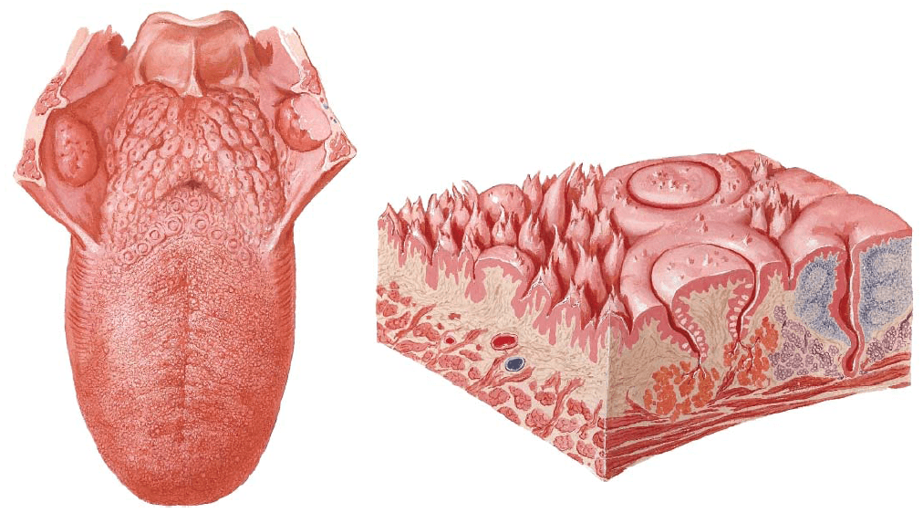
Small projections located on the tongue’s surface where taste buds are located are known as:
papillae
gustatory cells
olfactory epithelium
mucous producing glands
NOT a primary taste sensation
bitter
salty
sweet
peppery

Which of the following papillae do not contain taste buds:
fungiform
filiform
circumvallate
all of the above
What sensory receptor reacts to light?
Chemoreceptor
Nociceptor
Photoreceptor
Gravoreceptor
What receptor reacts to physical forces?
Chemoreceptor
Mechanoreceptor
Electroreceptor
Thermoreceptor
What receptor detects pain?
Thermoreceptor
Mechanoreceptor
Proprioceptor
Nocioreceptor
What receptor indicates change in body positions? (Hint: helps us with balance.)
Geogravity receptors
Proprioreceptors
Chemoreceptors
Photoreceptors
What receptor helps us identify changes in smell and taste?
Thermoreceptors
Proprioreceptors
Electroreceptors
Chemoreceptors
What is a thermoreceptor?
Responds to body position
Responds to changes in temperature
Responds to physical forces including touch and pressure
Responds to squirrels running outside
What receptor allows you to see a rainbow?
Photoreceptor
Thermoreceptor
Mechanoreceptor
Gravitonreceptor
Which receptor helps you hear your favorite band?
Mechanoreceptor
Bandreceptor
Thermoreceptor
Idon'tknowwhatIamthinking receptor

