
 Worksheets
Benchmark #2 Review: Integumentary, Skeletal, Muscular Systems
Total questions: 125
Worksheet time: 1hrs 29mins
Long bones have a _____ that makes up most of bone's length and is composed of compact bone
epiphyseal plate
yellow marrow
diaphysis
articular cartilage
Which of the following bones is NOT part of the axial skeleton?
pectoral girdle
skill
vertebrae
sternum
Which of the following bones is INCORRECTLY matched with its type?
ribs--irregular
carpals--short
ulna--long
cranial bones--flat
Red bone marrow functions to
produce fat
produce blood cells
store cells
produce new bone cells
Yellow marrow functions to ______.
produce new bone cells
store calcium
store fat
produce calcium
Which of the following structures would not be visible when looking at a slide of bone tissue under the microscope?
Haversian canals
canaliculi
osteocytes
epiphysis

Area that serves as storage for fat and is known as the medullary cavity
C
B
E
D
Spongy bone
C
B
E
D
Periosteum
C
F
E
B
Shaft, composed of compact bone and is called the diaphysis
C
F
A
B
Area that causes the lengthwise growth
D
F
A
B
Cartilage that provides a smooth surface that reduces friction
D
F
A
B


What bone is highlighted?
Vertebrae
Femur
Cranium
Ribs
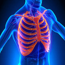
What bone is highlighted?
Ribs
Cranium
Clavicle
Femur

What is the place where two bones meet?
bones
tendons
ligament
joint

What is the name for the connector that attaches muscle to bone?
ligament
tendon
joint
bones
Articular cartilage is found in what part of the long bone?
inside of the medullary cavity
on the outer surface of the diaphysis
the outer surface of the epiphyses
in the spaces of the spongy bone
Mature bone cells are known as
Lacunae
Osteocytes
Collagen
Osteon
The function of Osteoclasts is
To breakdown calcified matrix of bone
Secrete acids that dissolve the inorganic component of calcified matrix
Phagocytize components of the bony matrix
All the above
The skull bones, hyoid bone and vertebral column bones are found in which skeleton
Axial Skeleton
Appendicular Skeleton


Name the bone identified as I.


What is A?
Lamellae
Osteon
Canaliculi
Central (Haversian) Canal
Skin is divided into three main layers. The top main layer of the skin is called the _________________.
hypodermis
subcutaneous
The coloring in the skin; found in the epidermis is due to this pigment.
chlorophyll
keratin
sebaceous

stratum basale of the epidermis
reticular layer of the dermis
Which layer of the skin is avascular (lacks blood vessels)?
What cell produces a protein and allows for waterproofing of the skin, nails, and hair?
What cell produces a dark brown pigment called melanin?
What layer of epidermis does cell division occur (mitosis)?
The oldest and mostly dead cells of the epidermis will be found here
The function of the integumentary system includes all of these except what?
Homeostasis
Cooling system
Synthesis of Vitamin A
Excretion of wastes
The epidermis is composed of what kind of cells?
Simple squamous epithelial
Stratified cuboidal epithelial
Stratified squamous epithelial
Transitional epithelial
What layer of the skin contains sweat glands?
Reticular layer of the Dermis
Stratum lucidum layer of the Epidermis
Papillary layer of the Dermis
Subcutaneous
The clear layer only present in the thick skin (palms of hands and soles of feet) is called:
encloses the lungs
pleura
pericardium
peritoneum
encloses the heart
pleura
pericardium
peritoneum
encloses the abdominopelvic region
pleura
pericardium
peritoneum
clings to the wall of the cavity, outside layer
serous
visceral
parietal
clings to the organs external surface, inside layer
serous
visceral
parietal
Which membrane has a parietal and visceral layer?
cutaneous
serous
mucous
synovial
membrane that surrounds and protects organs like the heart and the lungs
muscular
cutaneous
synovial
serous
Which of the following membranes is your body's outer covering and is the only dry membrane?
Cutaneous Membrane
Mucous Membrane
Serous Membrane
Synovial Membrane
Which of the following membranes in your body lines open cavities that are exposed to the outside world and are adapted for secretion and absorption?
Cutaneous Membrane
Mucous Membrane
Serous Membrane
Synovial Membrane
Which body membrane is found at movable joints like the shoulder, hip and knee, and works to protect the area where two bones come together?
Cutaneous Membrane
Mucous Membrane
Serous Membrane
Synovial Membrane
Which is the most common skin cancer?
Basal Cell Carcinoma
Squamous Cell Carcinoma
Melanoma
When diagnosing melanoma, which of the following does C stand for in ABCDE?
color change
cyanosis
cold to the touch
carotene present
When diagnosing melanoma, which of the following does B stand for in ABCDE?
bigger than dime
Border
blackish in color

Identify the hair follicle in the diagram
F
G
K
None of these

Which layer is the stratum basale?
A
B
C
D
E
Which layer is the stratum corneum?
A
B
C
D
E
Which layer includes the papillary and reticular layers of the dermis?
F
E
D
C
A
Which of the following is the lower dermal region that contains sweat and oil glands, along with blood vessels & deep pressure receptors?
stratum basale
papillary layer
reticular layer
subcutaneous layer
What is the difference between cancer that is benign and cancer that is malignant?
Benign cancer does not spread, but malignant cancer moves to other parts of the body.
Benign cancer is dangerous, but malignant cancer is not.
Malignant cancer can easily be removed, but benign cancer cannot.
Malignant cancer is the best kind to have.
The type of muscle tissue that is found in the walls of blood vessels is
voluntary muscle tissue
cardiac muscle tissue
skeletal muscle tissue
smooth muscle tissue
Muscles move bones by
pushing
pulling
both pushing and pulling
Muscles contract when
thin filaments slide past thin filaments
thick filaments slide past thick filaments
thin filaments slide past thick filaments
Which connective tissue surrounds fasicles?
epimysium
endomysium
perimysium
Which connective tissue surrounds the whole muscle?
epimysium
endomysium
perimysium
Which connective tissue surrounds individual muscle fibers?
epimysium
endomysium
perimysium
Which type of muscle cell is striated, voluntary and has multiple nuclei located at the periphery?
Cardiac
Smooth
Skeletal
Which type of muscle cell has one nucleus, is striated and has intercalated disks?
Skeletal
Cardiac
Smooth
Which of the following muscle cells are not striated, involuntary, and have one nucleus per cell?
Skeletal
Cardiac
Smooth

Which of the following puts the structures of the muscle in order from largest to smallest?
Whole muscle, muscle fiber, actin & myosin, myofibril, muscle fasicle
Whole muscle, muscle fasicle, muscle fiber, myofibril, actin & myosin
Whole muscle, muscle fiber, muscle fasicle, myofibril, actin & myosin
Whole muscle, muscle fasicle, muscle fiber, actin & myosin, myofibril

Identify the highlighted muscle
Hamstrings
Deltoid
Gluteus Maximus
Oblique

The sarcolemma is:
a material that fills up the space inside a muscle cell
the membrane that surrounds a muscle fiber
the smallest unit of a myofibril
the organelle that makes up a muscle fiber
Calcium's purpose in muscle contraction is to
cross the neuromuscular junction and stimulate the sarcolemma
travel down the T-tubule to reach interior myofibrils
bind to troponin, move tropomyosin, and expose actin's binding sites
attach to actin, forming a crossbridge
The contracting unit of a muscle is the _______________.
sarcomere
myofibril
muscle fiber
muscle cell
Movement of the arm/leg away from the midline
adduction
eversion
inversion
abduction
Body part movement in a circular or conical motion
ovicular
pronation
rotation
circumduction
Movement of the forearm/hand so palm faces downward or backwards
supination
pronation
palmar
flexion

extension
flexion
circumduction
adduction
abduction

extension
flexion
circumduction
adduction
abduction

supination
pronation
abduction
adduction
opposition

supination
pronation
abduction
adduction
opposition

inversion
plantar flexion
extension
dorsiflexion
circumduction

What is muscle J?
Rectus femoris
Fibularis longus
Vastus lateralis
Gastrocnemius
What is muscle A?
Rectus femoris
Fibularis longus
Vastus lateralis
Gastrocnemius
What is muscle H?
Vastus medialis
Fibularis longus
Vastus lateralis
Adductor muscle
What is muscle F?
Vastus medialis
Sartorius
Vastus lateralis
Tibialis anterior

What is muscle A?
Frontalis
Orbicularis oculi
Zygomaticus
Orbicularis oris
What is muscle B?
Frontalis
Orbicularis oculi
Zygomaticus
Orbicularis oris
What is muscle E?
Platysma
Sternocleidomastoid
Pectoralis major
Intercostals
What is muscle F?
Platysma
Sternocleidomastoid
Pectoralis major
Intercostals
What is muscle I?
Rectus abdominis
External oblique
Internal oblique
Transverse abdominus
What is muscle J?
Rectus abdominis
External oblique
Internal oblique
Transverse abdominus
What is muscle M?
Temporalis
Masseter
Deltoid
Triceps brachii
What is muscle N?
Temporalis
Masseter
Deltoid
Triceps brachii
What is muscle O?
Biceps brachii
Brachialis
Flexor carpi radialis
Brachioradialis

Which label is pointing to the endomysium?
A
B
C
D

What is another name #2? (Hint: It is a muscle layer)
Skeletal muscle
Epimysium
Perimysium
Endomysium
What is another name #5? (Hint: It is a muscle layer)
Skeletal muscle
Epimysium
Perimysium
Endomysium
What is another name #6? (Hint: It is a muscle layer)
Skeletal muscle
Epimysium
Perimysium
Endomysium

