
NEW
Font size
 Worksheets
Year 9 Body Systems Big Quiz of the Term!
Total questions: 58
Worksheet time: 39mins
What is the digestive system?
A system to take out the bad stuff that's in your body
A system where a certain group of organs work together to convert food into energy to feed the body
A system where certain organs in the body digest the food you eat
A system that the organs in the stomach make food for the body to digest.

Which is the esophagus?
A
B
C
D
E

Which parts of the diagram is the small and large intestine?
A~ small intestine
E~ large intestine
D~ small intestine
E~ large intestine
C~ large intestine
B~ small intestine
A~ large intestine
C~ small intestine
E~ small intestine
D~ large intestine

esophagus
liver
stomach
pancreas

stomach
pancreas
large intestine
small intestine

Mechanical Digestion
Chemical Digestion
Mechanical digestion
Chemical digestion
Digests food in the mouth.
Digests food in the stomach
Protects the stomach from digesting iteself
Absorbs nutrients from food so the body has energy.

waves of muscle contractions (squeezing)
stomach
small intestine
peristalsis

Digestive organ where most chemical digestion and absorption of food takes place
pancreas
small intestine
liver

absorbs water into bloodstream
gall bladder
large intestine
liver
Repair Tissue
Main source of energy for cells
Hydrate the body
create struc tures body structures
build structures and provide quick energy
repair tissue and store long-term energy
build structures/ hormones/ transport/ enzymes
provide energy/ transport
provide quick energy/ insulation
repair tissues
2nd source of energy/ insulates
build struc tures
give permission for the reaction
raise activation energy
speed up reactions
control how many reactions occur
voltage-gated channels
surface receptor
calcium channels
active site
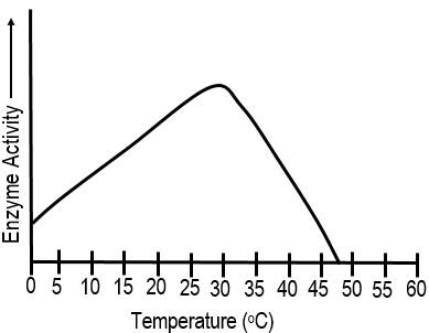
15 C
40 C
30 C
47 C

Substrate
Enzyme
Product
Complex

Substrate
Enzyme
Substrate
Product
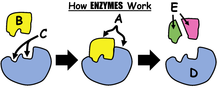
active site
enzyme
substrate
products

Label the diagram using vocab words. Number 1
Substrate
Enzyme
Active Site

Label Number 2
Substrate
Enzyme
Active Site

In reference to the image above, how would the absorption of a person with Celiac disease be affected ?
No effect on absorption.
The villi are flattened and therefore their absorption of nutrients would increase.
The villi are flattened and therefore their absorption of nutrients would decrease.
The villi are enlarged and absorption would increase.

Identify structure A in the diagram
muscle
nerve
capillary (blood vessel)

The villi shown in this diagram absorb the products of digestion by?
dialysis
diffusion
osmosis
breathing
Move materials around inside the cell
Move materials in and out of the cell
Communicate to other cells
Divide

The surface area was too larger to get through
The volume of the other cells was too large for the dye to get through
There was not enough surface area for the dye to get through
The volume was too low for the dye to stay in the square
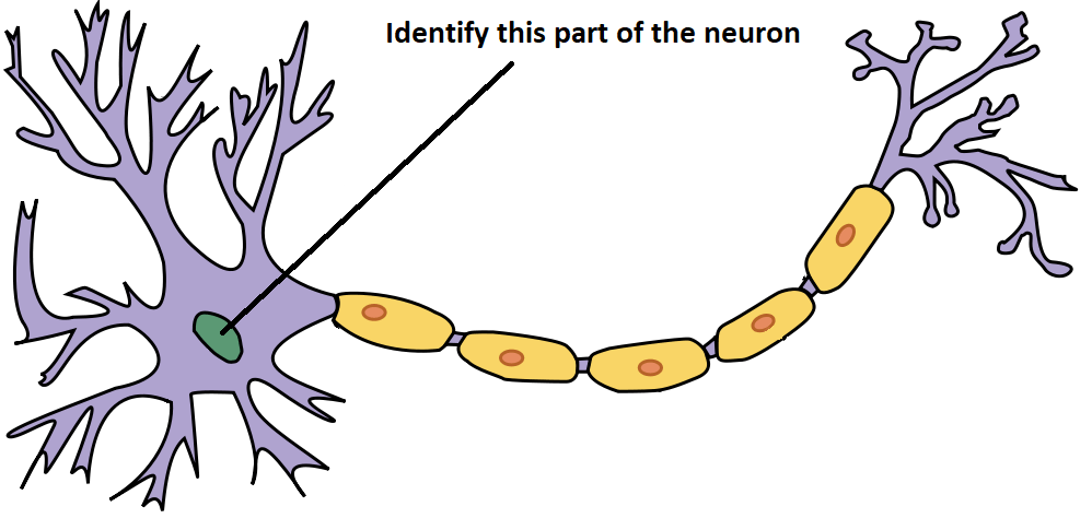
Nucleus
Cell Body
Axon
Node of Ranvier
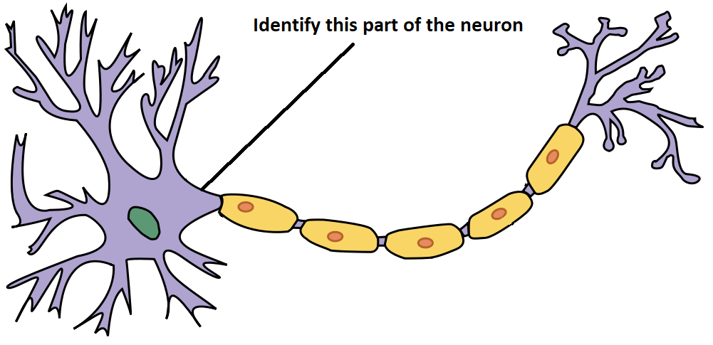
Nucleus
Cell Body
Axon
Node of Ranvier
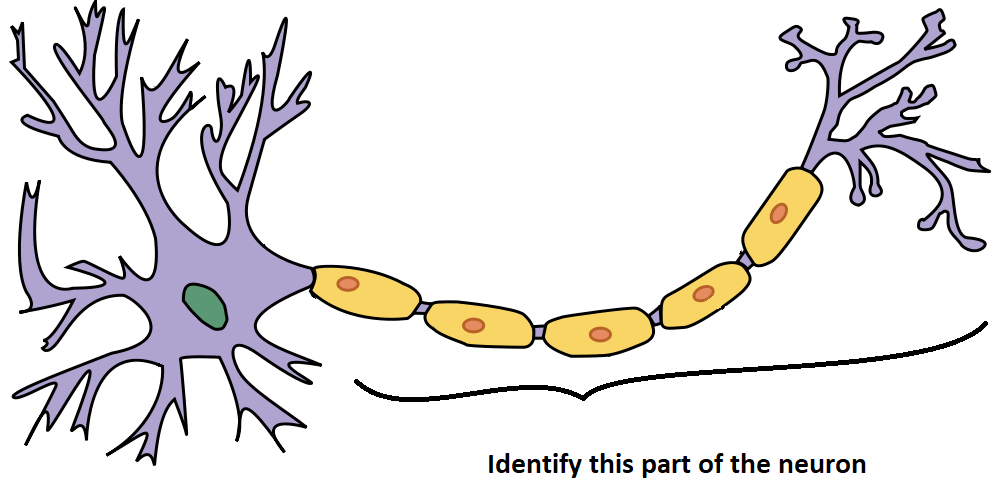
Nucleus
Cell Body
Axon
Node of Ranvier
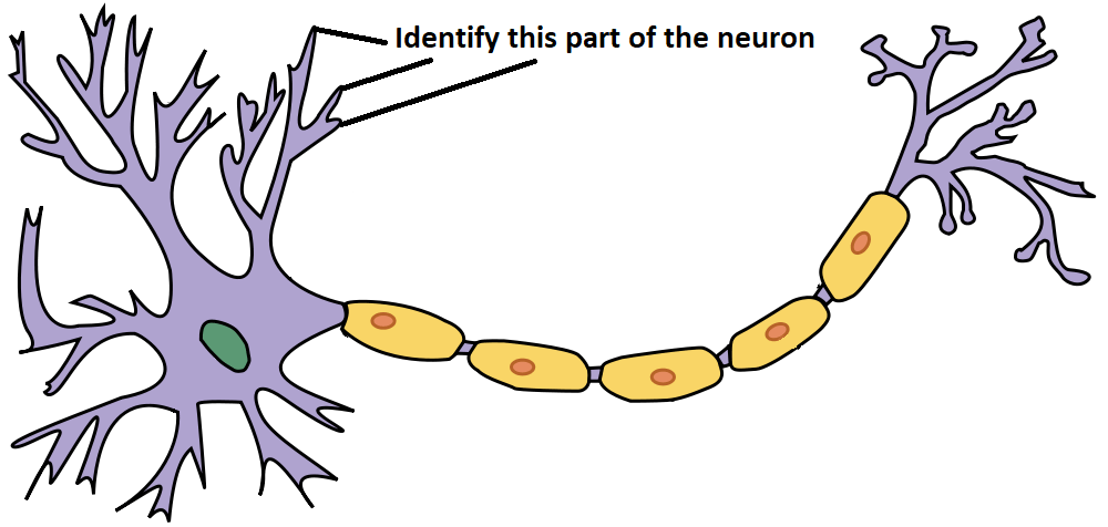
Dendrites
Axon Terminals
Myelin Sheath
Schwann Cells
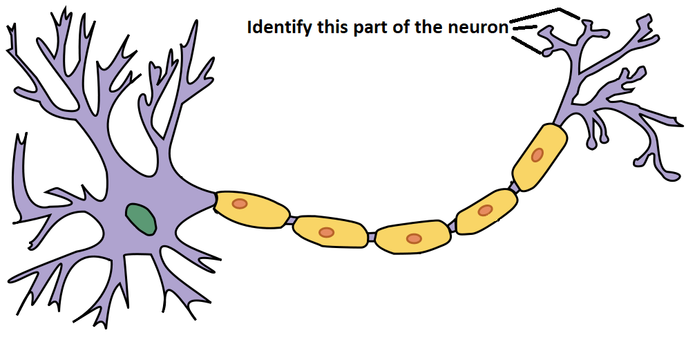
Dendrites
Axon Terminals
Myelin Sheath
Schwann Cells
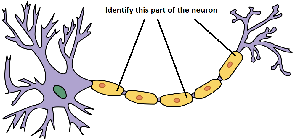
Dendrites
Axon Terminals
Myelin Sheath
nuclei
What does the neuron's Myelin Sheath do?
Controls the actions of the neuron
Produces food for the neuron
Carries information from the cell body to other neurons
Surrounds and insulates the axon
What do Dendrites do?
Receives information from other neurons via neurotransmitters
Produces fatty tissue for the Myelin Sheath
Sends information to the next cell
Controls the speed of electrical signal through the axon
What do Axon Terminals do?
Receives information from other neurons
Produces fatty tissue for the Myelin Sheath
Sends information to the next cell via release of neurotransmitters
Controls the speed of electrical signal through the axon
The space between the axon terminal of the sending neuron and the dendrite or cell body of the receiving neuron.
axon
synapse
dendrite
cell body
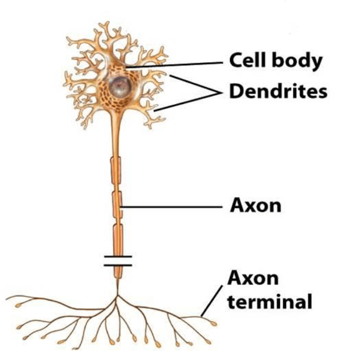
What type of neuron is represented on the image?
Motor Neuron
Interneuron
Sensory Neuron
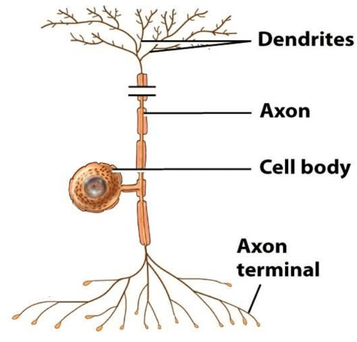
What type of neuron is represented on the image?
Motor Neuron
Interneuron
Sensory Neuron

What type of neuron is represented on the image?
Motor Neuron
Interneuron
Sensory Neuron
Type of neuron that perceive information about your environment.
Motor Neuron
Interneuron
Sensory Neuron
Type of neuron located only in the brain and spinal cord that pass impulses from one neuron to another.
Motor Neuron
Interneuron
Sensory Neuron
Type of neuron that sends nerve impulses to muscles and glands, by the command of other neurons.
Motor Neurons
Interneurons
Sensory Neurons

If I step on a nail...what type of neuron takes that signal TO the CENTRAL NERVOUS SYSTEM?
interneurons
motor
sensory
receptors

What type of neuron is A?
Sensory
Inter
Motor
What type of neuron is C?
Sensory
Inter
Motor
Which statement best describes a reflex?
Slow response that you have to think about
Rapid response to protect body from harm

_____ neurons carry messages away from the central nervous system and out to muscles or glands. The RED neuron in diagram.
sensory
motor
inter
central

Where would the nerve impulse start in this reflex arc?
A
C
D
E

Which number represents the stimulus?
1
2
3
4

Frontal
Parietal
Temporal
Occipital

Frontal
Parietal
Temporal
Occipital

Frontal
Parietal
Temporal
Occipital

Cerebrum
Cerebellum
Brain Stem
Diencephalon
Diencephalon
Frontal Lobe
Brain Stem
Cerebellum

Occipital Lobe
Parietal Lobe
Temporal Lobe
Frontal Lobe

Occipital Lobe
Parietal Lobe
Temporal Lobe
Frontal Lobe

The kinds of chemicals that control feedback loops
hormones
homeostasis
disease

