
 Worksheets
Anatomy SMS 2 Final Review 2024
Total questions: 110
Worksheet time: 4hrs 40mins
The teeth and tongue of the oral cavity break foodstuff down physically. This is an example of
mechanical digestion
chemical digestion
absorption
peristalsis
Which of the following answers correctly sequences the pathway food takes from the beginning to end?
mouth, esophagus, stomach, small intestine, large intestine
mouth, esophagus, stomach, large intestine, small intestine
mouth, stomach, esophagus, small intestine, large intestine
mouth, stomach, esophagus, large intestine, small intestine
Which organ makes bile to help break down fats?
liver
gallbladder
pancreas
What organ is responsible for the absorption of water and movement of waste?
large intestine
small intestine
stomach
oral cavity
Which of the following helps digest carbohydrates in the mouth...
salivary amylase
lysozymes
mucin
bolus

What are the fingerlike structures located in the small intestine that absorbs nutrients?
Duodenum
Villi
Bile
Pancreas


Waste foods are separated from solids and liquids in the __________________.
small intestine
large intestine
liver
stomach
The only freely moveable bone in the skull is the ______.
frontal
maxilla
occipital
mandible
Long bones have a _____ that makes up most of bone's length and is composed of compact bone
epiphyseal plate
yellow marrow
diaphysis
articular cartilage
Red bone marrow functions to
produce fat
produce blood cells
store cells
produce new bone cells
Yellow marrow functions to ______.
produce new bone cells
store calcium
store fat
produce calcium
How many vertebrae are in the neck?
2
4
7
12
Which is the correct order of ribs from superior to inferior?
floating, true, false
true, false, floating
true, floating, false
false, floating, true
Muscles can only _______ they cannot _____.
pull;push
push;pull
expand;increase
increase;expand
Muscles that you have no control over are called ___________ muscles.
Involuntary
Voluntary
Evolutionary
Revolutionary
Muscles that you can control are called ___________ muscles.
Involuntary
Voluntary
Evolutionary
Revolutionary
What are the three types of muscle tissues?
Skeletal, Red, White
Cardiac, Smooth, Skeletal
Red, White, Blue
Cardiac, Skeletal, Red
When your body receives signals from an external environment and transmits the information to the brain, which body system is being used
Nervous
Integumentary
Digestive
Skeletal
This muscle is found in the heart and never gets tired.
Cardiac
Smooth
Skeletal
Strong
Muscles are connected to bones by an elastic-like (stretchy) strap called a ______
Joint
Bone
Tendon
Muscle
What type of muscle makes of the walls of our internal organs?
smooth muscles
voluntary muscle
striated muscle
cardiac muscle
About how many muscles do you have in your body?
600
50
1,000
100
The back of your thigh is called your
Hamstring
Deltoid
Gastrocnemius
Tibialis Anterior
Quadriceps
The front of your thigh is called your
Hamstring
Deltoid
Gastrocnemius
Tibialis Anterior
Quadriceps
Your calf muscle is called your
Hamstring
Deltoid
Gastrocnemius
Tibialis Anterior
Quadriceps
The muscle in the back (posterior) of your upper arm is called your...
Pectoralis Major/Minor
Rectus Abdominus
Triceps Brachii
Biceps Brachii
Your chest muscle is called your...
Latissimus Dorsi
Pectoralis Major/Minor
Rectus Abdominus
Tibialis Anterior
Your Orbicularis Oculi is located around your
Eyes
Kneecaps
Wrists
Mouth
Your Orbicularis Oris is located around your
Nose
Ears
Mouth
Eyes
Your mandible (jaw) muscle that allows you to chew is called your:
Sternocleidomastoid
Gastrocnemius
Masseter
Infraspinatus
The bottom of the heart; points towards the left hip and rests on the diaphragm
aorta
base
apex
artery
The top of the heart; broad surface; great vessels emerge from here; points towards right shoulder
aorta
base
apex
artery
Receiving chambers of the heart
septum
Ventricles
Atria
valves
Discharging chambers of the heart
septum
Ventricles
Atria
valves
Divides left and right side of the heart
septum
Ventricles
Atria
valves
Carries blood away from the heart
artery
vein
septum
cardium
Carries blood towards from the heart
artery
vein
septum
cardium

What is 9?
right ventricle
left atrium
right atrium
left ventricle
What is 1?
superior vena cava
inferior vena cava
pulmonary veins
pulmonary artery
What is 2?
superior vena cava
inferior vena cava
pulmonary veins
pulmonary artery
What is 10?
superior vena cava
aorta
septum
pulmonary artery
produces enzymes that break down food
liver
pancreas
gall bladder
stomach
The pancreas, liver, and gallbladder are known as...
accessory organs
the digestive tract
acid makers
digestion helpers
Which organ produces bile that breaks down food?
pancreas
liver
gall bladder
stomach

Accurate facts about the bone(s) are
Zygomatic, 2 irregular
Lacrimal, 2, flat
Nasal bone, 1 irregular bone
Nasal bones, 2 flat bones

Zygomatic
Temporal
Parietal
Maxilla

Mandible
Zygomatic
Temporal
Maxilla

Maxilla
lacrimal
Mandible
zygomatic

Zygomatic
Vomer
Ethmoid
Lacrimal

Identify the bone highlighted in the image.
occipital
parietal
temporal
frontal
maxilla

Identify the bone highlighted in the image.
occipital
parietal
temporal
frontal
maxilla

Identify the bone highlighted in the image.
occipital
parietal
temporal
frontal
maxilla

Identify the bone highlighted in the image.
occipital
parietal
temporal
frontal
maxilla

suture highlighted in red
coronal
lamboidal
sagittal
metopic

suture highlighted in red
lamboidal
frontal
sagittal
squamosal

occipital bone
foramen magnum
coronal suture
fontanelle

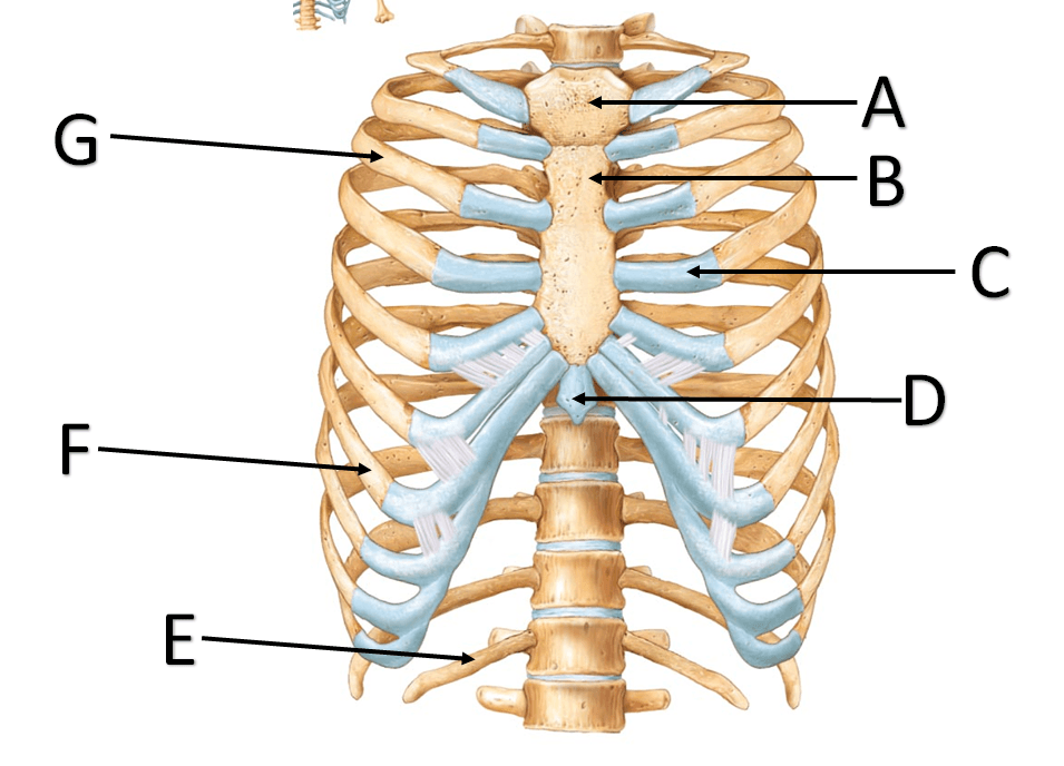
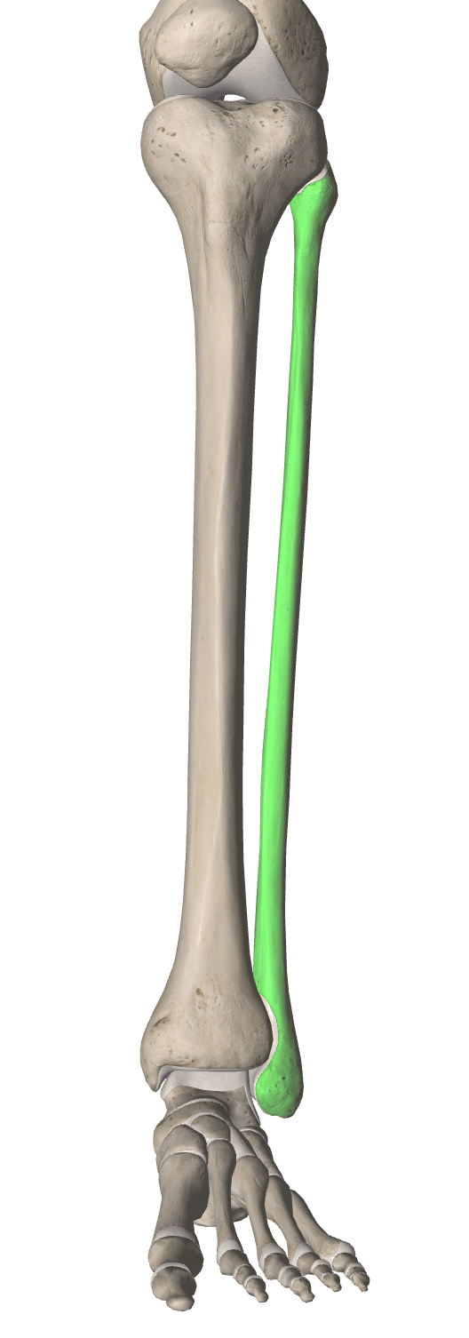
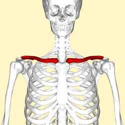
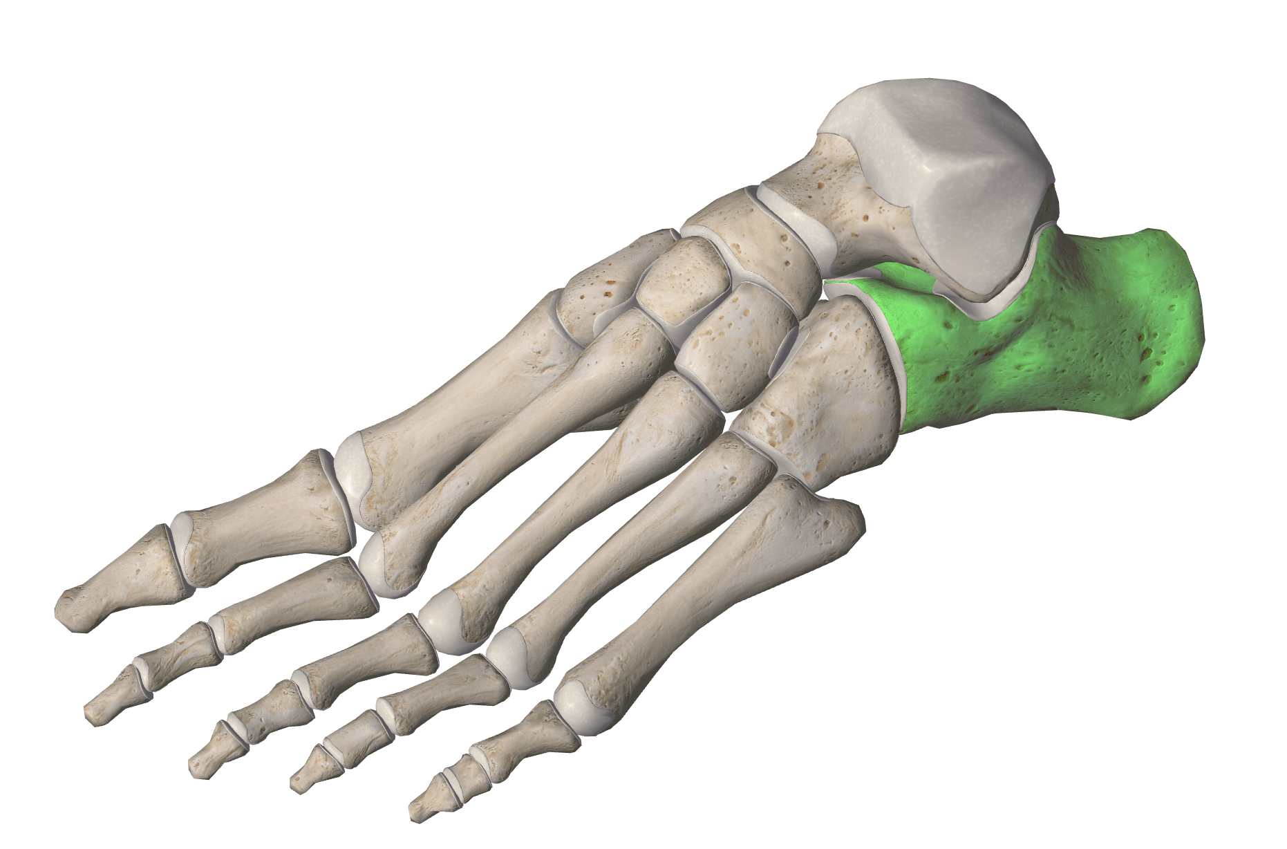
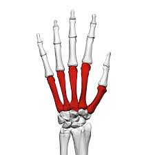
Which bones are highlighted
metacarpals
metatarsals
carpals
tarsals

Which bones are highlighted
carpals
tarsals
metacarpals
metatarsals
The anatomical name for the big toe is the
Popliteal
Pollex
Hallux
Patella

clavicle
scapula
humerus
radius
ulna

clavicle
scapula
humerus
radius
ulna

clavicle
scapula
humerus
radius
ulna

carpals
metacarpals
phalanges
ilium
ischium

coxal
femur
patella
fibula
tibia

?
pectoralis minor
diaphragm
pectoralis major
subclavius

?
diaphragm
rectus abdominis
external oblique
transversus abdominis

?
erector spinae
latissimus dorsi
scalenes
trapezius

?
infraspinatus
levator scapulae
latissimus dorsi
trapezius

Valve between the left atrium and the left ventricle.
tricuspid valve
mitral valve
pulmonary valve
aortic valve
Brings blood to the heart from the upper portion of the body.
pulmonary artery
superior vena cava
pulmonary vein
inferior vena cava
Thickest walled chamber of the heart.
left atrium
right ventricle
left ventricle
right atrium
Valve between the left ventricle and the aorta.
aortic valve
pulmonary valve
tricuspid valve
mitral valve
These vessels carry blood to each lung from the right ventricle.
superior vena cava
pulmonary arteries
pulmonary veins
aorta
Valve between the right atrium and the right ventricle.
mitral valve
pulmonary valve
aortic valve
tricuspid valve
Blood in the right atrium:
Oxygenated
Deoxygenated
Blood in the pulmonary artery:
Oxygenated
Deoxygenated
Blood in the pulmonary veins:
Oxygenated
Deoxygenated
Blood in the left atrium:
Oxygenated
Deoxygenated
Blood in the Inferior Vena Cava:
Oxygenated
Deoxygenated

