
 Worksheets
Unit 4 Exam Prep L4-L5
Total questions: 115
Worksheet time: 2hrs 30mins


What part is this?
Lens
Cornea
Optic Nerve
Auricle

What part is this?
Lens
Sclera
Pupil
Iris

What part is this?
Retina
Sclera
Optic Nerve
Anterior Chamber

What part is this?
Iris
Sclera
Retina
Pupil
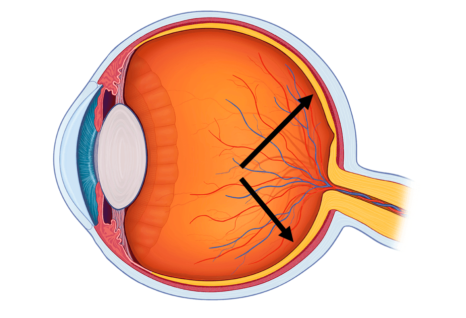
Photosensitive part of the eye that contains rods and cones.
Sclera
Retina
Optic Nerve

1
(a)
2
(a)
3
(a)
4
(a)
5
(a)
6
(a)
7
(a)
8
(a)
9
(a)
10
(a)
11
(a)
12
(a)
13
(a)
14
(a)


Which part is #1?
sclera
cornea
retina
lens
Which part is #2?
iris
cornea
retina
lens
Which part is #3?
sclera
cornea
retina
lens
Which part is #4?
pupil
iris
cornea
lens
Which part is #5?
pupil
iris
cornea
lens
Which part is #6?
sclera
cornea
retina
lens
Which part is #7?
sclera
optic nerve
cornea
retina

#3 Refers to the circular structure that surrounds the pupil and regulates the amount of light entering the eye.
Cornea
Pupil
Retina
Iris

#1 Refers to the clear tissue that covers the front of the eye
Retina
Cornea
Pupil
Iris

#2 refers to
optic nerve
cornea
iris
pupil

#7 Refers to the part of the eye that carried visual information from the eyeball to the brain
lens
optic nerve
retina
sclera

# 5 refers to the layer of receptor cells at the back of the eye in which the image is focused
Retina
Lens
Optic Nerve
Lens

Identify # 1
cornea
iris
lens
retina
Identify #4
cornea
iris
lens
pupil
Identify #5
cornea
iris
lens
retina
Identify #7
cornea
optic nerve
lens
retina
When light hits the retina, the _________ are stimulated.
sclera
optic disk
photoreceptors
choroid coat
Sensory neurons in the retina send ____________ which are routed to the visual cortex in the occipital lobe of the brain.
action potentials
blind spots
light waves
accomodations
In order to see clearly, light rays must be bent to hit the ______ of the retina.
blind spot
optic disk
fovea centralis
focal point
Light waves are bent first by the ______.
cornea
aqueous humor
lens
vitreous humor
If an object moves closer than the far point of vision, which is NOT one of the three reflexes that must occur to bring the image to proper focus?
Accommodation of the lens
Relaxing ciliary muscles
convergence of the eyes
constriction of the pupil
It lets light enter the eye
iris
retina
pupil
When you are looking at something up close, this reflex occurs to reduce the amount of light entering eye.
blind spot
Accommodation
pupil constriction
somatic reflex arc
Which of the following is not one of the three layers of the eye?
Vascular
Cartilage
Fibrous
Nervous
Which of the following extrinsic muscles is responsible for rotating the eye downward and towards the midline?
Superior Rectus
Inferior Rectus
Inferior Oblique
Superior Oblique
Which of the following muscles rotates the eye upward and away from the midline?
Superior Rectus
Inferior Rectus
Superior Oblique
Inferior Oblique

Identify the structure in the picture.
Retina
Choroid
Sclera
Tapetum Lucidum
The axons of retinal nerves form...
Optic Nerves
Optic Tracts
Optic Radiations
Optic Chiasma
Dim light causes the pupil to:
Dilate
Constrict
Stay the same
No
What are the six extraocular muscles that control the movement of the eye?
Rectus muscles (superior, inferior, medial, and lateral) and the oblique muscles (superior and inferior)
Front, back, left, and right muscles
Upper, lower, middle, and side muscles
Circular, spiral, diagonal, and vertical muscles
Which eye muscle is responsible for moving the eye inward towards the nose?
Lateral rectus muscle
Superior rectus muscle
Medial rectus muscle
Inferior rectus muscle
What is the function of the superior rectus muscle?
Elevating the eye and rotating it medially
Depressing the eye and rotating it laterally
Closing the eye and protecting it from foreign objects
Moving the eye in a circular motion
Which eye muscle is responsible for moving the eye downward and inward?
Inferior oblique muscle
Lateral rectus muscle
Superior oblique muscle
Superior rectus muscle
What is the role of the inferior oblique muscle in eye movement?
No role in eye movement
Upward and outward movement of the eye
Downward and inward movement of the eye
Side to side movement of the eye
What is the function of the lateral rectus muscle?
Moving the eye up and down
Widening the eye
Abducting the eye
Closing the eye
Which eye muscle is responsible for moving the eye upward and outward?
Medial rectus muscle
Inferior rectus muscle
Superior rectus muscle
Lateral rectus muscle
What is the function of the medial rectus muscle?
To move the eye up and down.
To move the eye outward away from the nose.
To move the eye inward towards the nose.
To control blinking.
What is the role of the inferior rectus muscle in eye movement?
Sideways movement of the eye
No role in eye movement
Downward movement and inward rotation of the eye
Upward movement and outward rotation of the eye
Which eye muscle is responsible for moving the eye upward and inward?
Medial rectus muscle
Inferior oblique muscle
Lateral rectus muscle
Superior rectus muscle
What is the correct path of light through the eye?
cornea - pupil - lens - retina - optic nerve - brain
retina - optic nerve - cornea - pupil - lens - brain
brain - optic nerve - retina - lens - pupil - cornea
cornea - pupil - retina - lens - optic nerve - brain
What is the correct order of layers of the eye from the outside to inside?
sclera - retina - choroid coat
choroid coat - sclera - retina
retina - choroid coat - sclera
sclera - choroid coat - retina
A light - sensitive layer of tissue at the back of the eye that turns light into
electrical signals.
Retina
Macula
Photoreceptors
Optic Nerve
Lens
Receives light from the lens, converting it to neural signals and transmitting them to the brain for visual recognition.
Pupil
Retina
Cornea
Sclera
Changes the direction of the light rays in which light enters the eye
Pupil
Iris
Lens
Cornea
Sclera
Regulates the amount of light entering the eye. Focuses light rays on the retina.
Retina
Sclera
Iris
Macula
Focuses the light rays that pass through it and changes focal distance of the eye
Lens
Sclera
Choroid
Changes the shape of the lens when the eye focuses. It also makes the clear fluid that fills the space between the cornea and the iris.
Ciliary Body
Vitreous
Macula
Choroid
Maintains shape of eye, ensures it's attached to retina, also helps with vision clarity
Vitreous
Macula
Optic Nerve
In the human eye, rods are to _________, as cones are to ________.
light/ color
color/ light
monochromat/ dichromat
dichromat/ monochromat
lens/ iris
What three primary colors of light are detected by your cones?
Green, red, and blue
Black, gray, and white
Blue, red, and yellow
Blue, green, and purple
Cyan, magenta, and yellow
Which part of the retina is responsible for the highest visual acuity?
Optic disc
Fovea
Peripheral retina
Macula
What is the role of optical pigments in photoreceptors?
To transmit nerve signals
To absorb light and initiate a chemical change
To provide structural support
To supply nutrients
How do rods and cones differ in their connection to the optic nerve?
Rods connect directly, cones do not
Cones connect directly, rods do not
Rods share connections, cones have individual connections
Both have the same type of connections
What is the main function of the optic nerve in vision?
To focus light onto the retina
To transmit visual information to the brain
To adjust the size of the pupil
To supply blood to the retina
purple pigment found in rods
glaucoma
rhodopsin
melanin
carotene
The process when the lens changes shape to focus close or far away
sensory adaptation
accommodation
Transduction
globe luxation

What happens to the iris muscles when there is dim light?
The radial muscles contract & the circular muscles relax
The circular muscles contract and the radial muscles relax

What happens to the iris muscles when there is a high light intensity?
The radial muscles contract and the circular muscles relax
The circular muscles contract and the radial muscles relax
The rods and cones contain optical pigments that absorb light and then break down.
Rod cells contain __________?
Cone cells contain _________?
Rod cells contain iodopsin and cones contain rhodopsin
Rod cells contain rhodopsin and cones contain iodopsin
The change in direction of a wave as it changes speed while moving from one medium to another.
refraction
redirection
absorption
reflection

A concave lens has an inward curved shape. It refracts light outward from the lens. They light rays spread apart as they exit the lens. Which represents a concave lens?
A
B
A and B
neither A or B
In order to see clearly, light rays must be bent to hit the ______ of the retina.
blind spot
optic disk
fovea centralis
focal point
If an object moves closer than the far point of vision, which is NOT one of the three reflexes that must occur to bring the image to proper focus?
Accommodation of the lens
Relaxing ciliary muscles
convergence of the eyes
constriction of the pupil
When you are looking at something up close, this reflex occurs to reduce the amount of light entering eye.
blind spot
Accommodation
pupil constriction
somatic reflex arc
When the brain detects that the tension in the eye muscles is changing as the eyes rotate inwards
convergence
Monocular depth cue
accommodation
retinal disparity
To focus on objects up close, what must the eye do?
accommodation
convergence
pupil constriction
pupil dilation

Which muscle will allow the eye to look down and away?
#1, the superior oblique
#4, the superior oblique
#1, the superior rectus
#4, the superior rectus
Assuming this is the right eye, identify #1.
superior rectus
inferior rectus
medial rectus
lateral rectus
Select all of the protective features of they eye.
eyebrows
eyelids
eyelashes
lacrimal glands
bony orbit
This defect is caused due to elongation of eye ball.
Hyperopia
Astigmatism
Presbyopia
This defect of the eye is caused by hardening or loss of elasticity of the lens due to age.
Myopia
Hypermetropia
Presbyopia
Astigmatism
This problem arises due to a build-up of fluid in the eyeball which puts pressure on the optic nerve.
glaucoma
myopia
cataract
hypermetropia


