
 Worksheets
Reproductive OSPE
Total questions: 210
Worksheet time: 4hrs 30mins

Histology
The following image represents? (red colored)
Ovaries
Testis
Epididymis
Testicular Lobe
Seminiferous Tubules
Histology
The following image represents? (green colored)
Ovaries
Testis
Epididymis
Testicular Lobe
Seminiferous Tubules
Histology
The following image represents? (yellow colored)
Ovaries
Testis
Epididymis
Testicular Lobe
Seminiferous Tubules
Histology
The following image represents? (blue colored)
Ovaries
Testis
Epididymis
Testicular Lobe
Seminiferous Tubules

Histology
The following image represents?
Ovaries
Testis
Epididymis
Testicular Lobe
Seminiferous Tubules

Histology
The following image represents? (purple labeled)
Cavity of tunica vaginalis
Tunica albuginea
Convoluted seminiferous tubules
Seminiferous epithelium
Interstitial tissue
Histology
The following image represents? (red labeled)
Cavity of tunica vaginalis
Tunica albuginea
Convoluted seminiferous tubules
Seminiferous epithelium
Interstitial tissue
Histology
The following image represents? (green labeled)
Cavity of tunica vaginalis
Tunica albuginea
Convoluted seminiferous tubules
Seminiferous epithelium
Interstitial tissue
Histology
The following image represents? (yellow labeled)
Cavity of tunica vaginalis
Tunica albuginea
Convoluted seminiferous tubules
Seminiferous epithelium
Interstitial tissue
Histology
The following image represents? (blue labeled)
Cavity of tunica vaginalis
Tunica albuginea
Convoluted seminiferous tubules
Seminiferous epithelium
Interstitial tissue

Histology
The following image represents?
Testis Stroma
Testis Parenchyma
Ovaries Stroma
Ovaries Parenchyma
Cavities
Histology
The following image represents? (red labeled)
Seminiferous tubules
Interstitial tissue
Testis lobules
Testis
Ovary
Histology
The following image represents? (yellow labeled)
Seminiferous tubules
Interstitial tissue
Testis lobules
Testis
Ovary

Histology
The following image represents?
Testis
Testicular lobule
Seminiferous tubules
Interstitial cells
Tunica albuginea
Histology
The following image represents? (red labeled)
Spermatogenic cells
Sertoli cells
Myoid cells
Leydig cells
Basement membrane
Histology
The following image represents? (yellow labeled)
Spermatogenic cells
Sertoli cells
Myoid cells
Leydig cells
Basement membrane

Histology
The following image represents? (red labeled)
Spermatogenic cells
Sertoli cells
Myoid cells
Leydig cells
Basement membrane
Histology
The following image represents? (yellow labeled)
Spermatogenic cells
Sertoli cells
Myoid cells
Leydig cells
Basement membrane
Histology
The following image represents? (green labeled)
Spermatogenic cells
Sertoli cells
Myoid cells
Leydig cells
Basement membrane

Histology
The following image represents?
Testis
Epididymis
Vas Deferens
Ejaculatory Duct
Seminal Vesicle
Histology
The following image represents? (red labeled)
Pseudostratified columnar epithelium with stereocilia
Stratified columnar epithelium with stereocilia
Smooth muscle fibers
Spermatozoa
Stereocilia
Histology
The following image represents? (yellow labeled)
Pseudostratified columnar epithelium with stereocilia
Stratified columnar epithelium with stereocilia
Smooth muscle fibers
Spermatozoa
Stereocilia
Histology
The following image represents? (green labeled)
Pseudostratified columnar epithelium with stereocilia
Stratified columnar epithelium with stereocilia
Smooth muscle fibers
Spermatozoa
Stereocilia
Histology
The following image represents? (blue labeled)
Pseudostratified columnar epithelium with stereocilia
Stratified columnar epithelium with stereocilia
Smooth muscle fibers
Spermatozoa
Stereocilia
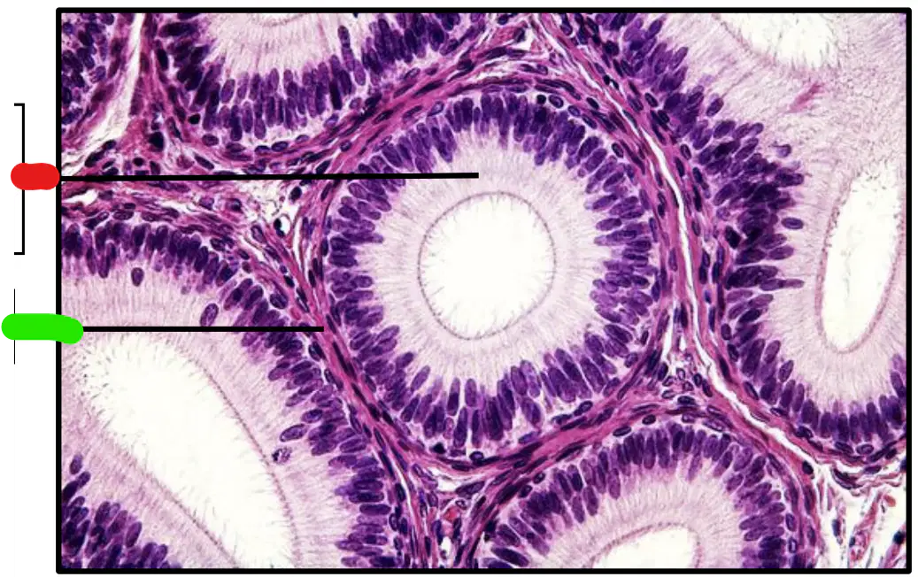
Histology
The following image represents? (red labeled)
Pseudostratified columnar epithelium with stereocilia
Stratified columnar epithelium with stereocilia
Smooth muscle fibers
Spermatozoa
Stereocilia
Histology
The following image represents? (green labeled)
Pseudostratified columnar epithelium with stereocilia
Stratified columnar epithelium with stereocilia
Smooth muscle fibers
Spermatozoa
Stereocilia

Histology
The following image represents? (red labeled)
Pseudostratified columnar epithelium with stereocilia
Stratified columnar epithelium with stereocilia
Smooth muscle fibers
Spermatozoa
Stereocilia
Histology
The following image represents? (yellow labeled)
Pseudostratified columnar epithelium with stereocilia
Stratified columnar epithelium with stereocilia
Smooth muscle fibers
Spermatozoa
Stereocilia
Histology
The following image represents? (green labeled)
Pseudostratified columnar epithelium with stereocilia
Stratified columnar epithelium with stereocilia
Smooth muscle fibers
Spermatozoa
Stereocilia
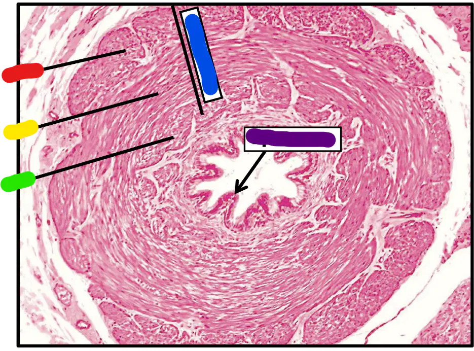
Histology
The following image represents?
Testis
Epididymis
Vas Deferens
Ejaculatory Duct
Seminal Vesicle
Histology
The following image represents? (red labeled)
Outer Longitudinal Layer
Middle Circular Layer
Inner Longitudinal Layer
Musculosa
Pseudostratified columnar with
fewer stereocilia
Histology
The following image represents? (yellow labeled)
Outer Longitudinal Layer
Middle Circular Layer
Inner Longitudinal Layer
Musculosa
Pseudostratified columnar with
fewer stereocilia
Histology
The following image represents? (green labeled)
Outer Longitudinal Layer
Middle Circular Layer
Inner Longitudinal Layer
Musculosa
Pseudostratified columnar with
fewer stereocilia
Histology
The following image represents? (blue labeled)
Outer Longitudinal Layer
Middle Circular Layer
Inner Longitudinal Layer
Musculosa
Pseudostratified columnar with
fewer stereocilia
Histology
The following image represents? (purple labeled)
Outer Longitudinal Layer
Middle Circular Layer
Inner Longitudinal Layer
Musculosa
Pseudostratified columnar with
fewer stereocilia

Histology
The following image represents? (red labeled)
Outer Longitudinal Layer
Middle Circular Layer
Inner Longitudinal Layer
Musculosa
Pseudostratified columnar with
fewer stereocilia
Histology
The following image represents? (yellow labeled)
Outer Longitudinal Layer
Middle Circular Layer
Inner Longitudinal Layer
Musculosa
Pseudostratified columnar with
fewer stereocilia
Histology
The following image represents? (green labeled)
Outer Longitudinal Layer
Middle Circular Layer
Inner Longitudinal Layer
Musculosa
Pseudostratified columnar with
fewer stereocilia
Histology
The following image represents? (blue labeled)
Outer Longitudinal Layer
Middle Circular Layer
Inner Longitudinal Layer
Musculosa
Pseudostratified columnar with
fewer stereocilia

Histology
The following image represents?
Seminal Vesicle
Prostate
Bulbourethral Gland
Testis
Vas Deferens
Histology
The following image represents? (red labeled)
Mucosal Folds
Musculosa
Serosa
Simple columnar or Pseudostratified
columnar
Simple cubical or Pseudostratified
cubical
Histology
The following image represents? (green labeled)
Mucosal Folds
Musculosa
Serosa
Simple columnar or Pseudostratified
columnar
Simple cubical or Pseudostratified
cubical
Histology
What is the lining epithelium?
Mucosal Folds
Musculosa
Serosa
Simple columnar or Pseudostratified
columnar
Simple cubical or Pseudostratified
cubical

Histology
What is the lining epithelium?
Mucosal Folds
Musculosa
Serosa
Simple columnar or Pseudostratified
columnar
Simple cubical or Pseudostratified
cubical
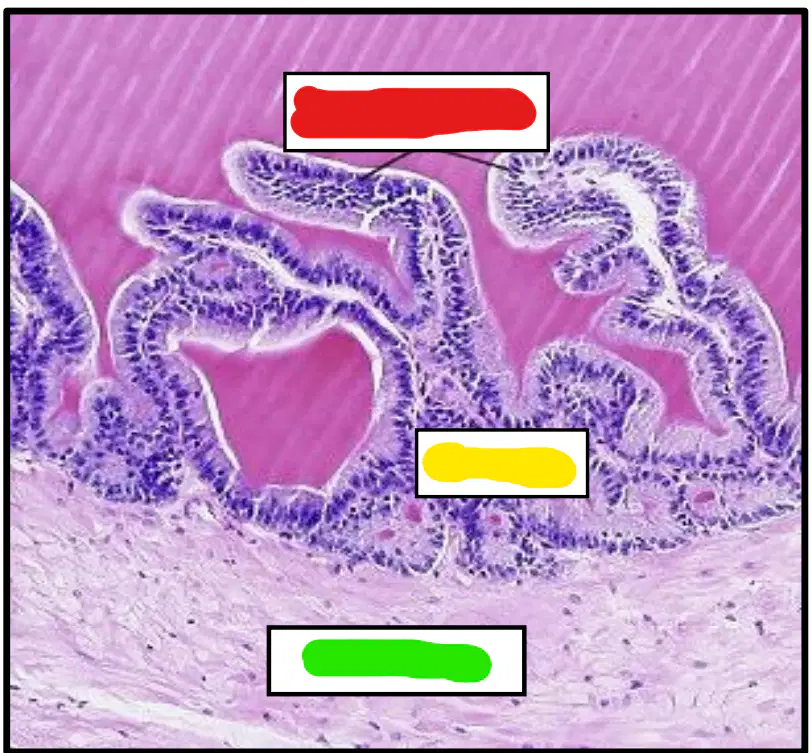
Histology
The following image represents?
Seminal Vesicle
Prostate
Bulbourethral Gland
Testis
Vas Deferens
Histology
The following image represents? (red labeled)
Muscosal fold
Mucosa
Submucosa
Musculosa
Serosa
Histology
The following image represents? (yellow labeled)
Muscosal fold
Mucosa
Submucosa
Musculosa
Serosa
Histology
The following image represents? (green labeled)
Muscosal fold
Mucosa
Submucosa
Musculosa
Serosa
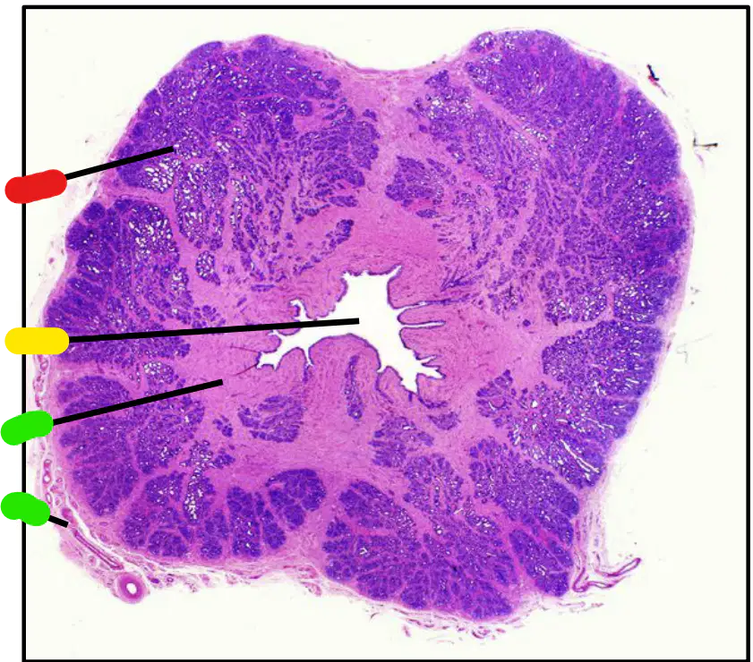
Histology
The following image represents?
Seminal Vesicle
Prostate
Bulbourethral Gland
Testis
Vas Deferens
Histology
The following image represents? (red labeled)
Prostatic acini
Prostatic urethra
Fibromuscular stroma
Epithelium
Capsule
Histology
The following image represents? (yellow labeled)
Prostatic acini
Prostatic urethra
Fibromuscular stroma
Epithelium
Capsule
Histology
The following image represents? (green labeled)
Prostatic acini
Prostatic urethra
Fibromuscular stroma
Epithelium
Capsule

Histology
The following image represents? (red labeled)
Prostatic acini
Prostatic urethra
Fibromuscular stroma
Epithelium
Capsule
Histology
The following image represents? (yellow labeled)
Prostatic acini
Prostatic urethra
Fibromuscular stroma
Epithelium
Capsule
Histology
The following image represents? (green labeled)
Prostatic acini
Prostatic urethra
Fibromuscular stroma
Epithelium
Capsule
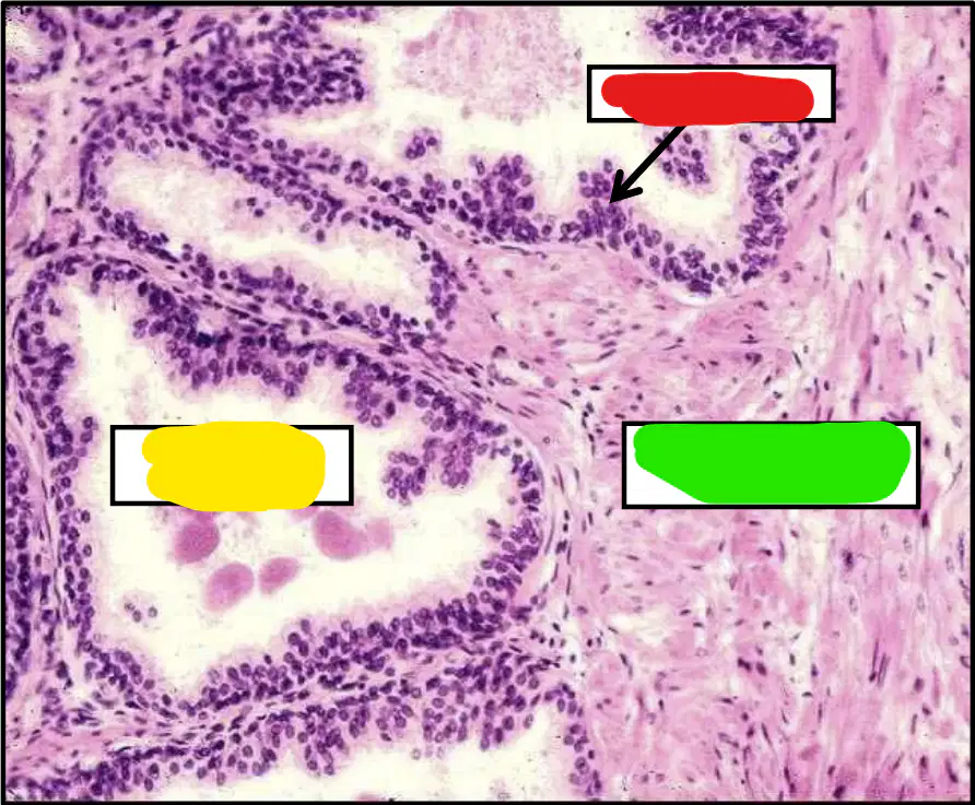
Histology
The following image represents? (red labeled)
Prostatic acini
Prostatic urethra
Fibromuscular stroma
(Cubical, Columnar or
Pseudostratified columnar)
Capsule
Histology
The following image represents? (yellow labeled)
Prostatic acini
Prostatic urethra
Fibromuscular stroma
(Cubical, Columnar or
Pseudostratified columnar)
Capsule
Histology
The following image represents? (green labeled)
Prostatic acini
Prostatic urethra
Fibromuscular stroma
(Cubical, Columnar or
Pseudostratified columnar)
Capsule

Histology
The following image represents?
Ovary
Fallopian Tube
Uterus
Cervix
Vagina
Histology
The following image represents? (red labeled)
Primary follicle
Secondary follicle
Covering (germinal) epithelium
Graffian follicle
Medulla
Histology
The following image represents? (yellow labeled)
Primary follicle
Secondary follicle
Covering (germinal) epithelium
Graffian follicle
Medulla
Histology
The following image represents? (green labeled)
Primary follicle
Secondary follicle
Covering (germinal) epithelium
Graffian follicle
Medulla
Histology
The following image represents? (blue labeled)
Primary follicle
Secondary follicle
Covering (germinal) epithelium
Graffian follicle
Medulla
Histology
The following image represents? (purple labeled)
Primary follicle
Secondary follicle
Covering (germinal) epithelium
Graffian follicle
Medulla

Histology
The following image represents? (red labeled)
Primary follicle
Secondary follicle
Primordial follicle
Graffian follicle
Tertiary follicle
Histology
The following image represents? (yellow labeled)
Primary follicle
Secondary follicle
Primordial follicle
Graffian follicle
Tertiary follicle
Histology
The following image represents? (green labeled)
Primary follicle
Secondary follicle
Primordial follicle
Graffian follicle
Tertiary follicle
Histology
The following image represents? (blue labeled)
Primary follicle
Secondary follicle
Primordial follicle
Graffian follicle
Tertiary follicle
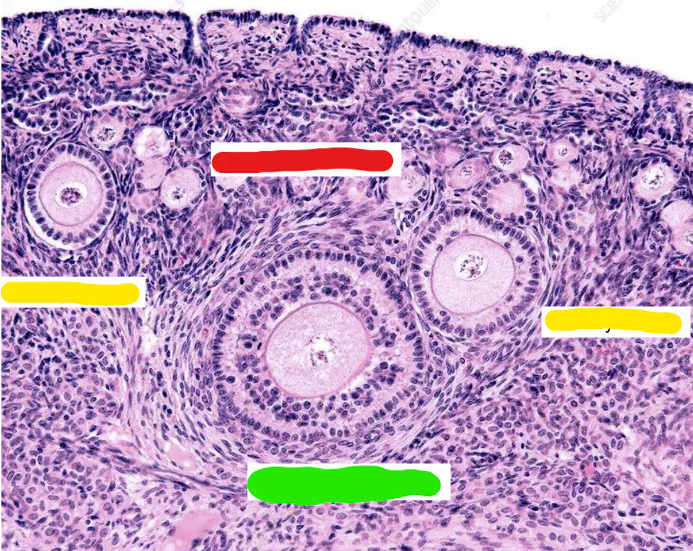
Histology
The following image represents? (red labeled)
Primary follicle
Secondary follicle
Primordial follicle
Graffian follicle
Tertiary follicle
Histology
The following image represents? (yellow labeled)
Primary follicle
Secondary follicle
Primordial follicle
Graffian follicle
Tertiary follicle
Histology
The following image represents? (green labeled)
Primary follicle
Secondary follicle
Primordial follicle
Graffian follicle
Tertiary follicle

Histology
The following image represents? (red labeled)
Primary follicle
Secondary follicle
Primordial follicle
Graffian follicle
Tertiary follicle
Histology
The following image represents? (yellow labeled)
Primary follicle
Secondary follicle
Primordial follicle
Graffian follicle
Tertiary follicle

Histology
The following image represents? (red labeled)
Primary follicle
Secondary follicle
Primordial follicle
Graffian follicle
Tertiary follicle
Histology
The following image represents? (yellow labeled)
Primary follicle
Secondary follicle
Primordial follicle
Graffian follicle
Tertiary follicle

Histology
The following image represents? (red labeled)
Primary follicle
Secondary follicle
Primordial follicle
Graffian follicle
Tertiary follicle
Histology
The following image represents? (yellow labeled)
Primary follicle
Secondary follicle
Primordial follicle
Graffian follicle
Tertiary follicle
Histology
The following image represents? (green labeled)
Primary follicle
Secondary follicle
Primordial follicle
Graffian follicle
Tertiary follicle
Histology
The following image represents? (blue labeled)
Primary follicle
Secondary follicle
Primordial follicle
Graffian follicle
Medulla
Histology
The following image represents? (purple labeled)
Primary follicle
Secondary follicle
Primordial follicle
Graffian follicle
Medulla
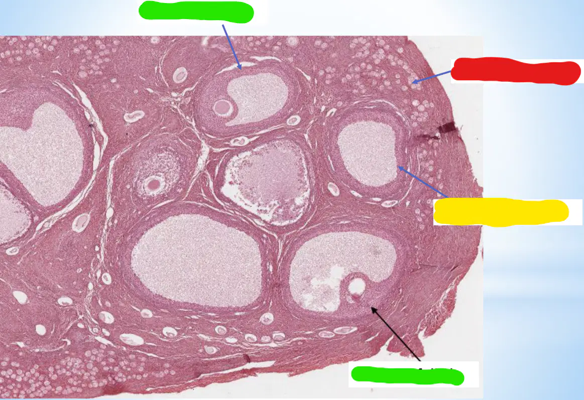
Histology
The following image represents? (red labeled)
Primary follicle
Secondary follicle
Primordial follicle
Graffian follicle
Medulla
Histology
The following image represents? (yellow labeled)
Primary follicle
Secondary follicle
Primordial follicle
Graffian follicle
Medulla
Histology
The following image represents? (green labeled)
Primary follicle
Secondary follicle
Primordial follicle
Graffian follicle
Medulla
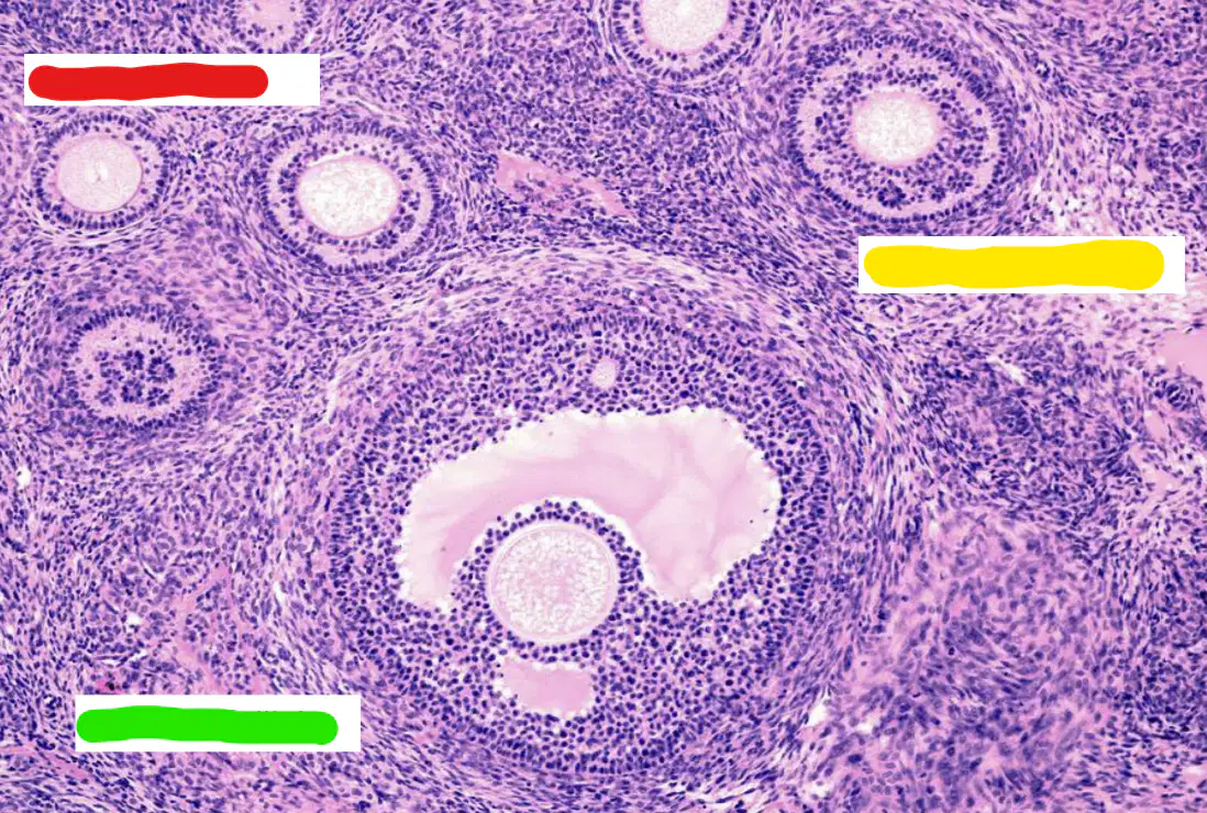
Histology
The following image represents? (red labeled)
Primary follicle
Secondary follicle
Primordial follicle
Graffian follicle
Medulla
Histology
The following image represents? (yellow labeled)
Primary follicle
Secondary follicle
Primordial follicle
Graffian follicle
Medulla
Histology
The following image represents? (green labeled)
Primary follicle
Secondary follicle
Primordial follicle
Graffian follicle
Medulla
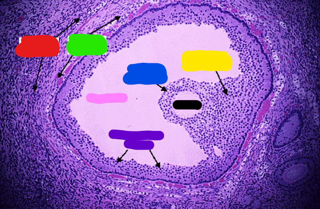
Histology
The following image represents?
Primary follicle
Secondary follicle
Primordial follicle
Graffian follicle
Medulla
Histology
The following image represents? (red labeled)
Theca Interna
Theca Externa
Corona Radiata
Ovum
Cumulus Oophorus
Histology
The following image represents? (green labeled)
Theca Interna
Theca Externa
Corona Radiata
Ovum
Cumulus Oophorus
Histology
The following image represents? (yellow labeled)
Antrum
Follicular cells
Corona Radiata
Ovum
Cumulus Oophorus
Histology
The following image represents? (blue labeled)
Antrum
Follicular cells
Corona Radiata
Ovum
Cumulus Oophorus
Histology
The following image represents? (pink labeled)
Antrum
Follicular cells
Corona Radiata
Ovum
Cumulus Oophorus
Histology
The following image represents? (purple labeled)
Antrum
Follicular cells
Corona Radiata
Ovum
Cumulus Oophorus
Histology
The following image represents? (black labeled)
Antrum
Follicular cells
Corona Radiata
Ovum
Cumulus Oophorus
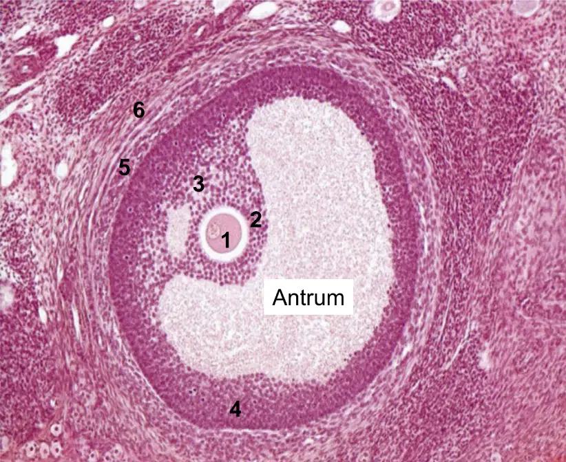
Histology
The following image represents?
Primary follicle
Secondary follicle
Primordial follicle
Graffian follicle
Medulla
Histology
The following image represents? (No. 1)
Corona Radiata
Secondary oocyte
Granulosa cells
Theca interna
Theca externa
Histology
The following image represents? (No. 2)
Corona Radiata
Cumulus oophorus
Granulosa cells
Theca interna
Theca externa
Histology
The following image represents? (No. 3)
Corona Radiata
Cumulus oophorus
Granulosa cells
Theca interna
Theca externa
Histology
The following image represents? (No. 4)
Corona Radiata
Cumulus oophorus
Granulosa cells
Theca interna
Theca externa
Histology
The following image represents? (No. 5)
Corona Radiata
Cumulus oophorus
Granulosa cells
Theca interna
Theca externa
Histology
The following image represents? (No. 6)
Corona Radiata
Cumulus oophorus
Granulosa cells
Theca interna
Theca externa
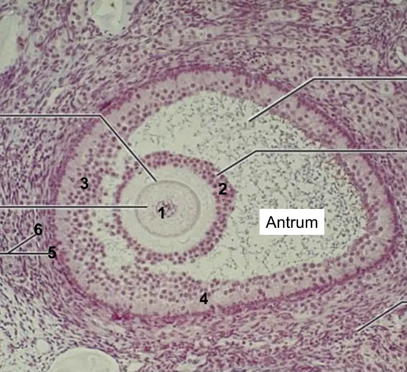
Histology
The following image represents?
Primary follicle
Secondary follicle
Primordial follicle
Graffian follicle
Medulla

Histology
The following image represents? (No. 1)
Corona Radiata
Secondary oocyte
Granulosa cells
Theca interna
Theca externa
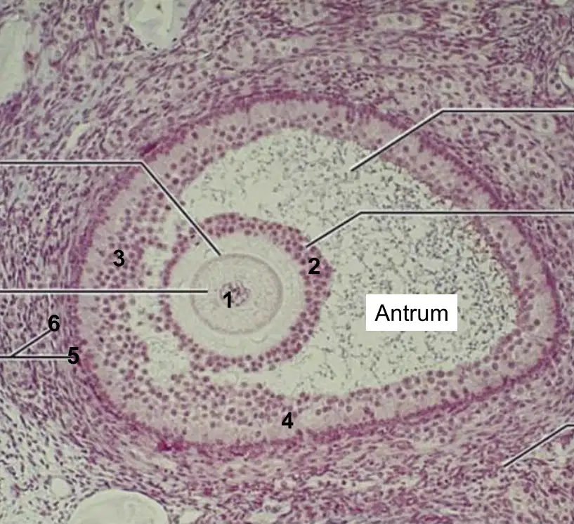
Histology
The following image represents? (No. 2)
Corona Radiata
Cumulus oophorus
Granulosa cells
Theca interna
Theca externa
Histology
The following image represents? (No. 3)
Corona Radiata
Cumulus oophorus
Granulosa cells
Theca interna
Theca externa
Histology
The following image represents? (No. 4)
Corona Radiata
Cumulus oophorus
Granulosa cells
Theca interna
Theca externa
Histology
The following image represents? (No. 5)
Corona Radiata
Cumulus oophorus
Granulosa cells
Theca interna
Theca externa
Histology
The following image represents? (No. 6)
Corona Radiata
Cumulus oophorus
Granulosa cells
Theca interna
Theca externa

Histology
The following image represents?
Primary follicle
Secondary follicle
Primordial follicle
Graffian follicle
Medulla
Histology
The following image represents? (red labeled)
Corona Radiata
Zona Pellucida
Cumulus Oophorus
Theca Interna
Secondary Oocyte
Histology
The following image represents? (yellow labeled)
Corona Radiata
Zona Pellucida
Cumulus Oophorus
Theca Interna
Secondary Oocyte
Histology
The following image represents? (green labeled)
Corona Radiata
Zona Pellucida
Cumulus Oophorus
Theca Interna
Secondary Oocyte
Histology
The following image represents? (blue labeled)
Corona Radiata
Zona Pellucida
Cumulus Oophorus
Theca Interna
Secondary Oocyte
Histology
The following image represents? (purple labeled)
Corona Radiata
Zona Pellucida
Cumulus Oophorus
Theca Interna
Secondary Oocyte
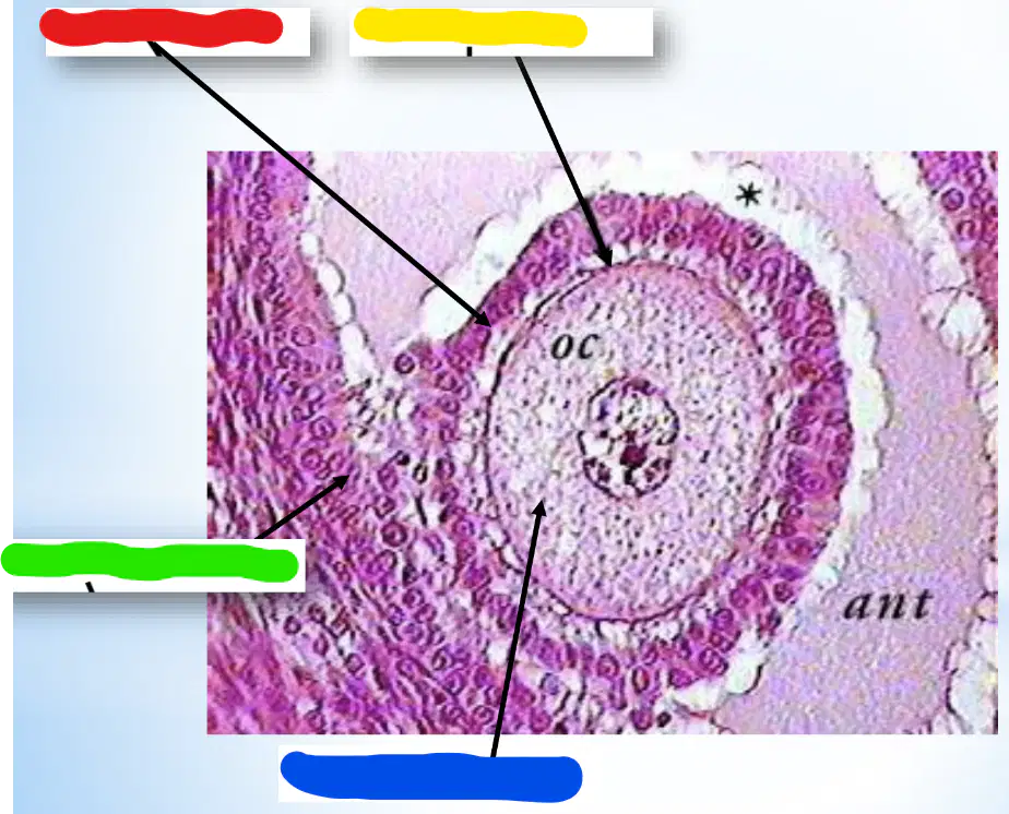
Histology
The following image represents? (red labeled)
Corona Radiata
Zona Pellucida
Cumulus Oophorus
Theca Interna
Secondary Oocyte
Histology
The following image represents? (yellow labeled)
Corona Radiata
Zona Pellucida
Cumulus Oophorus
Theca Interna
Secondary Oocyte
Histology
The following image represents? (green labeled)
Corona Radiata
Zona Pellucida
Cumulus Oophorus
Theca Interna
Secondary Oocyte
Histology
The following image represents? (blue labeled)
Corona Radiata
Zona Pellucida
Cumulus Oophorus
Theca Interna
Secondary Oocyte
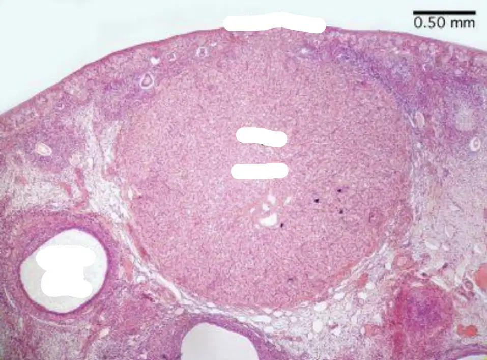
Histology
The following image represents?
Graffian Follicle
Corpus Luteum
Corpus Albicans
Primary Follicle
Secondary Follicle
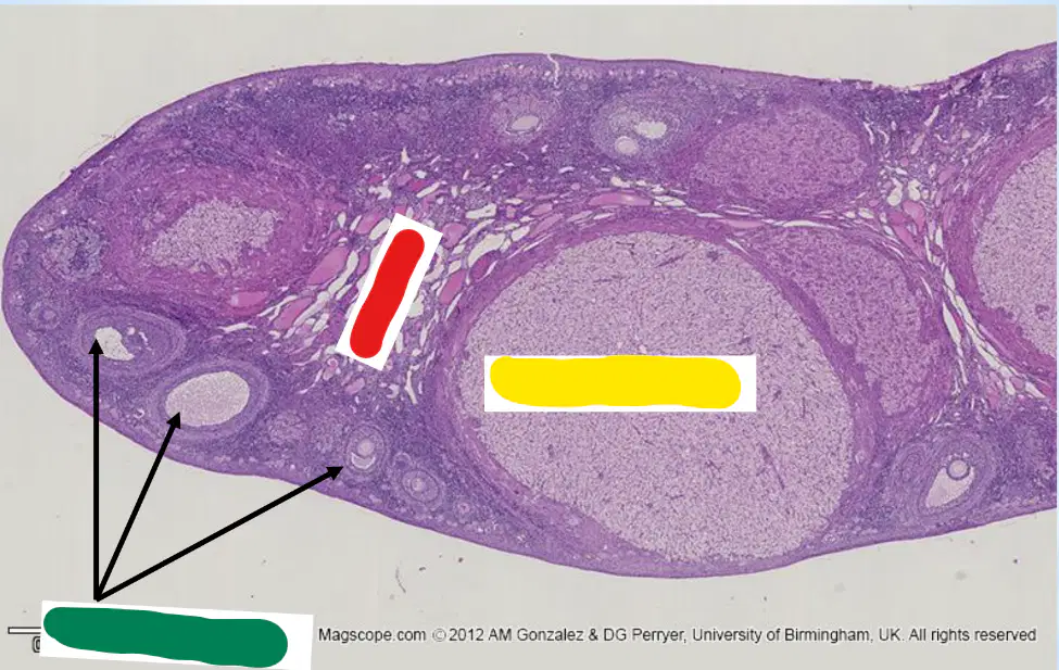
Histology
The following image represents? (red labeled)
Cortex
Medulla
Ovarian Follicle
Corpus Luteum
Tunica Vaginalis
Histology
The following image represents? (yellow labeled)
Cortex
Medulla
Ovarian Follicle
Corpus Luteum
Tunica Vaginalis
Histology
The following image represents? (green labeled)
Cortex
Medulla
Ovarian Follicle
Corpus Luteum
Tunica Vaginalis
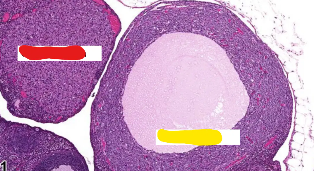
Histology
The following image represents? (red labeled)
Cortex
Medulla
Ovarian Follicle
Corpus Luteum
Tunica Vaginalis
Histology
The following image represents? (yellow labeled)
Cortex
Medulla
Ovarian Follicle
Corpus Luteum
Tunica Vaginalis
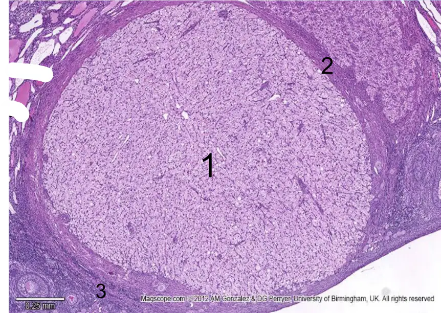
Histology
The following image represents?
Cortex
Medulla
Ovarian Follicle
Corpus Luteum
Tunica Vaginalis
Histology
The following image represents? (No. 1)
Granulosa Lutein cells
Theca Lutein cells
Follicular cells
Fibrous Capsule
Ovarian Stroma
Histology
The following image represents? (No. 2)
Granulosa Lutein cells
Theca Lutein cells
Follicular cells
Fibrous Capsule
Ovarian Stroma
Histology
The following image represents? (No. 3)
Granulosa Lutein cells
Theca Lutein cells
Follicular cells
Fibrous Capsule
Ovarian Stroma
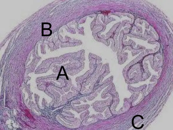
Histology
The following image represents?
Fallopian Tube
Uterus
Cervix
Vagina
Ovary
Histology
The following image represents? (A)
Mucosa
Submucosa
Musculosa
Serosa
Muscularis Mucosa
Histology
The following image represents? (B)
Mucosa
Submucosa
Musculosa
Serosa
Muscularis Mucosa
Histology
The following image represents? (C)
Mucosa
Submucosa
Musculosa
Serosa
Muscularis Mucosa
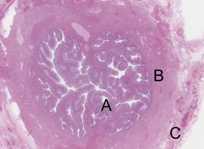
Histology
The following image represents?
Fallopian Tube
Uterus
Cervix
Vagina
Ovary
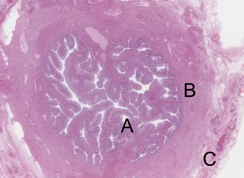
Histology
The following image represents? (A)
Mucosa
Submucosa
Musculosa
Serosa
Muscularis Mucosa
Histology
The following image represents? (B)
Mucosa
Submucosa
Musculosa
Serosa
Muscularis Mucosa
Histology
The following image represents? (C)
Mucosa
Submucosa
Musculosa
Serosa
Muscularis Mucosa
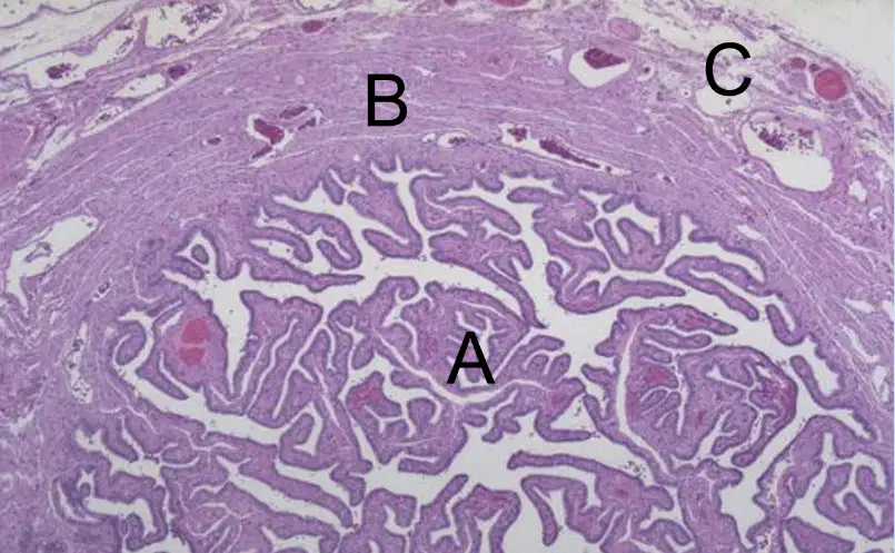
Histology
The following image represents?
Fallopian Tube
Uterus
Cervix
Vagina
Ovary
Histology
The following image represents? (A)
Mucosa
Submucosa
Musculosa
Serosa
Muscularis Mucosa
Histology
The following image represents? (B)
Mucosa
Submucosa
Musculosa
Serosa
Muscularis Mucosa
Histology
The following image represents? (C)
Mucosa
Submucosa
Musculosa
Serosa
Muscularis Mucosa
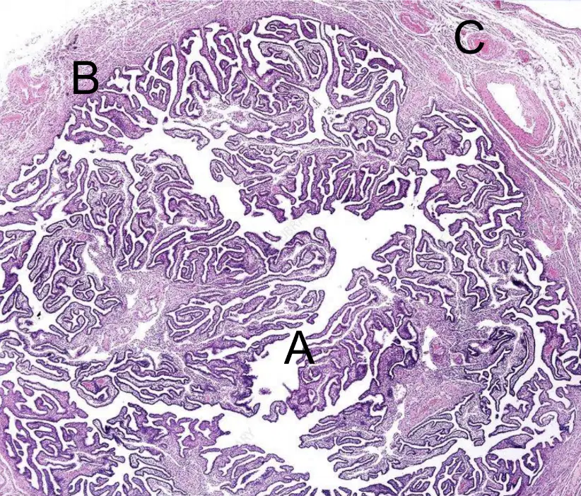
Histology
The following image represents?
Fallopian Tube
Uterus
Cervix
Vagina
Ovary
Histology
The following image represents? (A)
Mucosa
Submucosa
Musculosa
Serosa
Muscularis Mucosa
Histology
The following image represents? (B)
Mucosa
Submucosa
Musculosa
Serosa
Muscularis Mucosa
Histology
The following image represents? (C)
Mucosa
Submucosa
Musculosa
Serosa
Muscularis Mucosa
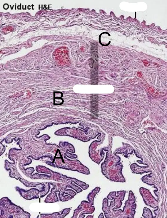
Histology
The following image represents?
Fallopian Tube
Uterus
Cervix
Vagina
Ovary
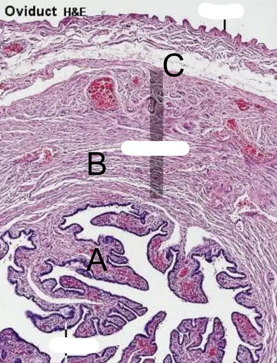
Histology
The following image represents? (A)
Mucosa
Submucosa
Musculosa
Serosa
Muscularis Mucosa
Histology
The following image represents? (B)
Mucosa
Submucosa
Musculosa
Serosa
Muscularis Mucosa
Histology
The following image represents? (C)
Mucosa
Submucosa
Musculosa
Serosa
Muscularis Mucosa
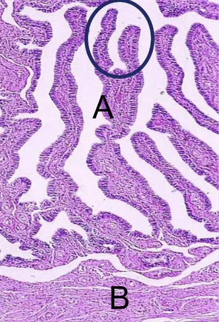
Histology
The following image represents?
Fallopian Tube
Uterus
Cervix
Vagina
Ovary
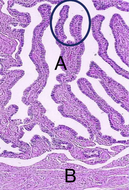
Histology
The following image represents? (A)
Mucosa
Submucosa
Musculosa
Serosa
Muscularis Mucosa
Histology
The following image represents? (B)
Mucosa
Submucosa
Musculosa
Serosa
Muscularis Mucosa

Histology
The following image represents?
Fallopian Tube
Uterus
Cervix
Vagina
Ovary
Histology
The following image represents? (1)
Endometrium (Mucosa)
Myometrium (Muculosa)
Perimetrium (Serosa)
Mucularis Mucosa
Stratum Vaculare
Histology
The following image represents? (2)
Endometrium (Mucosa)
Myometrium (Muculosa)
Perimetrium (Serosa)
Mucularis Mucosa
Stratum Vaculare
Histology
The following image represents? (3)
Endometrium (Mucosa)
Myometrium (Muculosa)
Perimetrium (Serosa)
Mucularis Mucosa
Stratum Vaculare

Histology
The following image represents?
Fallopian Tube
Uterus
Cervix
Vagina
Ovary
Histology
The following image represents? (red labeled)
Endometrium (Mucosa)
Myometrium (Musculosa)
Perimetrium (Serosa)
Muscularis Mucosa
Stratum Vasculare
Histology
The following image represents? (yellow labeled)
Endometrium (Mucosa)
Myometrium (Musculosa)
Perimetrium (Serosa)
Muscularis Mucosa
Stratum Vasculare
Histology
The following image represents? (green labeled)
Endometrium (Mucosa)
Myometrium (Musculosa)
Perimetrium (Serosa)
Muscularis Mucosa
Stratum Vasculare
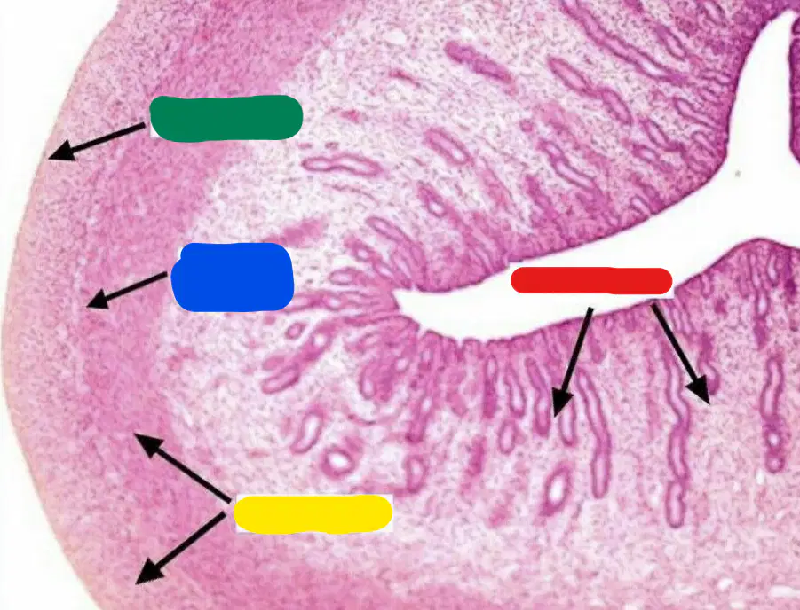
Histology
The following image represents?
Fallopian Tube
Uterus
Cervix
Vagina
Ovary
Histology
The following image represents? (red labeled)
Endometrium (Mucosa)
Myometrium (Musculosa)
Perimetrium (Serosa)
Muscularis Mucosa
Stratum Vasculare
Histology
The following image represents? (yellow labeled)
Endometrium (Mucosa)
Myometrium (Musculosa)
Perimetrium (Serosa)
Muscularis Mucosa
Stratum Vasculare
Histology
The following image represents? (green labeled)
Endometrium (Mucosa)
Myometrium (Musculosa)
Perimetrium (Serosa)
Muscularis Mucosa
Stratum Vasculare
Histology
The following image represents? (blue labeled)
Endometrium (Mucosa)
Myometrium (Musculosa)
Perimetrium (Serosa)
Muscularis Mucosa
Stratum Vasculare
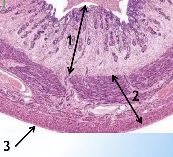
Histology
The following image represents?
Fallopian Tube
Uterus
Cervix
Vagina
Ovary
Histology
The following image represents? (1)
Endometrium (Mucosa)
Myometrium (Musculosa)
Perimetrium (Serosa)
Muscularis Mucosa
Stratum Vasculare
Histology
The following image represents? (2)
Endometrium (Mucosa)
Myometrium (Musculosa)
Perimetrium (Serosa)
Muscularis Mucosa
Stratum Vasculare
Histology
The following image represents? (3)
Endometrium (Mucosa)
Myometrium (Musculosa)
Perimetrium (Serosa)
Muscularis Mucosa
Stratum Vasculare
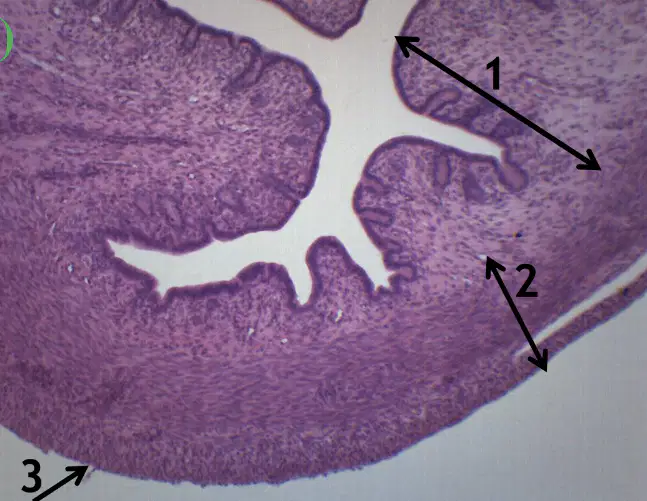
Histology
The following image represents?
Fallopian Tube
Uterus
Cervix
Vagina
Ovary
Histology
The following image represents? (1)
Endometrium (Mucosa)
Myometrium (Musculosa)
Perimetrium (Serosa)
Muscularis Mucosa
Stratum Vasculare
Histology
The following image represents? (2)
Endometrium (Mucosa)
Myometrium (Musculosa)
Perimetrium (Serosa)
Muscularis Mucosa
Stratum Vasculare
Histology
The following image represents? (3)
Endometrium (Mucosa)
Myometrium (Musculosa)
Perimetrium (Serosa)
Muscularis Mucosa
Stratum Vasculare
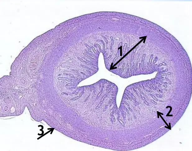
Histology
The following image represents?
Fallopian Tube
Uterus
Cervix
Vagina
Ovary
Histology
The following image represents? (1)
Endometrium (Muscosa)
Myometrium (Musculosa)
Perimetrium (Serosa)
Muscularis Mucosa
Stratum Vasculare
Histology
The following image represents? (2)
Endometrium (Muscosa)
Myometrium (Musculosa)
Perimetrium (Serosa)
Muscularis Mucosa
Stratum Vasculare
Histology
The following image represents? (3)
Endometrium (Muscosa)
Myometrium (Musculosa)
Perimetrium (Serosa)
Muscularis Mucosa
Stratum Vasculare

Histology
The following image represents which phase?
Follicular Phase
Ovulatory Phase
Luteal Phase
Menstrual Phase
Secretory Phase
Histology
The following image represents which phase?
Follicular Phase
Ovulatory Phase
Luteal Phase
Menstrual Phase
Secretory Phase
Histology
The following image represents which phase?
Follicular Phase
Ovulatory Phase
Luteal Phase
Menstrual Phase
Secretory Phase

Histology
The following image represents?
Ovary
Uterus
Fallopian Tube
Placenta
Mammary Gland

Histology
The following image represents? (red labeled)
Chorionic Villi
Intervillous space
Intravillous space
Decidua basalis
Placental barrier
Histology
The following image represents? (green labeled)
Chorionic Villi
Intervillous space
Intravillous space
Decidua basalis
Fetal capillaries

Histology
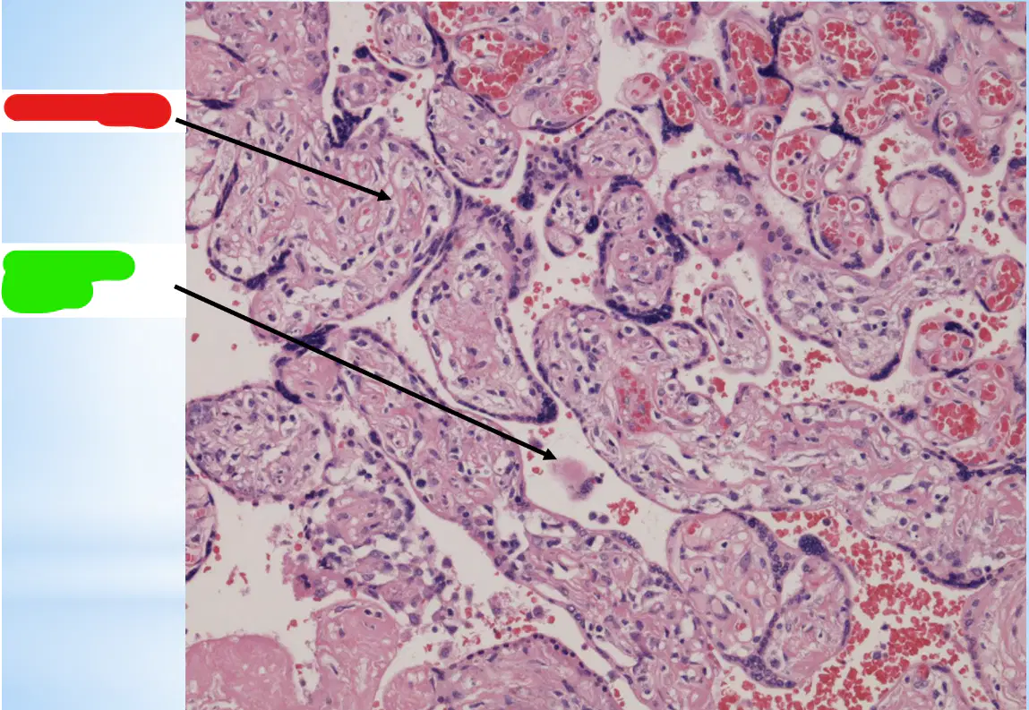
The following image represents?
Ovary
Uterus
Fallopian Tube
Placenta
Mammary Gland
Histology
The following image represents? (red labeled)
Chorionic Villi
Intervillous space
Intravillous space
Decidua basalis
Fetal capillaries
Histology
The following image represents? (green labeled)
Chorionic Villi
Intervillous space
Intravillous space
Decidua basalis
Fetal capillaries

Histology
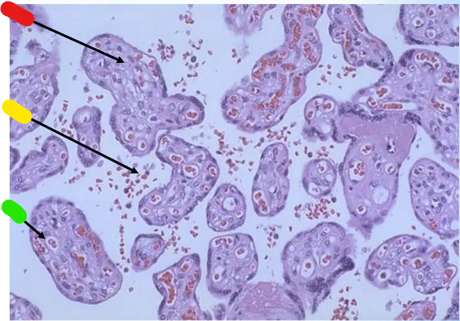
The following image represents?
Ovary
Uterus
Fallopian Tube
Placenta
Mammary Gland
Histology
The following image represents? (red labeled)
Chorionic Villi
Intervillous space
Intravillous space
Decidua basalis
Fetal capillaries
Histology
The following image represents? (yellow labeled)
Chorionic Villi
Intervillous space
Intravillous space
Decidua basalis
Fetal capillaries
Histology
The following image represents? (green labeled)
Chorionic Villi
Intervillous space
Intravillous space
Decidua basalis
Fetal capillaries

Histology
The following image represents?
Resting Mammary Gland
Mammary Gland at Pregnancy
Lactating Mammary Gland
Placenta
Fetal & Maternal Capillaries
Histology
The following image represents? (red labeled)
Septa
Duct
Alveoli
Milk secretion
Fetal & Maternal Capillaries
Histology
The following image represents? (yellow labeled)
Septa
Duct
Alveoli
Milk secretion
Fetal & Maternal Capillaries
Histology
The following image represents? (green labeled)
Septa
Duct
Alveoli
Milk secretion
Fetal & Maternal Capillaries

Histology
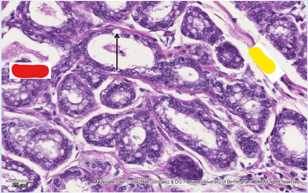
The following image represents?
Resting Mammary Gland
Mammary Gland at Pregnancy
Lactating Mammary Gland
Placenta
Fetal & Maternal Capillaries

Histology
The following image represents? (red labeled)
Septa
Duct
Alveoli
Milk secretion
Fetal & Maternal Capillaries
Histology
The following image represents? (yellow labeled)
Septa
Duct
Alveoli
Milk secretion
Fetal & Maternal Capillaries
Histology
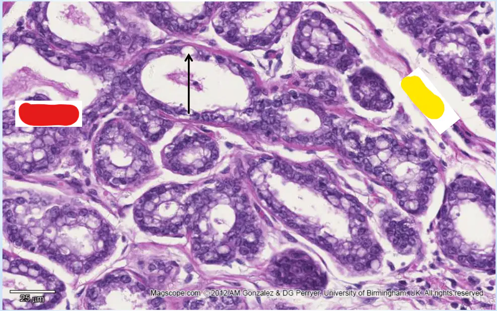
The following image represents?
Resting Mammary Gland
Mammary Gland at Pregnancy
Lactating Mammary Gland
Placenta
Fetal & Maternal Capillaries
Histology
The following image represents? (red labeled)
Septa
Duct
Alveoli
Milk secretion
Fetal & Maternal Capillaries
Histology
The following image represents? (yellow labeled)
Septa
Duct
Alveoli
Milk secretion
Fetal & Maternal Capillaries

Histology
The following image represents?
Resting Mammary Gland
Mammary Gland at Pregnancy
Lactating Mammary Gland
Placenta
Fetal & Maternal Capillaries

Histology
The following image represents? (red labeled)
Septa
Duct
Alveoli
Milk secretion
Fetal & Maternal Capillaries
Histology
The following image represents? (yellow labeled)
Septa
Duct
Alveoli
Milk secretion
Fetal & Maternal Capillaries
Histology
The following image represents? (green labeled)
Septa
Duct
Alveoli
Milk secretion
Fetal & Maternal Capillaries
Histology
The following image represents? (blue labeled)
Septa
Duct
Alveoli
Milk secretion
Fetal & Maternal Capillaries

Histology
The following image represents?
Resting Mammary Gland
Mammary Gland at Pregnancy
Lactating Mammary Gland
Placenta
Fetal & Maternal Capillaries

Histology
The following image represents? (red labeled)
Septa
Duct
Alveoli
Milk secretion
Fetal & Maternal Capillaries
Histology
The following image represents? (yellow labeled)
Septa
Duct
Alveoli
Milk secretion
Fetal & Maternal Capillaries
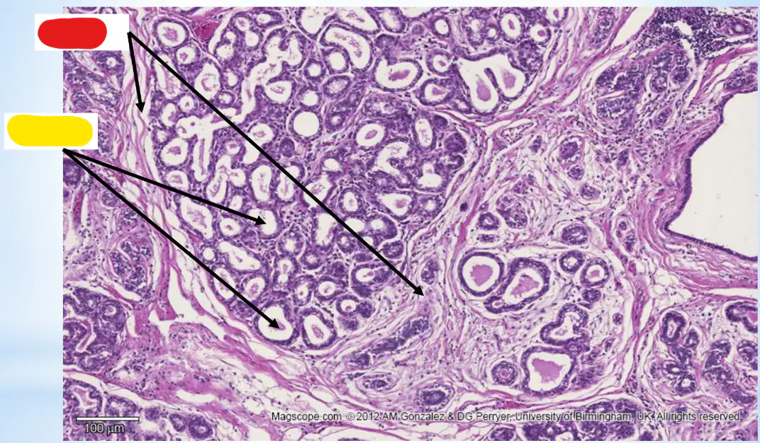
Histology
The following image represents?
Resting Mammary Gland
Mammary Gland at Pregnancy
Lactating Mammary Gland
Placenta
Fetal & Maternal Capillaries

Histology
The following image represents? (red labeled)
Septa
Duct
Alveoli
Milk secretion
Fetal & Maternal Capillaries
Histology
The following image represents? (yellow labeled)
Septa
Duct
Alveoli
Milk secretion
Fetal & Maternal Capillaries

