
 Worksheets
Spleen
Total questions: 76
Worksheet time: 1hrs 6mins
(a) covered entirely with peritoneum except for a small area at its hilum, where the vascular structures and lymph nodes are located.
The spleen is considered a (a) organ
The spleen is part of the (a) system.
Which system has the responsibility for phagocytosis?
Intraperitoneal
Retorperitoneal
Reticuloendothelial
Digestive
(a) is engulfing and destroying of damaged or old cells and their debris, foreign materials, and pathogens taking them out of the circulating blood
The spleen is considered the (a) lymphoid organ
The spleen is primarily composed of which type of tissue?
Epithelial
Connective
Dense
Lymph
Although crucial to the immune system, is it possible to remove the spleen without causing adverse effects or death?
Yes
NO
Which abdominal region does the spleen lie within?
Left hypochondrium
Epigastrium
Right hypochondriac
Hypogastrium
The spleen is (a) to the body and fundus of the stomach
The spleen is (a) to the tail of the pancreas
The spleen is (a) to the left colic flexure
The spleen is (a) to the diaphragm, the left lung, and the 8th, 9th, 10th, and 11th ribs
The left kidney is located __ and __ to the spleen
(a)
As part of the (a) system, the spleen's main function is to help remove old cells, debris, pathogens, and foreign substances from circulation
Which of the following best describes the main function of the spleen?
Help deoxygenate blood to return to the inferior vena cava
Remove old cells, debris, pathogens, and foreign materials from circulation
Creation lymph cells that can be used towards the immune system
The four major functions of the spleen are:
Defense
Hemotpyosis
Hematopoiesis
Red blood cell and platelet destruction
Service as a blood reservoir
The spleen is _ to _ cm thick
8-13cm
7-8cm
3-4cm
1-2cm
The spleen is __ to. __ cm in length
8-13cm
3-4cm
7-8cm
14-19cm
The spleen is __ to __ in AP diameter
3-4cm
7-8cm
8-13cm
9-10cm
The (a) is a portion of the spleen located medially
The (a) is where the vasculature enters and exits
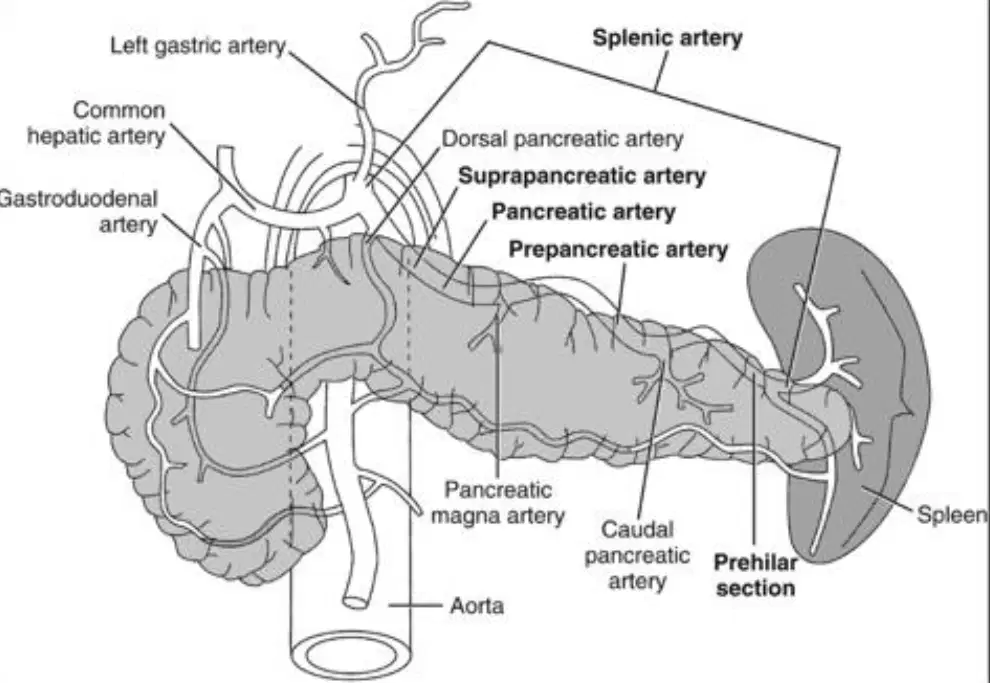
The spleen lies on the (a) end of the pancreas

What is labeled A?
(a)
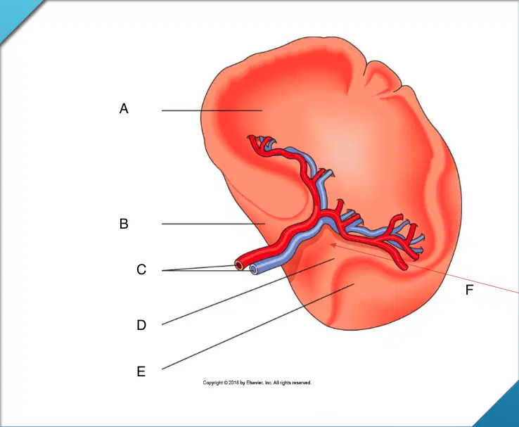
What is labeled B?
(a)

What structures are labeled C?
(a)

What is labeled D?
(a)

What is labeled E
(a)

What is labeled F
(a)
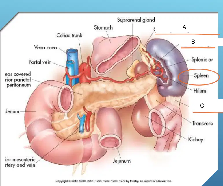
What is labeled A
(a)

What is labeled B
(a)
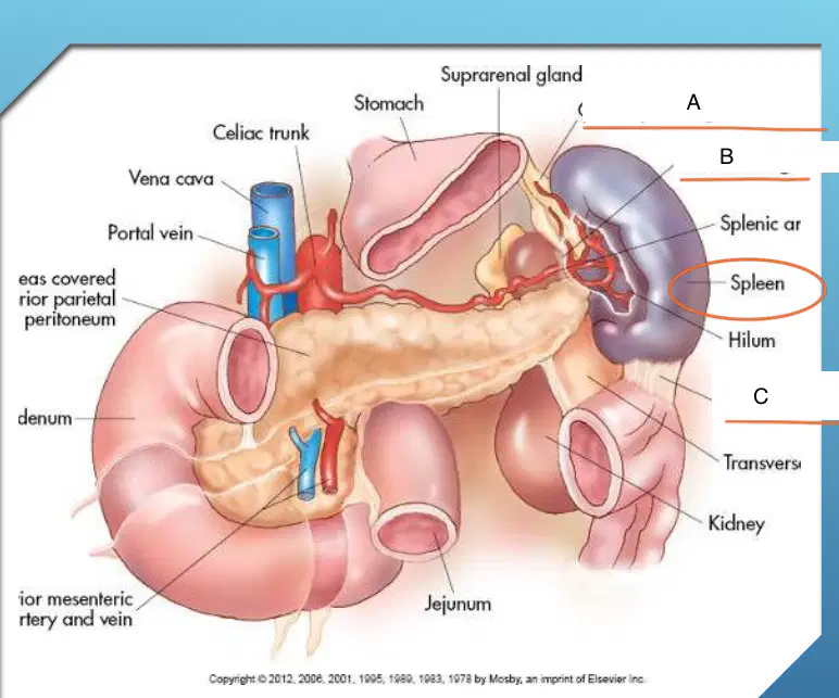
What is labeled C
(a)
Spleen is held in place by several (a) .
The splenic (a) are folds of peritoneum that connect the spleen to surrounding structures
Which of the following are splenic ligaments?
Ligamentum venosum
Gastrosplenic ligament
Splenorenal ligament
Phrenocolic ligament
Lienorenal ligament
Which of the following is NOT a splenic ligament?
Gastrosplenic
Splenorenal
Splenopancreatic
Lienorenal
Splenic artery
Splenic vein
Tail of pancreas
These apply to which ligament?
(a)
Which ligament is posteromedial to the spleen
(a)
The splenic flexure is (a) to the spleen
Left gastro-epiploic
Short gastric arteries
These apply to which ligament?
(a)
Which ligament is closer to the pancreas tail?
(a)
Which ligament is closer in relation to the stomach?
(a)
The outer covering of the spleen is called the (a)
The (a) is a relatively thin and delicate structure making the spleen very fragile and susceptible to rupture or injury
How many layers does the splenic capsule have?
1
2
3
4
The (a) layer of the splenic capsule is in contact with surrounding tissues
The (a) trabecular sheath supports the splenic tissue
Which of the following are functions of the spleen?
Supports the spleen
Contains internal structures
Contacts outside structures
Allows for its expansion and contraction to discharge blood when needed
The __ __ is one of the terminal branches of the celiac artery/trunk
(a)
Which direction does the splenic artery pass from the celiac axis?
Right
Left
The splenic artery passes left from the celiac axis across the __ crus of the diaphragm
Left
Right
The splenic artery passes left from the celiac axis across the left crus of the diaphragm and __ psosas muscle
left
Right
(a) runs superior to the pancreas
On entering the splenic hilum, the splenic artery immediately branches into how many smaller arteries?
1
2
5
6
What is the function of the six smaller arteries within the spleen?
supply the organ without deoxygenated blood to profuse the splenic parenchyma.
supply the organ without oxygenated blood to profuse the splenic parenchyma.
supply the organ with deoxygenated blood to profuse the splenic parenchyma.
supply the organ with oxygenated blood to profuse the splenic parenchyma.
What is the splenic vein formed by?
the union of smaller veins at the hilum of the spleen.
the union of larger veins at the hilum of the spleen.
the union of medium sized veins at the hilum of the spleen.
Splenic vein leaves the hilum in a (a) direction
(a) joins the Superior mesenteric vein (SMV) to form the Portal vein (PV
The splenic vein courses __ usually __ to the pancreas
(a)
What is the purpose of the splenic vein?
Drains blood from spleen, parts of stomach pancreas, and hindgut
Pumps blood into the spleen and parts of the stomach
The splenic blood is delivered to the liver via the __ __ for processing by multiple branches in the spleen
(a)
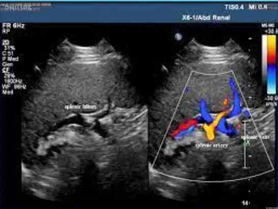
The (a) originate from the splenic hilum
(a) pass through other lymph nodes along the course of the splenic artery
(a) drain into the celiac nodes
(a) transport fluid and cells from the spleen to the celiac lymph nodes for filtration and immune surveillance
(a) supplying the spleen, accompany the splenic artery o derived from the celiac plexus
The splenic parenchyma should have a fine uniform (a) mid- to low-level echo pattern, as is seen within the liver parenchyma
The spleen is scanned in a __ scanning plane
Sagittal
Coronal
Transverse
How should the patient be positioned for a splenic ultrasound?
Prone
Supine
Right lateral decubitus
Left lateral decubitus
The transducer should be placed in the superior (a) intercostal margin

What is labeled 1?
(a)

What is labeled 2?
(a)

This image shows the (a)
What can a spleen x-ray show us?
Size, shape, position
Splenic torsion
Splenic enlargement
Length, width, height
X-ray, ultrasound, nuclear medicine, CT scan, and MRI are all
(a)
(a) uses magnetic fields and radio waves to create detailed images of internal organs, soft tissues, and blood vessels

