
 Worksheets
Ultrasonography and Ovarian Normal & Pathology I
Total questions: 80
Worksheet time: 40mins
Emma and Anika are undergoing fertility treatment, and their doctor uses sonography. What is one important clinical use of sonography in fertility treatment?
To monitor follicular growth
To measure blood pressure
To assess bone density
To evaluate lung function
About how many times does a woman ovulate in her lifetime?
400 times
1,000 times
200 times
600 times
A character visits a doctor for pelvic discomfort. The doctor finds a fluid-filled pelvic mass. What is the most likely origin?
Cystic
Solid
Fibrous
Muscular
In a cartoon hospital, what is the main role of sonography for suspected ovarian problems?
To indicate the need for surgical or medical intervention
To provide a precise diagnosis without further tests
To replace all other diagnostic methods
To treat ovarian carcinoma directly
What is the typical shape of the ovaries?
Almond-shaped
Circular
Triangular
Rectangular
If the uterus is the castle's main hall, where are the ovaries located in relation to it?
On each side of the uterus, close to the lateral pelvic wall
Directly above the uterus
Below the uterus, near the cervix
In front of the uterus, near the bladder
When the uterus is retroverted, what happens to the position of the ovaries?
Ovaries tend to be lateral and superior, near the uterine fundus
Ovaries move inferiorly and medially
Ovaries remain in the same position
Ovaries move posteriorly and inferiorly
After a hysterectomy, where are the ovaries positioned?
More medially and directly superior to the vaginal cuff
More laterally and inferior to the vaginal cuff
In the same position as before the hysterectomy
Posterior to the vaginal cuff
Which anatomical reference point is used to locate the ovaries during a pelvic ultrasound?
Internal iliac vessels
Uterine fundus
Vaginal cuff
Lateral pelvic wall
Which three phases of the menstrual cycle can be identified by ultrasound?
Proliferative phase, ovulation, and secretory phase
Follicular phase, ovulation, and luteal phase
Menstrual phase, ovulation, and luteal phase
Early proliferative phase, follicular phase, and luteal phase
Which hormone helps stimulate follicular growth, similar to the early proliferative phase of the menstrual?
Estrogen
Follicle-stimulating hormone (FSH)
Progesterone
Luteinizing hormone (LH)
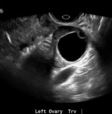
Dr. Rivera sees the following ovarian structure during Luna's ultrasound. What does this mean?
The presence of multiple follicles
Imminent ovulation
The beginning of the menstrual cycle
The end of the luteal phase
What is the size range of the dominant follicle during ovulation?
1.0 to 1.5 cm
2.0 to 2.5 cm
3.0 to 3.5 cm
0.5 to 1.0 cm
During the luteal phase, what is the classic sonographic appearance of a mature corpus luteum?
A large hyperechoic structure with smooth walls
A small hypoechoic or isoechoic structure with crenulated walls and low-level echoes
A highly echogenic structure with no internal echoes
A small anechoic structure with irregular walls
Imagine the corpus luteum as a character who stays in the ovary only if fertilization happens. What happens if fertilization does not occur?
It remains active throughout the menstrual cycle
It undergoes involutional changes and disappears shortly before or with the onset of menstruation
It grows larger and becomes a permanent structure in the ovary
It transforms into a follicle for the next ovulation cycle
Why is it hard to see the ovary on ultrasound in postmenopausal women?
It becomes highly echogenic
It is obscured by surrounding tissues
It becomes smaller in size and lacks discrete follicles
It is located deeper in the pelvic cavity
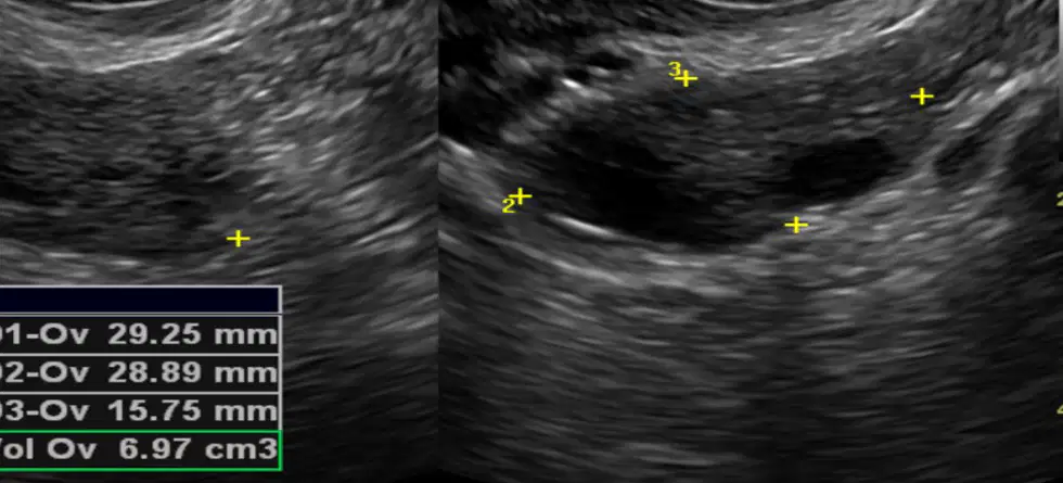
Using the image given, what is the mean ovarian volume in this female?
9.8 ± 5.8 cc
22 ± 5.8 cc
8.0 ± 5.8 cc
15.75 ± 5.8 cc
What ovarian volume is considered abnormal in a postmenopausal patient?
More than 22 cc
More than 8.0 cc
More than 15.75 cc
More than 6.97 cc
When is the volume of an ovary considered abnormal, regardless of its actual size?
When it is more than 22 cc
When it is more than 8.0 cc
When it is more than twice that of the opposite side
When it is less than 6.97 cc
How long does it usually take for a simple cystic mass to disappear when monitored with serial ultrasound?
4 weeks
6 weeks
8 weeks
10 weeks
When do doctors usually consider surgery for simple cystic masses?
When the cyst is greater than 4 cm and persists for more than 6 weeks
When the cyst is greater than 6 cm and persists for more than 8 weeks
When the cyst is greater than 5 cm and persists for more than 7 weeks
When the cyst is greater than 7 cm and persists for more than 9 weeks
How does a simple cystic mass appear on ultrasound?
Irregular with posterior shadowing
Smooth, well-defined with posterior enhancement
Rough, poorly defined with no enhancement
Irregular with no posterior enhancement
Which describes a complex mass on sonographic images?
Anechoic mass with internal echoes
Thick, irregular wall
Associated with ascites
Volume twice that of the opposite side
Which type of tumor is most likely to be malignant in a patient with ascites?
A significantly complex tumor
A simple cyst
A mass with no internal echoes
A tumor with a smooth wall
A character finds a solid object with thick, irregular walls. What could this mean?
Infection or malignancy
Normal tissue growth
Benign cyst
Fluid-filled cavity
When should a female with normal cycles schedule a Doppler ultrasound of her ovary?
The first 10 days of the cycle
Days 11-20 of the cycle
Days 21-28 of the cycle
Any day of the cycle
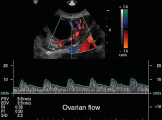
A gynecologist uses sonography to check blood flow in an adnexal lump. Which vessel feature would make the doctor most worried about it being malignant?
High-resistance flow and presence of a normal early diastolic notch
Low-resistance flow and absence of a normal early diastolic notch
High-resistance flow and absence of a normal early diastolic notch
Low-resistance flow and presence of a normal early diastolic notch
What is the cutoff value of the Resistive Index (RI) for a nonfunctioning ovary on Doppler ultrasound?
>0.2
>0.4
>0.6
>0.8
Which ovarian conditions in female can show abnormal Doppler waveform patterns?
Inflammatory lesions, metabolically active masses, and corpus luteum cyst
Normal ovarian function and corpus luteum cyst
Metabolically inactive masses and inflammatory lesions
Normal ovarian function and metabolically inactive masses
Which ovary Pulsatility Index (PI) value is considered abnormal in a Doppler ultrasound?
0.5
0.8
1.0
1.5
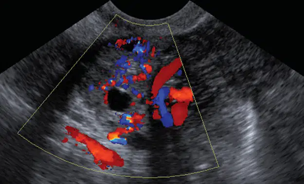
Why do sonographers use Doppler imaging to study ovaries?
To measure the size of the ovary
To assess blood flow in the ovarian vasculature
To detect ovarian cysts
To find the position of the ovary
A character finds bubbles in their garden that appear and disappear with the seasons. What are these bubbles most similar to if they were functional ovarian cysts?
Malignant masses that respond to cyclic hormonal stimulation
Benign cystic masses that respond to cyclic hormonal stimulation
Masses that do not respond to hormonal stimulation
Malignant masses that do not respond to hormonal stimulation
What is the most common cause of ovarian enlargement in young women?
Functional ovarian cysts
Hormonal therapy
Malignant tumors
Menstrual cycle irregularities
A female has a functional ovarian cyst. Which therapy is recommended to suppress the cyst?
Chemotherapy
Hormonal therapy
Radiation therapy
Antibiotic therapy
What is the usual size of most functional ovarian cysts?
Less than 3 cm in diameter
Less than 5 cm in diameter
Less than 7 cm in diameter
Less than 10 cm in diameter
How long does it usually take to observe changes in functional ovarian cysts during follow-up?
4 weeks
5 weeks
6 weeks
7 weeks
A young adult is diagnosed with a follicular cyst. What symptoms might she have?
Severe abdominal pain and fever
Dull adnexal pressure, pain, and menstrual irregularities
Nausea and vomiting
Sharp pelvic pain during ovulation
Which sonographic feature identifies a follicular cyst?
A solid, irregular mass with no enhancement
A well-defined, thin-walled, anechoic cystic mass with acoustic enhancement
A multilocular cyst with thick septations
A hyperechoic mass with calcifications
What is the usual size range of follicular cysts?
1-5 cm
3-10 cm
5-15 cm
10-20 cm
What is the main cause of a corpus luteum cyst forming?
Failure to resorb or excessive bleeding into it
Excessive production of hormones
Infection
Lack of blood supply
If a cartoon character like Minnie Mouse has a corpus luteum cyst, what symptoms would she most likely have?
Pain, enlarged tender ovary, nausea, and vomiting
Fever, chills, and abdominal swelling
Weight loss, fatigue, and dizziness
High blood pressure and rapid heartbeat
How long does a corpus luteum cyst usually take to resolve during pregnancy?
4-6 weeks
8-10 weeks
12-16 weeks
18-20 weeks
What sonographic features are seen in a corpus luteum cyst?
Thick wall and high-level echoes
Free fluid, low-level echoes, and a peripheral rim of color with a low-resistance waveform
Solid mass with irregular borders
High-resistance waveform and bilateral cysts
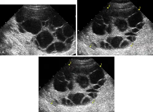
Which is the largest type of functional cyst seen in ultrasound images?
Theca-Lutein Cysts
Follicular Cysts
Corpus Luteum Cysts
Dermoid Cysts
Which condition is a risk factor for developing theca-lutein cysts?
Gestational trophoblastic disease
Polycystic ovary syndrome
Endometriosis
Ovarian cancer
Which symptom is most commonly associated with theca-lutein cysts?
Nausea and vomiting
Abdominal pain and fever
Irregular menstrual cycles
Weight loss and fatigue
What are the typical ultrasound findings in theca-lutein cysts?
Bilaterally enlarged ovaries with multiple multilocular cystic masses of varying sizes (3-20 cm)
A single large cyst with a smooth wall
Small, solid masses with irregular borders
Ovaries with no visible abnormalities
In which types of cysts can internal hemorrhage occur?
Follicular or corpus luteal cysts
Dermoid or sebaceous cysts
Epidermoid or ganglion cysts
Pilonidal or sebaceous cysts
A 28-year-old woman suddenly feels sharp lower abdominal pain while hiking. The doctor suspects a hemorrhagic cyst. What symptom is she most likely experiencing?
Acute onset of pelvic pain
Chronic back pain
Persistent headache
Sudden chest pain
Which sonographic appearance is typical for a hemorrhagic cyst?
Anechoic to complex or hyperechoic to anechoic
Uniformly hypoechoic
Completely anechoic
Uniformly hyperechoic
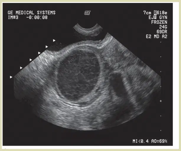
A middle-aged female is receiving an ultrasound, using the image given the radiologist suspects an ovarian cyst. Which additional finding would confirm this diagnosis?
Echogenic free intraperitoneal fluid in the cul-de-sac
Presence of calcifications in the cyst
Uniformly hypoechoic cyst appearance
Absence of blood flow in Doppler imaging
What is the main cause of Ovarian Hyperstimulation Syndrome (OHSS) when medicines are used to make ovaries work extra hard?
Genetic mutation
Iatrogenic complication of ovulation induction therapy
Viral infection
Hormonal imbalance
After treatment for Ovarian Hyperstimulation Syndrome, how long does it usually take for the syndrome to regress?
1 to 2 weeks
2 to 3 weeks
4 to 5 weeks
5 to 6 weeks
Which symptom is most likely in a cartoon character with mild Ovarian Hyperstimulation Syndrome?
Severe pelvic pain and abdominal distension
Pelvic discomfort without significant weight gain
Rapid weight loss
High fever
Which symptom or sonographic finding is expected in severe Ovarian Hyperstimulation Syndrome?
No significant weight gain
Ascites and pleural effusion
Normal ovarian size
Mild pelvic discomfort
Which symptom is most commonly seen in someone with Polycystic Ovary Syndrome (PCOS)?
Hirsutism
Jaundice
Bradycardia
Night sweats
On ultrasound, what finding in PCOS makes the ovaries look like they have a 'string of pearls'?
Solid mass with calcifications
Normal-sized ovary with no cysts
Multiple small peripheral follicles ('string of pearls' appearance)
Single large cyst
Which hormone is commonly elevated in PCOS?
Prolactin
Thyroid-stimulating hormone (TSH)
Luteinizing hormone (LH)
Follicle-stimulating hormone (FSH)
Which symptom could suggest endometriosis?
High fever
Frequent urination
Chronic pelvic pain
Heavy menstrual bleeding
Which hormone is most commonly responsible for the progression of endometriosis?
Testosterone
Progesterone
Estrogen
Thyroxine
Which ultrasound finding is most characteristic of an ovarian endometrioma?
Multilocular cyst with septations
Solid mass with calcifications
Anechoic cyst with thin walls
Homogeneous, ground-glass echogenicity within a cyst
Which symptom would most likely make a doctor order a pelvic ultrasound to check for endometriosis?
Frequent headaches
Skin rash
Chronic pelvic pain
Shortness of breath
If a paraovarian cyst is like a hidden treasure, where would it most likely be found?
Inside the uterine cavity
In the cervix
Between the ovary and the fallopian tube in the broad ligament
Within the ovarian stroma
Which sonographic feature identifies a typical paraovarian cyst?
Thick, irregular wall with internal echoes
Multiple septations and papillary projections
Solid mass with calcifications
Simple, thin-walled, anechoic cyst separate from the ovary
Which classic sonographic sign is seen in ovarian torsion?
Whirlpool sign
Snowstorm appearance
Target sign
Double bubble sign
What is the most common presenting symptom of ovarian torsion?
Chronic pelvic pain
Fever and chills
Acute onset of lower abdominal pain
Heavy menstrual bleeding
A female patient goes to her gynecologist for pelvic pain, who orders a sonogram to check if it's a hemorrhagic ovarian cyst. Which sonographic feature is classic for this cyst?
Lace-like internal echoes with possible fluid-fluid levels
Thick, calcified wall with shadowing
Completely anechoic with no internal echoes
Multiple solid nodules within the cyst
What is the most common clinical finding associated with a functional ovarian cyst?
Asymptomatic and found incidentally
Severe pelvic pain and fever
Rapid weight loss
Persistent abnormal uterine bleeding
Which Doppler parameter best distinguishes between benign and malignant ovarian masses?
Wall thickness
Resistive Index (RI)
Ovarian volume
Follicle count
Which sonographic finding most likely suggests ovarian torsion in a female patient with sudden lower abdominal pain?
Normal-sized ovary with central follicles
Enlarged ovary with peripheral follicles
Ovary with calcifications
Ovary with multiple cysts and increased vascularity
What is the role of Doppler ultrasound in diagnosing a suspected twisted ovary?
To visualize follicular development
To measure ovarian size only
To assess the absence or reduction of ovarian blood flow
To detect increased blood flow to the ovary
A teenage female patient present to the ER. which symptom is most suspicious for ovarian torsion?
Acute onset of lower abdominal pain
Frequent urination
Chronic pelvic pain
Heavy menstrual bleeding
Which ultrasound feature is most commonly seen in polycystic ovary syndrome (PCOS)?
Multiple small peripheral follicles ('string of pearls' appearance)
Single large ovarian cyst
Solid ovarian mass
Thickened endometrial lining
What is the minimum number of follicles per ovary that would make a doctor suspect PCOS?
At least 5 follicles
At least 12 follicles
At least 10 follicles
At least 20 follicles
Which of the following best describes the classic sonographic feature of a peritoneal inclusion cyst?
Multiple septations surrounding a normal ovary
Single, thick-walled cyst with papillary projections
Solid mass with calcifications
Completely anechoic cyst with no internal echoes
Which scenario is most likely to lead to peritoneal inclusion cysts?
Postmenopausal women with endometriosis
Men with a history of appendicitis
Young women with no history of abdominal surgery
Women of reproductive age with prior pelvic surgery or inflammation
On ultrasound, what clue helps distinguish a peritoneal inclusion cyst from an ovarian cyst?
Ovarian cysts are always bilateral
Peritoneal inclusion cysts typically envelop the ovary, which appears normal within the cystic mass
Peritoneal inclusion cysts are never surrounded by septations
An ovarian cyst typically contains solid components
After ovary removal surgery, what is a common symptom of ovarian remnant syndrome?
Frequent urination
Chronic pelvic pain
Persistent cough
Sudden weight loss
During the late follicular phase, what does the cumulus oophorus look like on a transvaginal ultrasound?
A thick-walled cyst with papillary projections
An anechoic cyst with no internal structures
A hyperechoic mural nodule within the dominant follicle
A solid mass with calcifications
Which statement about the cumulus oophorus and ultrasound is correct?
The cumulus oophorus is a sign of ovarian malignancy
Visualization of the cumulus oophorus suggests the oocyte is mature and ready for retrieval
The cumulus oophorus is always visible in every ovarian follicle on ultrasound
Ultrasound cannot detect the cumulus oophorus in any circumstance

