
 Worksheets
Doppler Effect Edelman (303-324)
Total questions: 77
Worksheet time: 1hrs 8mins
The doppler shift is also called (a)
(a) between the sound source and the receiver creates a frequency change
The (a) is a low frequency that rides on top of the much higher frequency transducer
(a) = reflected frequency - transmitted frequency
When are doppler shifts created?
When transmitted sound waves strike moving RBC's
When transmitted sound waves strike stationary RBC's
When transmitted sounds waves strike BBCs
When blood cells move towards the transducer the doppler shift is ___
Positive
Negative
When the blood cells move away from the transducer the doppler shift is __
Positive
Negative
Do doppler frequencies indicate SPEED or VELOCITY?
Velocity
Speed
Doppler shift = ____
2 x velocity of blood x transducer frequency x cos all divided by prop speed
Transducer frequency - relative frequency
2 x velocity of blood x transducer frequency x sin all divided by prop speed
What is the relationship between the velocity of blood and the doppler shift?
Inverse
Direct
Related
Unrelated
When velocity is halved, the doppler shift is (a)
Modern ultrasound systems actually measure the (a) between the received and transmitted sound waves
The x-axis of doppler spectrum represents ___ while the y-axis represents __
(a)
What is the relationship between the transmitted frequency and the doppler shift?
Proportional
Directly
Inverse
Unrelated
When the blood cells are moving (a) to the sound beam, the entire velocity is measured
If the sound beam and flow direction are not parallel, what determines how much of the velocity is measured?
Sine of the angle
Tangent of the angle
Cosine of the angle
(a) velocity = true velocity x cosine (angle)
What is the relationship between the cosine and the doppler shift?
Related
Inverse
Direct
Unrelated
When is the flow parallel to the sound beam?
0 to 180 degrees
0 to 360 degrees
0 to 270 degrees
0 to 90 degrees
Cosine 0 indicated movement (a) the transducer
Cosine 180 indicates flow (a) from the transducer
Can doppler shifts and velocities be measured with perpendicular incidence?
yes
no
What is the relationship between actual velocity and measured velocity when blood moves at a 60 degree angle?
One forth the actual velocity
Full actual velocity
One third the actual velocity
One half the actual velocity
When the blood travels at 2 m/s at a 60 degree angle to the sound beam, doppler reports the velocity at __
2 m/s
1 m/s
0.5 m/s
3 m/s
The most common clinical doppler devices are (a) , which distinguish the direction of flow toward or away from the transducer
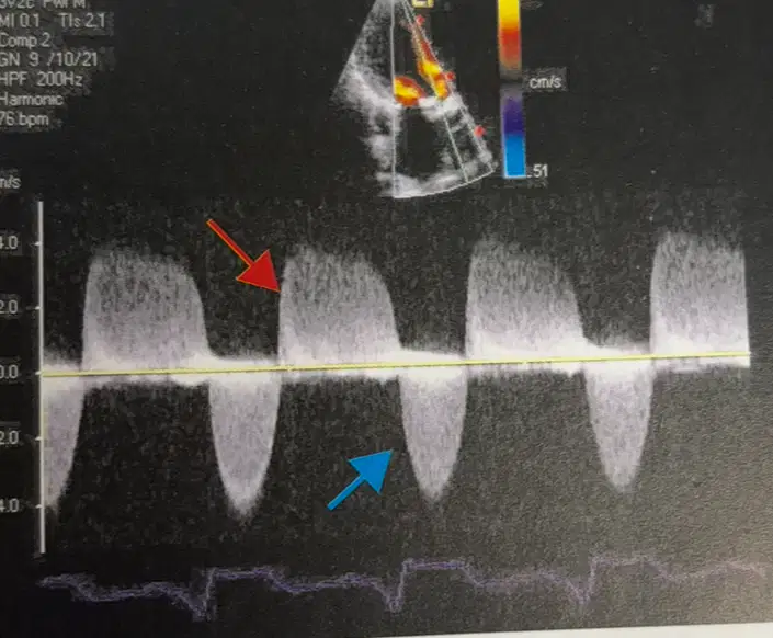
Bi directional spectral doppler tracing. Flow toward the transducer is (a) the baseline
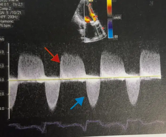
Bi directional spectral flow. Flow away from the transducer is (a) the baseline
(a) is a commonly used signal processing technique for bi directional doppler
Requires two crystals in the transducer
(a)
It has the ability to measure very high velocities
(a)
CW doppler: Exact location of the moving blood cells can not be determined. This is called (a)
Simultaneous anatomic imaging and doppler is called - (a)
Do Not use backing material
(a)
In a pulsed wave Doppler, how many crystals are necessary?
1
2
3
4
Range resolution, range specificity, freedom from range ambiguity artifact is great advantages of (a)
With (a) the sonographer positions a small marker, called sample volume or gate
Aliasing is a disadvantage of (a)
Regrading aliasing, the top of the image is (a)
Aliasing appears on the (a) of the spectrum
True or false: aliasing occurs only with pulsed doppler, never CW doppler
True
False
Aliasing occurs when the sampling rate is too (a) in comparison to the measured blood velocities
(a) = PRF/2
Is the highest doppler frequency or velocity that can be measured without the appearance of aliasing
(a)
When the sample volume is deep, the PRF is low and the nyquist limit is (a)
Deeper sample volumes have a lower PRF and creates (a)
When the sample volume is shallow, the PRF is high, velocity is sampled many times per second, and the nyquist limit is (a)
(a) Nyquist limit accurately measures the velocities without aliasing
Reduce doppler shift, raise the nyquist limit
(a)
(a) frequency transducers create more aliasing
Adjust scale to its maximum
(a)
2. Select a new ultrasonic view with a shallower sample volume
(a)
3. Select a new ultrasonic view with a shallower sample volume
(a)
3. Select a lower frequency transducer
(a)
4. Use baseline shift
(a)
5. Use CW doppler
(a)
Adjusting scale (a) nyquist limit
Shallower view, (a) nyquist limit
Lower frequency transducer, (a) doppler shift
Zero baseline shift, (a) remains but display more appealing
CW doppler. (a) aliases but range ambiguity
Range resolution, sample volume, sample volume, limited maximum velocity, aliasing
(a)
Range ambiguity, region of overlap, unlimited maximum velocity, no aliasing
(a)
Blood moving at a velocity of 2 m/s creates a doppler frequency of 3,000 Hz. What will the doppler shift be if the velocity increases to 4 m/s?
3,000 Hz
2 Hz
1500 Hz
6000 Hz
When using a 4MHz transducer, a doppler shift of 3,000 Hz is recorded. What will the doppler shift be when a 2MHz is used?
3,000 Hz
2 Hz
1500 Hz
6000 Hz
An 8 MHz transducer with a PRF of 5,000 Hz measures a doppler shift of 7 KHz. The study repeated with a 4MHz transducer. What doppler shift will be measured
3.5Hz
2KHz
3.5dB
3,500 Hz
Found in veins or slower-moving arterial blood.
Doppler shifts: 100 Hz to 1 kHz
(a)
Common in medium-sized arteries, such as renal or carotid arteries.
Doppler shifts: 1 kHz to 5 kHz
(a)
Found in high-speed arterial blood flow, such as in the aorta or stenotic vessels.
Doppler shifts: 5 kHz to 10 kHz or higher
(a)
Higher ultrasound frequencies (e.g., 5-10 MHz) used in superficial structures lead to (a) Doppler shifts.
Lower ultrasound frequencies (e.g., 2-5 MHz) used in deeper structures result in (a) Doppler shifts
A typical Doppler shift in the (a) can range from 1 kHz to 5 kHz, depending on the blood flow velocity and the ultrasound frequency used
A Doppler shift in the (a) might be in the range of 100 Hz to 1 kHz, due to slower blood flow velocities.
If the PRF is set at 10 kHz, the Nyquist limit is 5 kHz. If the Doppler shift exceeds 5 kHz, the system incorrectly interprets the frequency causing (a)
Involves mixing the received signal with two reference signals that are 90 degrees out of phase with each other (I/Q demodulation).
Provides information about both the magnitude and direction of the Doppler shift
(a)
Common in spectral Doppler.
Involves rectifying the Doppler signal to produce a positive-only signal and then applying a low-pass filter to extract the envelope of the signal, which represents the amplitude variations
(a)
Combines B-mode imaging (grayscale) with spectral Doppler to provide anatomical and flow information simultaneously
(a)
The reflected Doppler signals are processed using (a) to create a spectrum displaying velocities (frequency shifts) on the vertical axis and time on the horizontal axis. The intensity of the signal represents the number of blood cells moving at each velocity.

