wayground logo

Free Printable Worksheets

Font size

S
M
L
XL
Worksheets

BIO160 A&P Chapter 4-6 Study Guide

Total questions: 110

Worksheet time: 2hrs 50mins

Name
Class
Date
1.

What is the cutaneous membrane made of?

a)

Simple squamous epithelium and loose connective tissue

b)

Stratified squamous and dense connective tissue

c)

Transitional epithelium and adipose tissue

d)

Cuboidal epithelium and cartilage

2.

What is the mucous membrane made of?

a)

Dense connective tissue and keratinized epithelium

b)

Cartilage and cubodial epithelium

c)

Adipose tissue and transitional epithelium

d)

Epithelium and loose connective tissue

3.

The serous membrane is primarily made up of:

a)

Simple squamous epithelium and areolar connective tissue

b)

Transitional epithelium and dense connective tissue

c)

Stratified squamous epithelium and adipose tissue

d)

Cubodial epithelium and cartilage

4.

Where is the cutaneous membrane (skin) located?

a)

Lining the abdominal cavity

b)

Covering the ends of bones in joints

c)

Lining the respiratory and digestive tracts

d)

Covering the outside of the body

5.

Where is the mucous membrane located?

a)

Covering the outside of the body

b)

Lining closed ventral body cavities like thoracic and abdominal cavities

c)

Surrounding movable joints

d)

Lining body cavities that open to the exterior like the digestive, respiratory and urinary tracts

6.

Where is the serous membrane located?

a)

Lining cavities that open up to the exterior like the digestive, respiratory and urinary tract

b)

Covering the outside of the body

c)

Lining closed ventral body cavities, such as the thoracic and abdominal cavities

d)

Surrounding synovial joints like the knee and elbow

7.

What is the name of the organ system that consist of your skin and all its appendages?

a)
Skeletal system
b)
Muscular system
c)
Nervous system
d)
Integumentary system
8.

Which of the following is NOT a function of the integumentary system?

a)

Protection from injury and pathogens

b)

Vitamin D synthesis

c)

Blood cell production (hematopoisis)

d)

Sensory reception for touch, pressure and pain

9.

Where is the epidermis located and made of?

a)

Outermost layer of skin; keratinized stratified squamous epithelium

b)

Inner layer of skin; loose connective tissue

c)

Lining of joint cavities; specialized connective tissue

d)

Lining of the digestive tract; simple columnar epithelium

10.

Where is the dermis and what is it made of?

a)
The dermis is located above the epidermis and is made of muscle tissue.
b)
The dermis is found on the surface of the skin and consists of fat cells.
c)
The dermis is located within the bones and is made of cartilage.
d)

The dermis is located beneath the epidermis and is made of connective tissue

11.

Where is the hypodermis what is it made of?

a)
The hypodermis is situated on the surface of the skin and is composed of bone cells.
b)
The hypodermis is located beneath the dermis and is made of loose connective tissue and fat cells.
c)
The hypodermis is located within the dermis and consists of dense connective tissue.
d)
The hypodermis is found above the epidermis and is made of muscle tissue.
12.

What is the role of keratinocytes?

a)
Keratinocytes are involved in the regulation of blood flow in the skin.
b)
Keratinocytes are responsible for producing melanin.
c)
Keratinocytes help in the absorption of nutrients in the skin.
d)
Keratinocytes play a crucial role in forming the skin barrier and producing keratin.
13.

What is the role of Melanocytes?

a)
The role of Melanocytes is to produce melanin, which gives color to the skin and protects against UV radiation.
b)
Melanocytes are involved in the production of red blood cells.
c)
Melanocytes help in the digestion of food.
d)
Melanocytes are responsible for producing hormones in the body.
14.

What is the role Dendritic cells?

a)
Dendritic cells produce antibodies to fight infections.
b)
Dendritic cells activate T cells and help initiate immune responses.
c)
Dendritic cells are responsible for blood clotting.
d)
Dendritic cells primarily function as red blood cell producers.
15.

Which of the following lists the layers of epidermis in order from deepest to most superficial?

a)
stratum basale, stratum spinosum, stratum granulosum, stratum lucidum, stratum corneum
b)
stratum lucidum, stratum granulosum, stratum basale, stratum corneum, stratum spinosum
c)
stratum granulosum, stratum corneum, stratum lucidum, stratum basale, stratum spinosum
d)
stratum corneum, stratum lucidum, stratum granulosum, stratum spinosum, stratum basale
16.

Which of the epidermal layer is primarily responsible for continuous cell division to replace lost skin cells?

a)
Stratum lucidum
b)
Stratum granulosum
c)
Stratum basale
d)
Stratum corneum
17.

Which layer of epidermis is known for "spiny" cells connected by desmosomes and provides strength to the skin?

a)
stratum granulosum
b)
stratum spinosum
c)
stratum corneum
d)
stratum lucidum
18.

Which epidermal layer contains keratohyalin granules that help form tough, protective outer skin?

a)
Stratum basale
b)
Stratum granulosum
c)
Stratum corneum
d)
Stratum lucidum
19.

Which layer is found only in thick hairless skin (like palm and soles) and appears clear under a microscope?

a)
stratum granulosum
b)
stratum lucidum
c)
stratum corneum
d)
stratum spinosum
20.

Which epidermal layer forms the outermost, fully keratinized protective barrier of the skin?

a)
stratum corneum
b)
stratum spinosum
c)
stratum lucidum
d)
stratum granulosum
21.

Which epidermis layers have poor have poor nutrient reach?

a)
Stratum lucidum and stratum basale
b)
Stratum basale and stratum corneum
c)

Stratum corneum and stratum lucidum and stratnum granulosum

d)

Stratum granulosum, stratum basale

22.

What structure/layer is responsible for fingerprint pattern?

a)
Stratum corneum
b)
Dermal papillae
c)
Subcutaneous layer
d)
Epidermis
23.

what is the difference between the papillary and reticular layer of the dermis?

a)
The papillary layer is thin and contains loose connective tissue, while the reticular layer is thicker and made of dense irregular connective tissue.
b)
Both layers are identical in structure and function, with no significant differences.
c)
The papillary layer is thicker and made of dense connective tissue, while the reticular layer is thin and contains loose connective tissue.
d)
The papillary layer is found only in the palms and soles, while the reticular layer is present everywhere else.
24.

What does sebaceous glands produce?

a)
Hormones
b)
Sweat
c)
Enzymes
d)
Sebum
25.

What are the difference between sweat and sebaceous glands?

a)
Sebaceous glands help in temperature regulation.
b)
Sweat glands are found only on the face.
c)
Sweat glands produce sweat for temperature regulation, while sebaceous glands produce sebum for skin lubrication.
d)
Sweat glands produce oil for hair growth.
26.

Where are eccrine sweat glands located?

a)
Eccrine sweat glands are only found in the armpits.
b)
Eccrine sweat glands are primarily found in the stomach area.
c)
Eccrine sweat glands are located all over the body, especially on the palms, soles, and forehead.
d)
Eccrine sweat glands are located exclusively on the face.
27.

What is the role of eccrine sweat glands?

a)
To provide nutrients to hair follicles.
b)
The role of eccrine sweat glands is to regulate body temperature by producing sweat.
c)
To produce oil for skin hydration.
d)
To filter toxins from the blood.
28.

Where are apocrine sweat glands located?

a)
Armpits, groin, and around the nipples.
b)
Face and neck
c)
Feet and back
d)
Forehead and palms
29.

What is the role of apocrine sweat glands?

a)
The role of apocrine sweat glands is to produce a thicker secretion that may contribute to pheromone release and body odor.
b)
To produce a watery secretion for temperature regulation.
c)
To secrete oil for skin hydration.
d)
To filter toxins from the bloodstream.
30.

What is acne and what does it result from?

a)
Acne is a skin condition resulting from clogged hair follicles due to oil and dead skin cells.
b)
Acne is a viral infection that affects the hair follicles.
c)
Acne is a result of excessive sun exposure and dehydration.
d)
Acne is a type of bacteria that causes skin infections.
31.

What area/zone of the follicle is responsible for hair formation?

a)
Hair shaft
b)
Dermal papilla
c)
Outer root sheath
d)
Hair bulb
32.

What is the function of arrector pili?

a)
To produce sweat for cooling the body.
b)
To protect the scalp from UV rays.
c)
The function of arrector pili is to cause hair to stand up and assist in thermoregulation.
d)
To provide nutrients to hair follicles.
33.

What is a nail formed from and what is its function?

a)
Nails are formed from keratin and their function is to protect the tips of fingers and toes.
b)
Nails are composed of cartilage and serve as a sensory organ.
c)
Nails are formed from muscle tissue and assist in movement.
d)
Nails are made of bone and help in gripping objects.
34.

What is a nail bed?

a)
The skin beneath the nail plate that supports the nail.
b)
The part of the nail that grows outwards.
c)
The area where nail polish is applied.
d)
The cuticle surrounding the nail.
35.

What can burns be caused by?

a)
Food, beverages, air
b)
Water, ice, steam
c)
Dust, dirt, sand
d)

Heat, chemicals, electricity, radiation

36.

What is the rule of nines?

a)
A method for calculating body weight based on height.
b)
A guideline for determining the severity of fractures.
c)
A technique for assessing heart rate during exercise.
d)
The rule of nines is a method for estimating burn area by dividing the body into sections, each representing 9% of total body surface area.
37.

What is skin cancer?

a)
A rare disease affecting internal organs.
b)
Skin cancer is a type of cancer that originates in the skin cells.
c)
A condition caused by sunburn only.
d)
A type of skin treatment for acne.
38.

What is the difference between basal cell carcinoma, squamous cell carcinoma and malignant melanoma?

a)
Basal cell carcinoma is the most aggressive type of skin cancer.
b)
Malignant melanoma is the least serious and has a low risk of metastasis.
c)
Basal cell carcinoma is slow-growing and rarely metastasizes, squamous cell carcinoma is more aggressive with a higher metastasis risk, and malignant melanoma is the most serious, capable of rapid spread.
d)
Squamous cell carcinoma is slow-growing and rarely spreads.
39.

How is the ABCDE rule used to recognize melanoma?

a)
The ABCDE rule focuses on the size and shape of moles only.
b)
The ABCDE rule is used to recognize melanoma by assessing asymmetry, border irregularity, color variation, diameter, and evolution of moles.
c)
The ABCDE rule assesses the smell and feel of moles.
d)
The ABCDE rule is used to measure skin temperature and texture.
40.

What part of skeleton belong to the axial skeleton?

a)
Pelvis
b)
Humerus
c)
Tibia
d)
Skull, vertebral column, rib cage
41.

What parts belong to the appendicular skeleton?

a)
Cranial bones, thoracic cage, spine
b)
Shoulder girdle, upper limbs, pelvic girdle, lower limbs.
c)
Jaw, teeth, facial bones
d)
Skull, vertebral column, ribs
42.

What are five functions of bones?

a)
Energy production
b)
1. Support and structure 2. Protection of organs 3. Facilitation of movement 4. Mineral storage 5. Blood cell production
c)
Temperature control
d)
Hormone regulation
43.

What's the difference between compact and spongy bone?
a)
Compact bone is found only in the skull, while spongy bone is in the limbs.
b)
Spongy bone is denser than compact bone and provides more strength.
c)
Compact bone contains more blood vessels than spongy bone.
d)
Compact bone is dense and strong, while spongy bone is lighter and contains trabecular structures.
44.

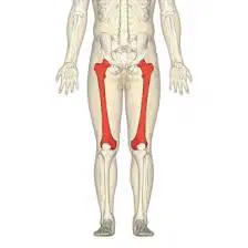
What type of bone is this (femur)?

a)

Long bone

b)

Flat bone

c)

Short bone

d)

Irregular bone

45.

What type of bone is this (scapula)?

a)
Short bone
b)
Irregular bone
c)
Long bone
d)
Flat bone
46.

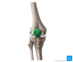
What type of bone is this (patella)?

a)

small bone

b)
flat bone
c)
long bone
d)
irregular bone
47.

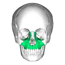
What type of bone is this (maxilla)?

a)

Small bone

b)
Irregular bone
c)
Flat bone
d)
Long bone
48.

What is the function epiphyseal plate?

a)
The function of the epiphyseal plate is to facilitate bone growth in length.
b)
To connect muscles to bones.
c)
To provide nutrients to the bone.
d)
To store fat for energy.
49.

Describe the macroscopic structure of a long bone.

a)
A long bone is made up of only cartilage and lacks a periosteum.
b)
A long bone consists only of the diaphysis and epiphyses.
c)
A long bone has a diaphysis, epiphyses, metaphysis, medullary cavity, and periosteum.
d)
A long bone has a hollow structure with no medullary cavity.
50.

What is the difference between red and yellow marrow? Where is each found?

a)
Yellow marrow is found in all bones of children; red marrow stores fat.
b)
Red marrow is found only in adults; yellow marrow produces blood cells.
c)
Red marrow produces blood cells; yellow marrow stores fat. Red marrow is found in most bones of children and in specific bones of adults.
d)
Red marrow is primarily found in the brain; yellow marrow is in the heart.
51.

Describe the microscopic structure of compact bone.

a)
Trabecular bone structure
b)
Bone marrow composition
c)
Cartilage cell arrangement
d)
The microscopic structure of compact bone includes osteons, concentric lamellae, Haversian canals, lacunae, osteocytes, and canaliculi.
52.

What does the central canal of each osteon contain?

a)
Lymphatic vessels
b)
Cartilage and fat
c)
Bone marrow cells
d)
Blood vessels and nerves
53.

What doe canaliculi contain and what is their function?

a)
Canaliculi contain blood vessels and their function is to transport oxygen in bone tissue.
b)
Canaliculi contain fat cells and their function is to store energy in bone tissue.
c)
Canaliculi contain extensions of osteocytes and their function is to facilitate communication and nutrient exchange in bone tissue.
d)
Canaliculi contain nerve endings and their function is to transmit pain signals in bone tissue.
54.

What tissue "models" of bones that are replaced with bone tissue in growing fetus?

a)
Muscle tissue
b)
Adipose tissue
c)
Epithelial tissue
d)
Cartilage
55.

What is the name of the bone formation process?

a)
Calcification
b)
Chondrogenesis
c)
Mineralization
d)
Ossification
56.

What role do osteoblast play in bone formation?

a)
Osteoblasts break down bone tissue.
b)
Osteoblasts play a crucial role in synthesizing and mineralizing bone.
c)
Osteoblasts are responsible for muscle contraction.
d)
Osteoblasts transport nutrients to bone cells.
57.

What is the difference between osteoblast and osteoclast?

a)
Osteoblasts and osteoclasts are the same type of cell.
b)
Osteoblasts break down bone, while osteoclasts build bone.
c)
Osteoblasts build bone, while osteoclasts break down bone.
d)
Osteoblasts are found in cartilage, while osteoclasts are found in muscle.
58.

What two factors are bones remodeled in response to?

a)
Nutritional intake and environmental factors
b)
Genetic predisposition and age
c)
Injury recovery and physical therapy
d)
Mechanical stress and hormonal changes
59.

What are the four major events of bone fracture repair in sequence

a)
Hematoma formation, Fibrocartilaginous callus formation, Bony callus formation, Bone remodeling
b)
Cartilage degeneration, Bone growth, Joint fusion, Muscle repair
c)
Inflammation, Osteoporosis, Ligament healing, Tendon repair
d)
Hematoma absorption, Cartilage calcification, Bone necrosis, Joint dislocation
60.

What two critical minerals are stored in bone?

a)
Zinc and copper
b)
Calcium and phosphorus
c)
Iron and magnesium
d)
Sodium and potassium
61.

Identify the following bones of the skull

a)

Frontal, parietal, temporal, occipital

b)
clavicle
c)
mandible
d)
maxilla
62.

Identify the Facial bones

a)
Frontal bone
b)
Temporal bone
c)
Occipital bone
d)

Maxilla, Mandible, Zygomatic bones, Nasal bones, Palatine bones

63.

What common name is coccyx known by?

a)
spine
b)
tailbone
c)
hipbone
d)
kneecap
64.

Difference between "true ribs" and "false ribs"

a)
True ribs attach directly to the sternum; false ribs do not.
b)
False ribs attach to the spine; true ribs do not.
c)
True ribs are found only in the upper chest.
d)
True ribs are longer than false ribs.
65.

How many pairs of ribs do humans have?

a)
10 pairs
b)
14 pairs
c)
8 pairs
d)
12 pairs
66.

Identify bones in pectoral girdle

a)
Clavicle and Scapula
b)
Femur and Tibia
c)
Pelvis and Sternum
d)
Humerus and Radius
67.

Identify bones in upper limbs

a)
Tibia
b)
Pelvis
c)
Femur
d)

Humerus, Radius, Ulna,

68.

Identify bones in hand

a)
Clavicle
b)
Scapula
c)
Carpals, Metacarpals, Phalanges
d)
Tarsals
69.

Identify bones in pelvic girdle

a)
Humerus
b)
Femur
c)
Tibia
d)
Ilium, Ischium, Pubis
70.

Difference between "true pelvis" and "false pelvis"

a)
The true pelvis is involved in childbirth, while the false pelvis supports abdominal organs.
b)
The false pelvis is involved in childbirth, while the true pelvis supports abdominal organs.
c)
The true pelvis is larger than the false pelvis.
d)
The true pelvis is only found in males, while the false pelvis is found in females.
71.

Identify bones thigh

a)
Tibia
b)
Humerus
c)
Femur
d)
Radius
72.

Identify bones in knee

a)

Patella

b)
Scapula
c)
Radius
d)
Humerus
73.

Identify bones in leg

a)
radius
b)

tibia, fibula

c)
humerus
d)
ulna
74.

What are joints?

a)
Joints are the connections between bones that allow for movement.
b)
Joints are the muscles that connect to bones.
c)
Joints are the types of cells found in bones.
d)
Joints are the fluid-filled sacs in the body.
75.

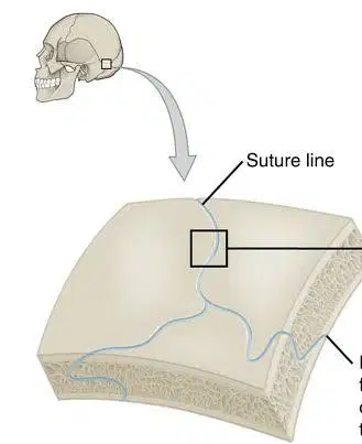
Difference between synarthrosis, amphiarthrosis and diarthrosis?
a)
Synarthrosis: freely movable; Amphiarthrosis: immovable; Diarthrosis: slightly movable.
b)
Synarthrosis: slightly movable; Amphiarthrosis: freely movable; Diarthrosis: immovable.
c)
Synarthrosis: immovable; Amphiarthrosis: slightly movable; Diarthrosis: freely movable.
d)
Synarthrosis: movable; Amphiarthrosis: immovable; Diarthrosis: slightly movable.
76.

What type of joint is this?

a)

Fibrous

b)

cartilaginous

c)

synovial

77.

What type of joint is this

a)

fibrous

b)

cartilaginous

c)

synovial

78.

What type of joint is this?

a)

Fibrous

b)

Cartilaginous

c)

Synovial

79.

Describe the four main feature of synovial joints?
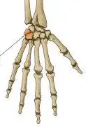
a)
The four main features of synovial joints are articular cartilage, synovial cavity, synovial fluid, and joint capsule.
b)
Synovial fluid, bone marrow, joint capsule, and periosteum
c)
Articular ligaments, synovial membrane, joint cavity, and cartilage
d)
Articular cartilage, synovial cavity, muscle fibers, and tendon sheath
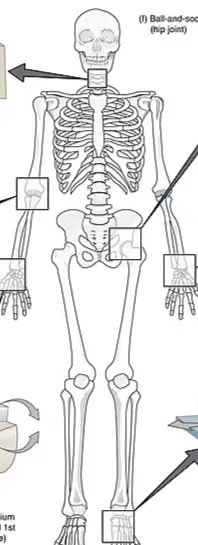
80.

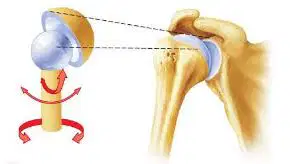
What classification does this synovial joint fall under?

a)

Plane joint

b)

Hinge joint

c)

Condylar Joint

d)

Ball and Socket Joint

81.

What classification does this synovial joint fall under?

a)
Ball-and-socket joint
b)

Plane joint

c)
Hinge joint
d)

Condylar Joint

82.

What classification does this synovial joint fall under?

a)

Plane joint

b)
Hinge joint
c)
Ball-and-socket joint
d)

Condylar joint

83.

What classification does this synovial joint fall under?

a)
Hinge joint
b)

Plane joint

c)

Condylar joint

d)
Ball-and-socket joint
84.

What is the essential function of a muscle

a)
To provide structural support to bones.
b)
The essential function of a muscle is to facilitate movement.
c)
To produce hormones for growth.
d)
To store energy for the body.
85.

Difference between skeletal, cardiac and smooth muscle?

a)
Skeletal muscle: non-striated, involuntary
b)
Cardiac muscle: striated, voluntary
c)
Smooth muscle: striated, voluntary
d)
Skeletal muscle: striated, voluntary; Cardiac muscle: striated, involuntary; Smooth muscle: non-striated, involuntary.
86.

Why are muscle cells called "muscle fiber? in skeletal and smooth muscle?

a)
Muscle cells are called muscle fibers because they are found only in the heart.
b)
Muscle cells are called muscle fibers due to their round shape.
c)
Muscle cells are called muscle fibers because they are primarily made of fat.
d)
Muscle cells are called muscle fibers due to their elongated, thread-like shape.
87.

Though the essential function of a muscle is to shorten, and therein to produce movement what additional functions to muscles have?

a)
Muscles primarily store energy for later use.
b)
Muscles are responsible for producing hormones.
c)
Muscles help in the digestion of food.
d)
Muscles maintain posture, stabilize joints, generate heat, and aid in blood circulation.
88.

What is a sarcomere?

a)
A nerve cell that transmits signals.
b)
A protein that helps in blood clotting.
c)
The basic unit of muscle contraction in striated muscle.
d)
A type of connective tissue in the body.
89.

How do myosin (thick filaments) and actin (thin filaments) interact to produce movement/shortening during muscle contraction?

a)
Myosin heads release actin, causing muscle to lengthen.
b)
Actin filaments push myosin heads away, preventing contraction.
c)
Myosin and actin do not interact during muscle contraction.
d)
Myosin heads bind to actin, pull actin filaments, and shorten the muscle during contraction.
90.

What is myosin and actin comprised of?

a)
Myosin and actin are comprised of proteins made of amino acids.
b)
Myosin and actin consist of nucleic acids.
c)
Myosin and actin are composed of lipids.
d)
Myosin and actin are made of carbohydrates.
91.

What is the sarcoplasmic reticulum and what is its function?

a)
The sarcoplasmic reticulum is a specialized organelle in muscle cells that stores and regulates calcium ions, crucial for muscle contraction and relaxation.
b)
A structure that transports oxygen in muscle cells
c)
An organelle that produces proteins for muscle growth
d)
A type of muscle fiber that generates energy
92.

What is the role of calcium ions in skeletal muscle contraction?

a)
Calcium ions are responsible for the relaxation of muscle fibers during contraction.
b)
Calcium ions facilitate muscle contraction by enabling the interaction between actin and myosin.
c)
Calcium ions inhibit muscle contraction by blocking actin and myosin interaction.
d)
Calcium ions are only involved in nerve signal transmission, not muscle contraction.
93.

What kind of molecule is acetylcholine (ACh)

a)
Acetylcholine is a carbohydrate.
b)
Acetylcholine is a protein.
c)
Acetylcholine is a nucleic acid.
d)

Acetylcholine is an neurotransmitter

94.

What are the three major pathways which muscle regenerates ATP to power muscle contractions?

a)
Lactic acid fermentation
b)
Beta-oxidation
c)
Phosphagen system, Anaerobic glycolysis, Aerobic respiration
d)
Protein catabolism
95.

When sufficient ACh binds to the muscle fiber’s sarcolemma (plasma membrane), Na+ diffuses in (more than K+ diffuses out) and this leads to an ‘action potential’ (i.e., an electrical stimulus). This signal is propagated over the sarcolemma of the plasma membrane which causes a release of ­what ion into the muscle fiber? What does the influx of this ion enable?


a)
Calcium ions (Ca2+)
b)
Chloride ions (Cl-)
c)
Potassium ions (K+)
d)
Sodium ions (Na+)
96.

Ultimately, what effect does acetylcholine (ACh) have on the skeletal muscle fiber?

a)
Acetylcholine causes relaxation of skeletal muscle fibers.
b)
Acetylcholine inhibits muscle contraction in skeletal muscle fibers.
c)
Acetylcholine causes muscle contraction in skeletal muscle fibers.
d)
Acetylcholine has no effect on skeletal muscle fibers.
97.

What is the role of creatine phosphate?

a)
Creatine phosphate is a hormone that regulates metabolism.
b)
Creatine phosphate acts as an energy reservoir in muscle cells, replenishing ATP.
c)
Creatine phosphate is primarily used for fat storage in the body.
d)
Creatine phosphate is a type of protein that builds muscle mass.
98.

As it pertains to the aerobic pathway vs anaerobic glycolysis, is the aerobic pathway relatively fast or slow?

a)
very fast
b)
moderate
c)
slow
d)
instantaneous
99.

When does the anaerobic pathway kick in and why is its role significant if it yields relatively low amounts of ATP per glucose?


a)
The anaerobic pathway is activated during high oxygen levels.
b)
The anaerobic pathway kicks in during low oxygen conditions, and it is significant for energy production when aerobic respiration is not possible.
c)
It is significant because it produces more ATP than aerobic respiration.
d)
The anaerobic pathway is only used in muscle cells during rest.
100.

  Distinguish between the origin and insertion of a muscle.

a)
The origin and insertion are interchangeable terms for muscle attachment points.
b)
The origin is the point where the muscle contracts, while the insertion is the point where it relaxes.
c)
The origin is the fixed point of attachment of a muscle, while the insertion is the movable point that the muscle pulls towards the origin.
d)
The origin is always located at the distal end of the muscle, while the insertion is at the proximal end.
101.

What does it mean for a muscle to be a ‘prime mover?’

a)
A prime mover is a muscle that is always active regardless of the movement.
b)
A prime mover is a muscle that stabilizes a joint during movement.
c)
A prime mover is a muscle that assists in a movement.
d)
A prime mover is the main muscle responsible for a specific movement.
102.
The muscle tissue shown is called ______.
a)
Skeletal
b)
Cardiac
c)
Smooth
103.
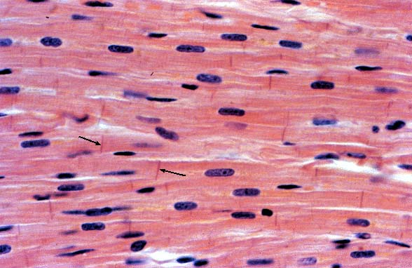
What tissue is this? (The arrows help you.)
a)
Cardiac muscle
b)
Skeletal muscle
c)
Smooth muscle
d)
Nervous tissue
104.

  Identify and know the primary movements of the following muscles of the head and neck:

a. Facial muscles

 i. Frontalis,

orbicularis oculi,

orbicularis oris,

buccinator,

masseter,

temporalis


a)
Frontalis (closes eyelids)
b)
Frontalis (raises eyebrows), orbicularis oculi (closes eyelids), orbicularis oris (purses lips), buccinator (compresses cheeks), masseter (elevates mandible), temporalis (elevates and retracts mandible)
c)
buccinator (elevates mandible)
d)
masseter (purses lips)
105.

  Identify and know the primary movements of the following muscles of the upper limbs

    i. Biceps brachii,

triceps brachii,

brachialis,

brachioradialis


a)
Biceps brachii: flexes elbow and supinates forearm; Triceps brachii: extends elbow; Brachialis: flexes elbow; Brachioradialis: assists in elbow flexion.
b)
Biceps brachii: flexes shoulder; Triceps brachii: flexes wrist; Brachialis: extends shoulder; Brachioradialis: flexes fingers.
c)
Biceps brachii: extends elbow; Triceps brachii: flexes elbow; Brachialis: extends elbow; Brachioradialis: assists in shoulder flexion.
d)
Biceps brachii: rotates forearm; Triceps brachii: stabilizes shoulder; Brachialis: rotates wrist; Brachioradialis: extends wrist.
106.

Identify the primary movements for the lower limbs

Lower limbs

  i. Gluteus maximus,

hamstring group (biceps femoris, semimembranosus, semitendinosus),

quadriceps femoris group,

gastrocnemius


a)
Hip abduction
b)
Knee rotation
c)
Hip extension, knee flexion, knee extension, plantarflexion
d)
Hip flexion
107.

  Identify and know the primary movements of the following muscles of the trunk:

 i. Pectoralis major,

intercostal muscles,

rectus abdominis,

trapezius,

latissimus dorsi,

deltoid


a)
Intercostal muscles: only rib elevation
b)
Pectoralis major: flexion, adduction, medial rotation; Intercostal muscles: rib elevation/depression; Rectus abdominis: flexion of the vertebral column; Trapezius: elevation, retraction, rotation of scapula; Latissimus dorsi: extension, adduction, medial rotation; Deltoid: abduction, flexion, extension of the arm.
c)
Rectus abdominis: rotation of the vertebral column
d)
Pectoralis major: extension, abduction, lateral rotation
108.

Describe muscular dystrophy

a)
Muscular dystrophy is a type of cancer that targets muscle tissue.
b)
Muscular dystrophy is a viral infection that causes muscle spasms.
c)
Muscular dystrophy is an autoimmune disease that affects the heart.
d)
Muscular dystrophy is a genetic disorder that causes progressive muscle weakness and degeneration.
109.

When a nerve impulse reaches the end of a neuron (colored in yellow), calcium enters the neuron. What event does the influx of calcium trigger?


a)
Inhibition of nerve signals
b)
Activation of muscle fibers
c)
Degradation of neurotransmitters
d)
Release of neurotransmitters
110.

Describe what is found in dark ‘A’ bands vs. light ‘I’ bands and be able to recognize these bands in an image of a sarcomere.

a)
Light 'I' bands contain both thick and thin filaments, while dark 'A' bands contain only thin filaments.
b)
Dark 'A' bands contain thick filaments (myosin) and overlapping thin filaments, while light 'I' bands contain only thin filaments (actin).
c)
Dark 'A' bands are lighter than light 'I' bands and contain no filaments.
d)
Dark 'A' bands contain only thin filaments (actin) and light 'I' bands contain thick filaments (myosin).