
Font size
 Worksheets
Joints and Broken Bones Test
Total questions: 96
Worksheet time: 1hrs 3mins
The majority of the joints of the body are:
Which type of joints are the suture joints in the skull?
Cartilaginous
Synovial
Fibrous
Cranial
The majority of the joints of the body are:
Which type of joints are the suture joints in the skull?
Cartilaginous
Synovial
Fibrous
Cranial
Why is the shoulder the joint which is most likely to be dislocated?
Because it is more mobile than it is stable
Because it does not contain bursae
Because the humeral head is often the site of fractures
Because it experience the most trauma of all of the synovial joints
Skull
Swing

Where is the pivot joint located ?
Neck
Knee
Thumb

Where is the hinge joint located
neck
elbow
knee

Where is the plane joint located?
Elbow
Between tarsal bones
Between c1 and c2 vertebrae

What type of joint is displayed in the picture?
Ellipsoid joint
Pivot joint
Synovial joint

What type of joint is displayed in the picture?
Pivot joint
Ellipsoid joint
Hinge joint

What type of joint is displayed in the picture?
Saddle joint
Ellipsoid joint
Pivot joint

What joint is displayed in picture
Hinge Joint
Saddle joint
Ball and socket joint

What joint is displayed in picture?
hinge joint
plane joint
pivot joint
Which is an example of a hinge joint
knee and elbow
shoulder and hip
neck
ankle and wrist


The skull is an example of what type of joint?
Cartilaginous (amphiarthrosis)
Synovial (diarthrosis)
Fibrous (synarthrosis)


Bones that have complex shapes are called...
Flat bones
Irregular bones
Sesamoid bones
Long bones
What is the purpose of synovial fluid in synovial joints?
To hold the joint together
To lubricate the joint
To prevent bone rubbing
To provide flexibility to the joint

ball-and-socket joint
hinge joint
pivot joit
planar joint

ball-and-socket joint
hinge joint
pivot joint
planar joint
gliding movement ONLY
ball-and-socket joint
hinge joint
pivot joint
planar joint
What connects bone to bone?
Tendons
Ligaments
Joint Capsule
Bursae
A clear and slippery liquid that lubricates the joint and stops the bones rubbing together, is ...
Bursae
Cartilage
Synovial Fluid
Joint Capsule

S-shaped separation of the bone. Occurs when the foot is firmly planted and body is suddenly rotated.
Greenstick Fracture
Oblique Fracture
Spiral Fracture
Transverse Fracture

Occurs in a straight line at right angle to the shaft of the bone. Usually caused by a direct blow.
Greenstick Fracture
Oblique Fracture
Spiral Fracture
Transverse Fracture

The xray shows a fracture that has bones broken into more than two pieces. What type of fracture is this?
Simple
Compound
Impacted
Comminuted
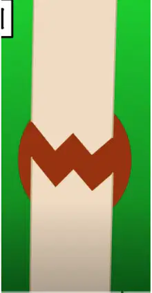
This is the first stage of bone healing.
Osteoclasts appear and take care of bone fragments.
Bleeding and then a blood clot is formed and it swells.
Phagocytes come and clean up the area by removing dead cells and any germs.
Chondroblast hold the broken ends together with a fibrocartilaginous tissue. It’s called a soft callus.
During this phase of bone healing the blood vessels in and around the bone are torn and bleeding occurs
Hematoma formation
Fibrocartilaginous callus formation
Bony callus formation
Remodeling
This is the stage in which collagen fibers start to span the break and connect the broken bones.
Hematoma formation
Fibrocartilaginous callus formation
Bony callus formation
Remodeling
At the end of this stage of bone healing the final structure resembles the original unbroken bone
Hematoma formation
Fibrocartilaginous callus formation
Bony callus formation
Remodeling
This acts as the body’s natural splint
Hematoma formation
Fibrocartilaginous callus formation
Bony callus formation
Remodeling
The initial even following a bone fracture is:
Hemorrhage and hematoma formation
Endochondral ossification
Remodeling
Ossification of internal callus
Formation of granulation tissue
Healing of a fracture, even after severe damage, depends on the survival of:
The circulatory supply and the cellular components of the Endosteum and Periosteum
The bone’s mineral strength and its resistance to stress
Osteoblasts within the spongy and compact bone
External callus which protects the fractured area

This cell lays down new bone and helps repair damage.
Osteoblast
Osteoclast
Osteocyte
Osteobuddies
The xray shows a fracture that has bones broken into more than two pieces. What type of fracture is this?
Simple
Compound
Impacted
Comminuted

What is happening in this stage of bone healing?
Bone is fractured
Hard callus forms, osteoblast cells create new bone
Remodeling, osteoclasts remove extra bone until its completely healed
Soft callus forms, chondroblast and fibroblasts produce the firbocartilaginous calus.

What is happening in this stage of bone healing?
Bone is fractured
Hard callus forms, osteoblast cells create new bone
Remodeling, osteoclasts remove extra bone until its completely healed
Soft callus forms, chondroblast and fibroblasts produce the fibrocartilaginous calus

What is happening in this stage of bone healing?
hematoma forms
Hard callus forms, osteoblast cells create new bone
Remodeling, osteoclasts remove extra bone until its completely healed
Soft callus forms, chondroblast and fibroblasts produce the fibrocartilaginous calus

What is happening in this stage of bone healing?
Bone is fractured
Hard callus forms, osteoblast cells create new bone
Remodeling, osteoclasts remove extra bone until its completely healed
Soft callus forms, chondroblast and fibroblasts produce the fibrocartilaginous calus

#2 - the "shaft" of the bone is known as the -
epiphysis
medullary cavity
diaphysis
articulate cartilage

#5 is made up of -
spongy bone
compact bone
disphysis bone
yellow marrow

#6, the outer lining of the bone is called the.
periosteum
spongy bone
epiphysis
compact bone

#7 Is the site of bone growth and is known as the
yellow marrow
periosteum
compact bone
epiphyseal plate

#9 contains
compact bone
spongy bone
yellow marrow
periosteum
This type of bone contains the medullary cavity.
Which of the following is filled with yellow marrow?
medullary cavity
cancellous bone
The wider ends of the long bone are anatomically referred to as the:

Identify 4, which covers both ends of the bone.
Cartilage
Proximal Epiphysis
Distal Epiphysis
Epiphyseal Line
Identify the thin plate labeled #6, which is also known commonly as the "growth plate".
Spongy Bone
Compact Bone
Articular Cartilage
Epiphyseal Line
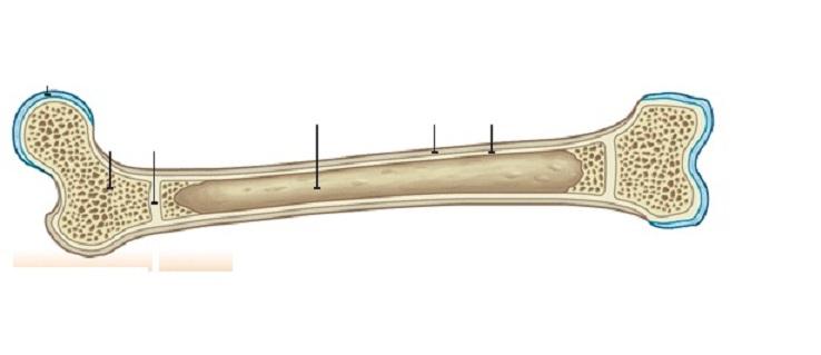

Yellow marrow is which letter?
E
A
H
J

What does I represent?
medullary canal
red marrow
diaphysis
periosteum

Diaphysis is which letter?
D
F
J
K
What are the 2 types of bone tissue found in a long bone?
spongy and compact
red and yellow
Which bone structure makes new blood cells?
Compact
Spongy
Red Marrow
Yellow Marrow
What is the function of yellow bone marrow?
To store energy as fat
To produce blood cells
To regulate blood calcium levels
To protect against glucose intolerance
Which type of bone are the femur and humerus?
Long bones
Flat bones
Short bones
Irregular bones
What is the function of compact bone?
To provide structural scaffolding for supporting weight.
To store energy as fat
To produce blood cells
To regulate blood calcium levels
What is the main function of bones in the human body?
Sensation, perception, and cognition.
Digestion, absorption, and elimination.
Support, protection, and structure
Movement, flexibility, and coordination.
What is the process called when a broken bone heals and reforms?
bone remodeling
bone regeneration
bone restructuring
bone reconstruction
What is the outer layer of a bone called?
periosteum
epiphysis
medullary cavity
endosteum
What is the function of osteoclasts in bone remodeling?
Bone formation
Bone growth
Bone resorption
Bone repair
What are the two main types of bone tissue?
dense bone and porous bone
hard bone and soft bone
compact bone and spongy bone
solid bone and hollow bone

The weight-bearing bone of the lower leg:
Femur
Tibia
Fibula
Fibia

This lower leg bone (red) is known as:
Femur
Tibia
Fibula
Tibula

The highlighted bones are called:
Tarsals
Metatarsals
Phalanges
Feets

