
 Worksheets
Muscle Review
Total questions: 123
Worksheet time: 2hrs 35mins
Which type of muscle tissue is voluntary?
Smooth
Cardiac
Skeletal
None
Which muscle tissue is found in the walls of hollow organs and blood vessels?
Skeletal
Smooth
Cardiac
None of the above
Which type of muscle tissue has striations and is involuntary?
Skeletal
Smooth
Cardiac
All of the above
Smooth muscle can best be described as:
Striated and voluntary
Striated and involuntary
Non-striated and involuntary
Non-striated and voluntary
Which muscle type contracts and relaxes rapidly when stimulated by a motor neuron?
Smooth
Cardiac
Skeletal
Visceral
Which muscle type contracts and relaxes slowly and can be self-exciting and rhythmic?
Skeletal
Smooth
Cardiac
Connective
Which type of muscle tissue forms a network of cells that contract as a unit?
Smooth
Cardiac
Skeletal
Voluntary
Which muscle type is self-exciting and rhythmic?
Cardiac and smooth
Skeletal only
Smooth only
Skeletal and cardiac
The ability of a muscle to respond to a stimulus is called:
Contractility
Extensibility
Excitability
Elasticity
The ability of a muscle to shorten or contract is called:
Extensibility
Elasticity
Excitability
Contractility
The ability of a muscle to stretch is called:
Excitability
Elasticity
Extensibility
Flexibility
The ability of a muscle to return to its original shape after stretching is:
Elasticity
Excitability
Extensibility
Contractility
Which of the following is NOT a function of the muscular system?
Movement
Heat production
Maintaining posture
Producing hormones
Which is a function of the muscular system?
Movement
Heat production
Maintaining posture
All of the above
The connective tissue that surrounds the entire skeletal muscle is the:
Perimysium
Endomysium
Epimysium
Sarcolemma
The connective tissue that surrounds bundles of muscle fibers (fascicles) is the:
Perimysium
Epimysium
Endomysium
Sarcolemma
The connective tissue that covers each individual muscle fiber is the:
Epimysium
Perimysium
Endomysium
Fascia
Skeletal muscles are attached to bones by:
Ligaments
Tendons
Cartilage
Fascia
Increasing the angle between two bones is called:
Flexion
Abduction
Extension
Adduction
Decreasing the angle between two bones is called:
Flexion
Extension
Adduction
Abduction
Movement of a bone away from the midline is:
Abduction
Adduction
Flexion
Rotation
Movement of a bone toward the midline is:
Abduction
Adduction
Extension
Rotation
Upward movement of a body part is:
Depression
Flexion
Elevation
Rotation
Downward movement of a body part is:
Depression
Extension
Rotation
Elevation
Movement that revolves a bone around an axis is:
Flexion
Rotation
Abduction
Elevation
The less movable end of a muscle is the:
Insertion
Origin
Agonist
Antagonist
The more movable end of a muscle is the:
Origin
Insertion
Agonist
Synergist
The muscle that performs the main action is called the:
Antagonist
Agonist (prime mover)
Synergist
Stabilizer
Muscles that assist the prime mover are:
Antagonists
Synergists
Origins
Insertions
Muscles that oppose an action are:
Agonists
Antagonists
Synergists
Flexors
During elbow flexion, which muscle acts as the antagonist?
Biceps brachii
Triceps brachii
Brachialis
Deltoid
An isotonic contraction involves:
Force generation without shortening
Muscle shortening with movement
No movement or tension
Only relaxation
An isometric contraction involves:
Muscle shortening
Muscle lengthening
Force generation without shortening
Relaxation
The smallest functional unit of a muscle fiber is the:
Sarcomere
Myofibril
Fascicle
Sarcolemma
During contraction, actin filaments:
Shorten
Slide over myosin filaments
Thicken
Remain stationary
During contraction, the Z lines:
Move closer together
Move apart
Stay the same
Disappear
During contraction, the H-zone:
Increases
Decreases
Stays the same
Disappears
The A-band remains:
The same
Longer
Shorter
Absent
The sliding filament theory describes:
How muscle fatigue occurs
How muscles contract and relax
How bones move joints
How tendons attach
The actin and myosin filaments during contraction:
Shorten
Slide past one another
Stay still
Detach permanently
The neuromuscular junction is where:
Two muscles meet
A neuron meets a muscle fiber
Two bones connect
A tendon meets bone
The neurotransmitter released at the neuromuscular junction is:
Dopamine
Acetylcholine
Serotonin
Epinephrine
Acetylcholine binds to receptors on:
The tendon
The muscle fiber membrane
The synaptic vesicle
The neuron
The binding of acetylcholine triggers:
Muscle contraction
Muscle relaxation
Muscle injury
Protein breakdown
Thick filaments are composed of:
Actin
Myosin
Tropomyosin
Troponin
Thin filaments are composed of:
Actin
Myosin
ATP
Collagen
The area that remains the same during contraction is the:
A band
I band
H zone
Z line
The Z lines mark the boundaries of a:
Fascicle
Sarcomere
Myofibril
Fiber
The part that decreases in size during contraction is the:
H zone and I band
A band only
Actin
Z line
The area containing only actin filaments is the:
H zone
I band
A band
Z line
Muscles move bones by:
Pushing
Pulling on bones at joints
Expanding
Vibrating
Joints allow muscles to:
Produce movement
Attach to tendons
Relax
Shorten only
Voluntary movement occurs when:
Muscles contract and pull on bones
Joints push on tendons
Ligaments shorten
Cartilage bends
A temporary loss of muscle strength and endurance is:
Muscle strain
Muscle fatigue
Muscle spasm
Rhabdomyolysis
A sudden, painful, involuntary contraction is:
Fatigue
Strain
Spasm or cramp
Dystrophy
Overstretching or tearing of muscle fibers is called:
Fatigue
Cramp
Strain
Dystrophy
Which action is performed by the Biceps Brachii?
Extends forearm at elbow
Flexes forearm at elbow and supinates the forearm
Abducts arm at shoulder
Extends arm at shoulder
Which of the following best describes the actions of the Deltoid muscle?
Flexes forearm and rotates wrist
Lateral portion abducts arm; anterior flexes; posterior extends arm at shoulder
Adducts and medially rotates arm
Extends and adducts arm at shoulder
The External Oblique muscle primarily functions to:
Compress the abdomen and rotate the vertebral column
Flex the thigh and rotate the hip outward
Extend the spine and stabilize posture
Rotate the arm medially and flex the shoulder
What is the main action of the Masseter?
Depresses the jaw
Elevates and protrudes the mandible
Opens the mouth and draws lips back
Rotates the head
The Orbicularis Oris muscle is responsible for:
Closing the eyes
Raising the corners of the mouth
Closing and protruding the lips
Elevating the eyebrows
Which muscle closes the eye?
Orbicularis Oris
Zygomaticus
Masseter
Orbicularis Oculi
The Pectoralis Major performs which of the following actions?
Flexes arm at shoulder and adducts it medially
Abducts arm and extends shoulder
Extends forearm and supinates hand
Compresses abdomen and flexes trunk
Which of the following best describes the action of the Rectus Femoris?
Flexes leg at knee and thigh at hip
Extends leg at knee and flexes thigh at hip
Flexes thigh and rotates leg outward
Extends thigh and leg at hip
The Sternocleidomastoid muscle works to:
Rotate and flex the head, and assist with inhalation
Extend the head backward
Elevate the jaw
Depress the shoulder
What movement is caused by the Tibialis Anterior?
Plantar flexion of the foot
Dorsiflexion and inversion of the foot
Flexion of the leg at the knee
Extension of the thigh at the hip
The Transversus Abdominis muscle functions to:
Rotate the spine
Compress the abdomen
Flex the trunk
Extend the back
Which muscle extends the leg at the knee and is located on the outer thigh?
Rectus Femoris
Vastus Medialis
Vastus Lateralis
Biceps Femoris
The Vastus Medialis muscle’s main action is to:
Flex thigh at hip
Extend leg at knee
Adduct thigh
Rotate leg inward
Which muscle elevates the corner of the mouth, helping you smile?
Zygomaticus
Orbicularis Oris
Masseter
Orbicularis Oculi
The Rectus Abdominis muscle primarily:
Compresses the abdomen and flexes the vertebral column
Extends the spine
Elevates the ribs during breathing
Rotates the torso laterally
What is the main function of the Adductor muscle group?
Extend the thigh
Flex the leg at the knee
Adduct the thigh; some flex or extend the thigh
Rotate the leg outward
The Biceps Femoris muscle acts to:
Extend leg at knee
Flex leg at knee and extend thigh at hip
Abduct thigh and rotate it medially
Flex thigh at hip only
Which muscle performs plantar flexion of the foot and flexion of the leg at the knee?
Tibialis Anterior
Gastrocnemius
Rectus Femoris
Biceps Femoris
The Gluteus Maximus primarily:
Flexes thigh at hip
Extends thigh at hip and laterally rotates it
Abducts thigh
Rotates thigh medially
Which action is performed by the Gluteus Medius?
Adducts thigh and flexes knee
Abducts and medially rotates thigh
Extends and laterally rotates thigh
Flexes leg at knee
The Latissimus Dorsi is responsible for:
Flexing and abducting the arm
Extending, adducting, and medially rotating the arm at the shoulder
Elevating the scapula
Extending the forearm at the elbow
The Semitendinosus muscle functions to:
Extend leg at knee
Flex leg at knee and extend thigh at hip
Abduct thigh at hip
Rotate arm inward
The Trapezius muscle acts to:
Rotate and retract the scapula
Flex arm at shoulder
Elevate the ribs
Extend leg at knee
The Triceps Brachii muscle performs which action?
Flexes forearm at elbow
Extends forearm at elbow
Rotates hand medially
Abducts arm at shoulder

What is this muscle?
masseter
zygomaticus
orbicularis oris
orbicularis oculi

Muscle A?
rectus abdominus
external obliques
internal obliques
transverse abdominus

What muscles are covered in red in the picture?
Biceps
Triceps
Deltoid
Lats


What muscles are covered in red in the picture?
Trapezius
Deltoid
Lats
Abs

What muscles are covered in red in the picture?
Bicep
Tricep
Quads
Hamstring


trapezius
latissimus dorsi
deltoid
biceps brachii

This muscle (E) closes the mouth and protrudes the lips. It is often called the "kissing" muscle.
Orbicularis oris
Buccinator
Zygomaticus
Orbicularis oculi


Identify the muscle labeled 13
tibialis anterior
tibialis posterior
fibularis
gastrocnemius
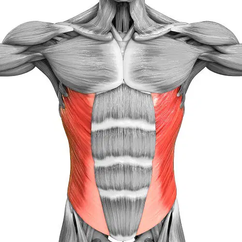
Name the muscle!
Abdominal external oblique
Trapezius
Quadriceps femoris
Deltoid


Identify the muscle
adductor
oblique
abdominals
quadricep
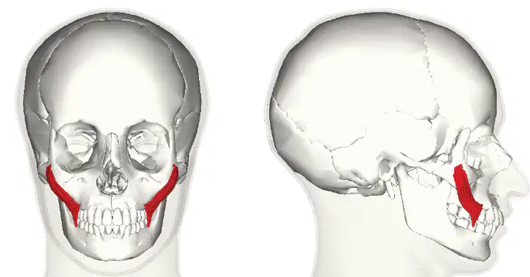
Name the muscle!
Zygomaticus
Orbicularis oculi
Nasalis
Frontalis
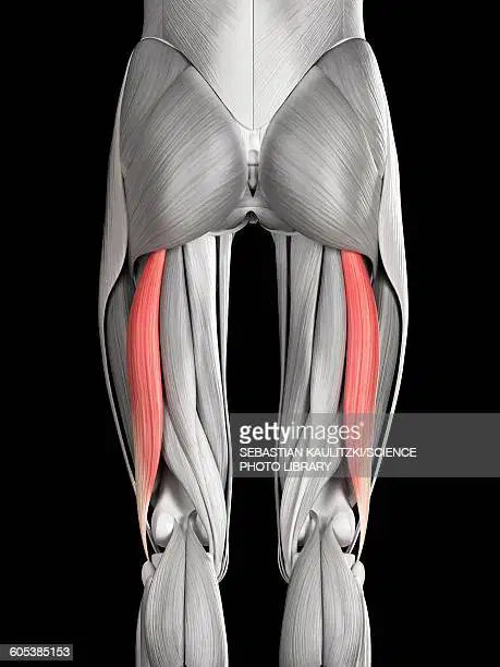
Name the muscle!
Quadriceps femoris
Sartorius
Abductor magnus
Biceps femoris
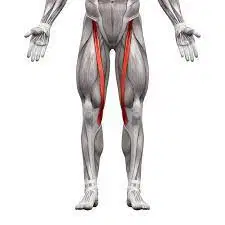
Name the muscle!
Sartorius
Quadriceps femoris
Deltoids
Gluteus maximus
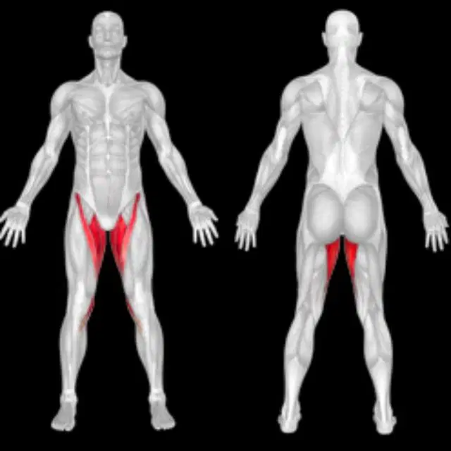
Name the muscle!
Abductor magnus
Latissimus dorsi
Quadriceps femoris
Trapezius
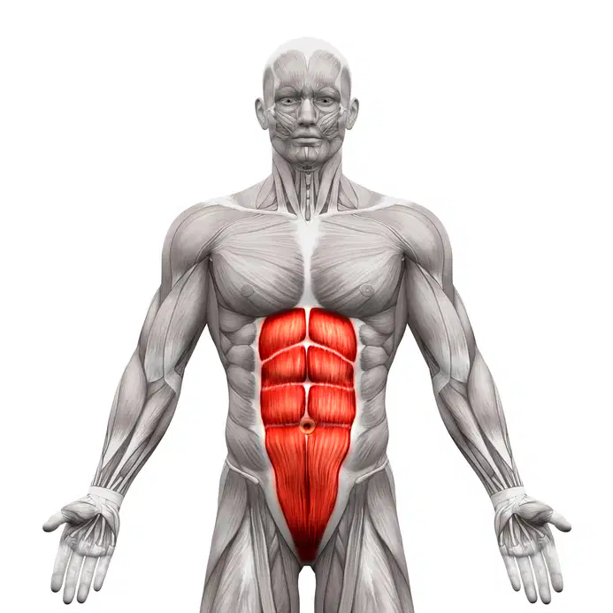
Name the muscle!
Rectus abdominus
Latissimus dorsi
Pectoralis major
Sartorius
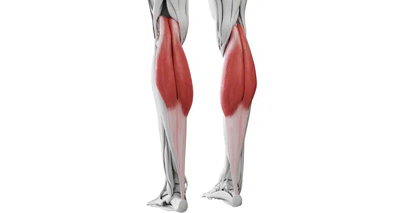
Name the muscle!
Gastrocnemius
Sartorius
Abductor magnus
Frontalis
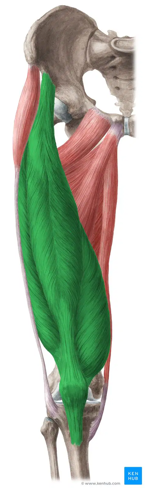
Name the muscle!
Trapezius
Deltoid
Gastrocnemius
Quadriceps femoris
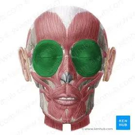
Name the muscle!
Orbicularis Oculi
Zygomaticus
Frontalis
Deltoid
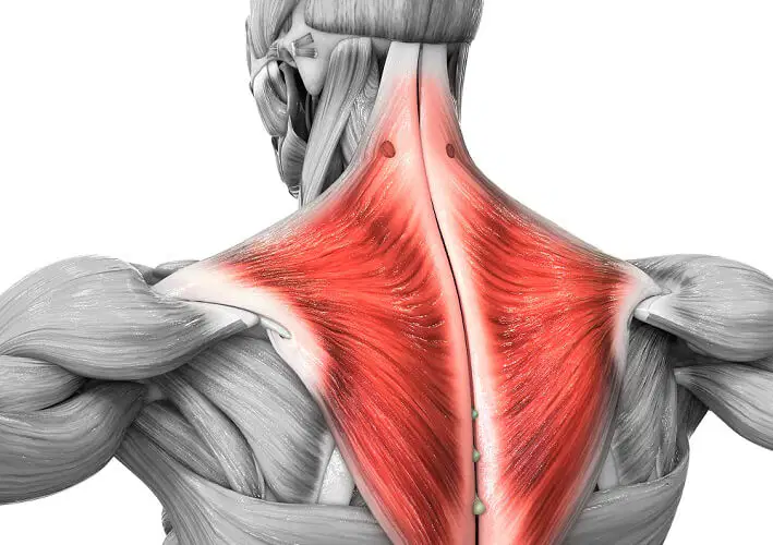
Name the muscle!
Pectoralis major
Trapezius
Zigomaticus
Nasalis
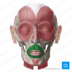
Name the muscle!
Orbicularis Oris
Zigomaticus
Trapezius
Frontalis

This muscle elevates the mandible (5)
frontalis
zygomaticus
temporalis
masseter

This muscle allows for dorsiflexion of the foot (Number 5)
tibialis anterior
gastrocnemius
quadriceps group
hamstrings

Identify the muscle being pointed to.

Identify the muscle highlighted in red.

These muscles that make up someone's "six pack" are really called what? (muscle highlighted in red)
extermal obliques
gluteus maximus
rectus abdominus
gastrocnemius

Identify the muscle highlighted in green.
flexor carpi radialis
sartorius
rectus femoris
peroneus longus

Identify the muscle highlighted in red.
gastrocnemius
semitendinosus
biceps femoris
semimembranosus

Identify the muscle highlighted in red.
gluteus medius
gluteus maximus
adductors
hamstring group

Identify the muscle highlighted in blue.
Vastus Medialis
Vastus Intermedius
Rectus Abdominus
Vastus Lateralis

Identify the muscle highlighted in blue.
Vastus Medialis
Vastus Intermedius
Rectus Abdominus
Vastus Lateralis

What muscles are highlighted in the picture?
Adductor Muscles
Tibialis Anterior
Iliopsoas
Quadriceps Femoris

