
 Worksheets
Nervous System Review for Test Fall 25 HSFD
Total questions: 123
Worksheet time: 2hrs 3mins
What is the insulating membrane that surrounds parts of the axon that helps to speed up action potential?
Axon
Dendrites
Node of Ranvier
Myelin Sheath

An electrical impulse within a single neuron is called a
synapse
membrane potential
action potential
cell body
The part of the neuron that receives incoming messages is the
dendrite
axon
cell body
axon terminal
The cell body contains the
dendrites
axon terminals
nucleus and many organelles
myelin sheath

What maintains the resting membrane potential in a neuron?
sodium-potassium pumps
sodium channels
potassium channels
gravity
Which part of a neuron TRANSMITS an electrical impulse?
(a)
The electrical impulse that travels down the axon of a neuron is called
action potential
signal
message
none of these
The difference in charge between the inside and outside of a cell is called:
depolarization
membrane potential
resting state
repolarization
A difference in concentration of ions on the inside versus the outside of a cell is called:
depolarization
action potential
concentration gradient
membrane potential
The sodium-potassium pump requires:
water
concentration gradient
ATP
calcium
When an action potential travels down the axon it:
ensures everything is the same
reverses the charge from positive to negative
reverses the charge from negative to positive
none of these
Functions of the Nervous System
sensory input
integration (choice making)
motor output
sensory input
motor output
integration
output
none of these
Cell responsible for insulating the CNS neurons
microglia
astrocyte
oligodendrocyte
schwann cell
Myelin sheath is the:
insulating wrap around neurons
covering of the brain
only structure that is on a neuron
all of these
The___________ of the brain is where choice making occurs.
cerebellum
cortex
parietal lobe
pons
There are ______ventricles of the brain.
2
3
4
5
The central canal along with the ventricles are filled with:
synovial fluid
myelin sheath
cerebral spinal fluid
all of these
The layers of protection for the brain are called:
meninges
myelin
schwann
CSF
In order from outside to inside the meninges are:
pia mater
arachnoid
dura mater
arachnoid
pia mater
dura mater
dura mater
arachnoid
pia mater
pia mater
dura mater
arachnoid
Structures that make up the Diencephalon:
Pituitary & Cerebellum
Thalamus & Hypothalamus
Frontal & Temporal Lobes
None of these
Hypothalamus Controls:
blood pressure, heart rate
body temperature, hunger
digestive tract, pupil of the eyes,
thirst, sleep
pain perception, pleasure, fear
All of These
The pathways that carry messages to the brain are known as:
efferent pathways
afferent pathways
Voluntary movements are called:
somatic
autonomic
Autonomic movements are:
involuntary
voluntary
The Autonomic Nervous System is divided into two parts:
sympathetic and parasympathetic
voluntary and somatic
Fight or Flight
sympathetic
parasympathetic
Restores the body to a state of calm after a stressful event.
sympathetic
parasympathetic
3 shapes of neurons:
unipolar
bipolar
multipolar
All of these
Cell bodies are housed OUTSIDE of the CNS in units known as a ganglion and are Always sensory:
bipolar neurons
multipolar neurons
unipolar neurons
The neurons that make up the CNS:
unipolar
bipolar
multipolar
The _________ lobe is the OUTPUT center of the brain
frontal
parietal
temporal
occipital
The _______lobe is the largest part of the cerebrum.
parietal
temporal
occipital
frontal
The ______ lobe receives images upside down and then breaks patterns down
into familiar items.
occipital
temporal
parietal
frontal
The ________lobe is responsible for hearing and balance
frontal
parietal
occipital
temporal
Controls coordination:
parietal lobe
cerebellum
frontal lobe
cervical plexus
The brain stem controls:
speech
hearing
heartbeat, breathing and blood pressure
None of these
The most common type of neuron:
unipolar
bipolar
multipolar
Association or interneurons transmit impulses:
outside the CNS
Within the CNS
outside of the spinal cord
_______is the largest part of a neuron and contains the nucleus.
cell body
dendrites
axon
Long, slender fiber that transmits signals from the cell
body to the axon terminals in a neuron.
dendrites
cell body
axon
Protects the axon of a neuron
myelin sheath
neurotransmitter
axon terminal
Projections from the cell body that receive signals in a neuron:
axon
dendrites
Cells that insulate neurons in the PNS:
schwann cells
oligidendrocytes
Carry impulses to and from the brain
spinal nerves
cranial nerves
Nerve fibers that carry impulses away from the central nervous system organs to effector organs (muscles and glands)
motor
somatic sensory
visceral sensory
The cortex of the brain is the_______ layer.
inner
outer
The pathways that carry messages to the brain are known as:
efferent pathways
afferent pathways
This pathway carries messages from the brain to the body is the _______pathway.
efferent
afferent
The dominate part of the Autonomic Nervous System
sympathetic
parasympathetic
Groups of nerves in the PNS are called:
plexus
cranial
Spinal cord ends at:
L5
Sacrum
L3
L1
If the spinal cord ends at L1, how do nerves get to the
abdominal area, back, pelvic region and legs?
saltatory conduction
cervical plexus
cauda equina
None of these
Understanding speech, recognition of objects, people, places, and events
and memory of events and outcome of choices are all controlled by the ______lobe.
frontal
occipital
parietal
temporal
The structures that make up the brain stem are:
thalamus, hypothalamus
cerebellum
midbrain, pons, medulla oblongata
None of these
Spinal nerves are made up of:
afferent neuron
efferent neuron
one afferent neuron and one efferent neuron
What cell is found in the CNS and connects neurons to blood vessels, forms scar tissue and creates the blood brain barrier?
Microglial
Oligodendrocytes
Astrocytes
Ependymal
Produces and regulates CSF, helping to maintain CNS health, and forming physical and immune barriers against pathogens.
Microglial
Oligodendrocytes
Astrocytes
Ependymal
What neuroglial cell is found in the PNS and forms individual myelin sheaths.
Oligodendrocytes
Schwann
Satellite
Ependymal
What type of neuroglial cell is found in the CNS and phagocytes bacteria and cellular debris?
Microglial
Oligodendrocytes
Astrocytes
Ependymal
The structures that make up the CNS:
Brain & Spinal Cord
Cervical plexus & Thoracic Plexus
Lumbar Plexus & Sciatic Nerve
Peripheral Nerves
The interbrain is the:
Diencephalon
Brain Stem
forebrain
hindbrain
Our personality, morals and ethics, ability to form words for speaking, and skeletal muscle movements are all controlled by the________lobe.
frontal
parietal
occipital
temporal

Breathing, heartbeat, blood pressure and swallowing.

This structure is involved in fear/panic response
Cerebellum
Hippocampus
Amygdala
Parietal Lobe
coordination of skeletal muscles and balance
What part of the brain regulates the sleep/wake cycles.
Hypothalamus
thalamus
pons
cerebrum
What is the function of the nerve cells?
They are responsable of your defense mechanism.
They transmit signals or messages to other neurons, organs or glands.
They transmit substances to your endocrine system to produce hormones.
What direction does an electrical signal move?
Dendrite, Cell body, Axon
Cell body, Axon, Dendrite
Cell body, Dendrite, Axon
Axon, Cell Body, Dendrite
What are the ridges on the cerebrum called?
Gyri
Sulci
Fissure
White matter
What are the grooves in the cerebrum called?
Gyri
Sulci
Fissures
Ridges
The _______ is an almond shaped structure in the brain's limbic system that encodes emotional messages to long-term storage.
Hippocampus
Amygdala
Hypothalamus
Cerebrum
When a neuron is at REST,
the inside of the neuron is negatively charged compared to the outside.
the inside of the neuron is positively charged compared to the outside.
there is no charge difference established
none of these is true
_________helps the impulses travel faster down the axon of a neuron.
boutons
dendrites
synaptic terminals
myelin sheath
Transmits impulses within the CNS between the sensory and motor neurons.
bipolar neurons
unipolar neurons
interneurons/
association neurons
none of these
Unipolar neurons are:
motor
sensory
__________nerves ALWAYS go to the CNS
motor
sensory
The hypothalamus, thalamus, and pituitary make up the:
brain stem
CNS
mid brain
diencephalon
White matter is located in the:
cortex of brain
interior of brain
only in brain stem
Grey matter is located _________of the brain.
interior
only in spinal canal
cortex
A pea-sized endocrine gland located at the base of the brain near the hypothalamus
pineal gland
pituitary gland
hypothalamus
corpus callosum
Converts short-term memories into long-term memories and is located within the temporal lobe.
hippocampus
hypothalamus
pituitary gland
thalamus
Located deep within the brain and regulates your emotions and behavior
pineal gland
occipital lobe
limbic system
RAS
Small, almond-shaped structure that controls fear and anxiety.
pineal gland
pituitary gland
thalamus
amygdala
Tiny pinecone shaped gland that secretes melatonin regulating the circadian cycle of sleep and wakefulness.
amygdala
pineal gland
hippocampus
pituitary gland
Regulates consciousness, wakefulness, and sleep-wake transitions
RAS
corpus callosum
hippocampus
pineal gland
produces, transports, and removes cerebrospinal fluid in the CNS
cerebellum
ventricles
RAS
limbic system
Bridge between cerebellum and cortex
medulla oblongata
pons
hippocampus
The spinal cord ends at L1 but the __________ supplies nerve innervation to the lower extremities and abdomen.
sacral plexus
cauda equina
thoracic plexus
lumbar plexus
A spinal nerve is made up of:
motor neuron
sensory neuron
both a sensory and a motor neuron
A ________is a group of spinal nerves
plexus
tract
Grey matter
conducts nerve impulses to and from the brain
contains cell bodies, dendrites and synapses to process information
stores neurotransmitters
has myelinated axons for faster transmission of impulses
Contains myelinated axons for faster transmission of impulses
white matter
grey matter
A selectively permeable membrane regulates the passage of large and small molecules into the microenvironment of the brain.
cerebellum
blood brain barrier
mucus membrane
sacral filium
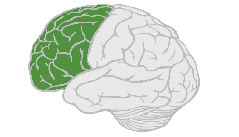
The highlighted lobe of this brain is the:
frontal lobe
temporal lobe
parietal lobe
occipital lobe
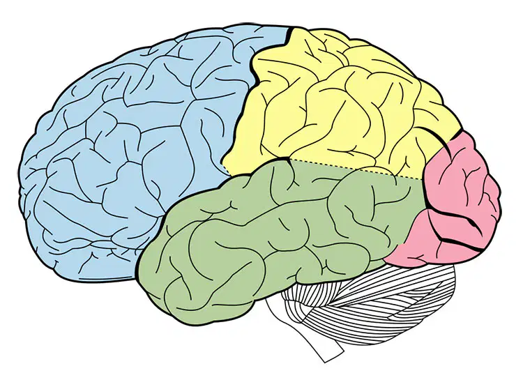
The pink (ish) lobe is the:
cerebellum
frontal lobe
occipital lobe
parietal lobe
Outnumber neurons 10:1 and support, insulate, and protect neurons.
neuroglial cells
spinal nerves
cranial nerves
What are reflexes?
Responds to motor output
Stimulus
Effector
Sensory Neuron
Motor Neuron
Interneuron
In which order is a signal sent from stimulus to response?
Sensory Organ, Sensory Neuron, Interneuron, Motor Neuron, Effector
Sensory Neuron, Sensory Organ, Interneuron, Motor Neuron, Effector
Interneuron, Sensory Neuron, Sensory Organ, Motor Neuron, Effector
Motor Neuron, Effector, Interneuron, Sensory Neuron, Sensory Organ
Effector, Motor Neuron, Interneuron, Sensory Neuron, Sensory Organ
What is a rapid, predictable and involuntary response to a stimuli known as?
reflex
synapse
neurotransmission
electrochemical
Which of the following is the correct order for a reflex arc?
Receptor -- Sensory Neuron -- Integration -- Motor Neuron -- Effector
Sensory Neuron -- Integration -- Motor Neuron -- Receptor -- Effector
Effector -- Motor Neuron -- Integration -- Sensory Neuron -- Receptor
Integration -- Sensory Neuron -- Motor Neuron -- Effector --Receptor
Carries sensory impulses to the brain and then carries the motor impulses to the muscles and glands.
vertebrae
spinal cord
saltatory conduction
none of these
Progressive disease that destroys motor neurons, leading to muscle weakness and paralysis.
TIA
meningitis
ALS
DMD
A degenerative brain disorder causing memory loss, confusion, and cognitive decline.
alzheimer’s Disease
cerebral palsy
gullian barre
CVA
A bacterial infection of the meninges
viral meningitis
bacterial meningitis
epstein barr
CP
A non-progressive motor disorder caused by early brain damage affecting movement and posture.
guiiain barre
cerebral palsy
multiple sclerosis
Neurological disorder with abnormal brain electrical activity causing seizures.
cerebral palsy
epilepsy
guillain barre
multiple sclerosis
Autoimmune attack on myelin that disrupts nerve signaling, causing weakness and vision problems
epilepsy
multiple sclerosis
guillain barre
DMD
Autoimmune disorder causing rapid ascending muscle weakness, often after an infection.
eptsein barr
epilepsy
guillain-barré syndrome
multiple sclerosis
Loss of dopamine-producing cells causing tremors, stiffness, and slowed movement.
multiple sclerosis
parkinson's disease
alzheimer’s disease
epilepsy
Interruption of blood flow to the brain causing sudden weakness, speech problems, or paralysis.
parkinson's disease
epilepsy
cerebral vascular accident
multiple sclerosis

