
 Worksheets
TEST yourself on the parts of a neuron
Total questions: 76
Worksheet time: 38mins
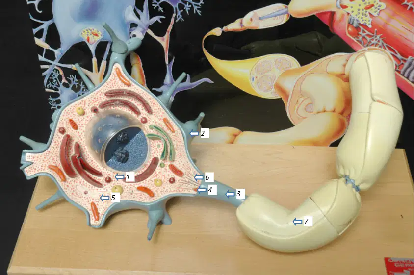
In the neuron model image, which structure is indicated by label 2?
Soma
Dendrite
Axon
Axon hillock
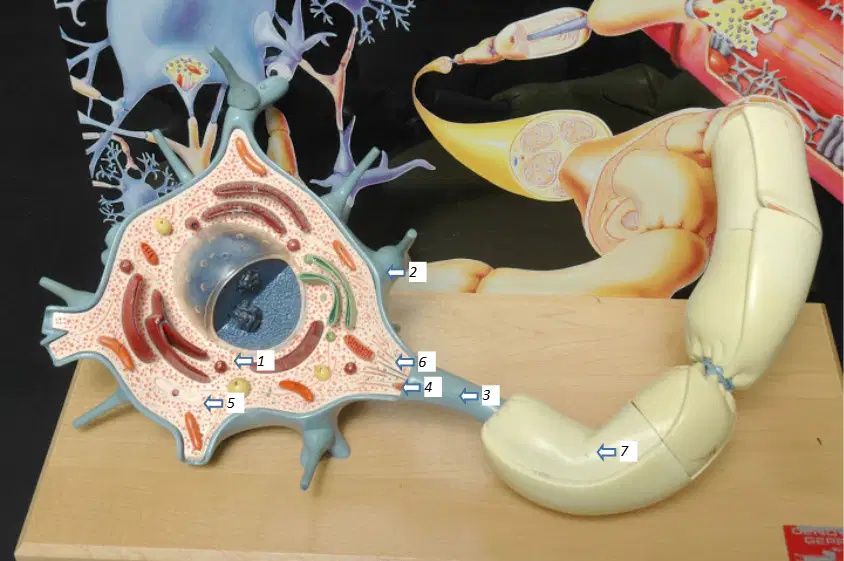
In the neuron model image, which structure is indicated by label 3?
Axon
Node of Ranvier
Schwann cell
Neurofibrils
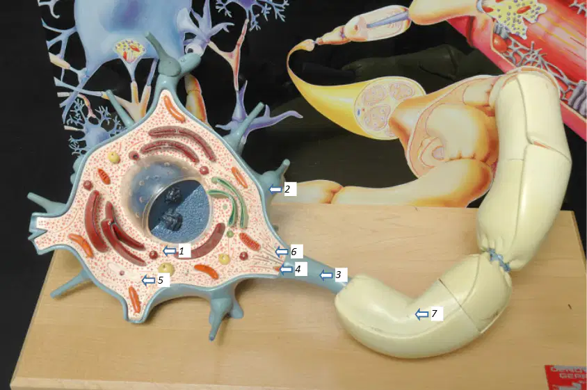
In the neuron model image, which structure is indicated by label 4?
Axon hillock
Nissl bodies
Axolemma
Myelin sheath
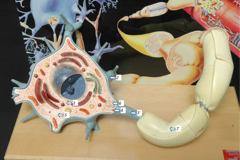
In the neuron model image, which structure is indicated by label 5?
Nissl bodies
Neurofibrils
Soma
Axoplasm
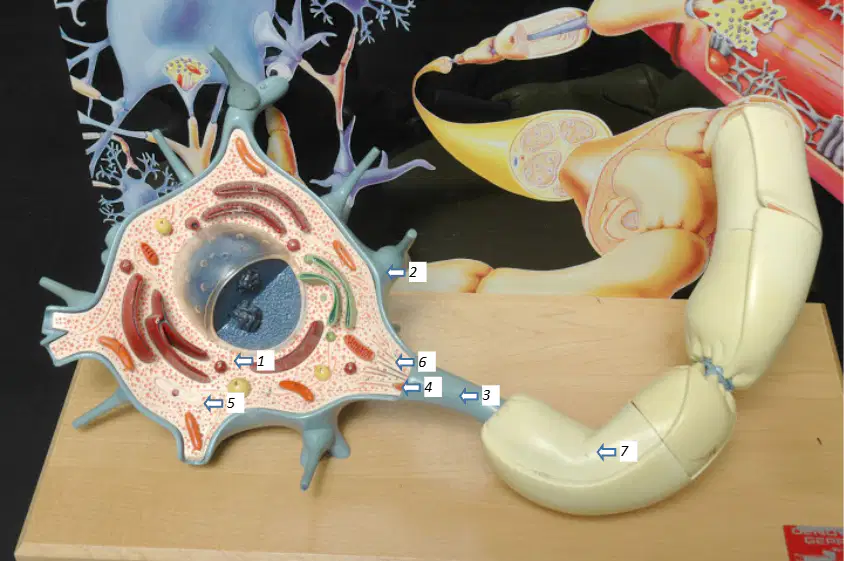
In the neuron model image, which structure is indicated by label 6?
Neurofibrils
Axolemma
Schwann cell
Node of Ranvier
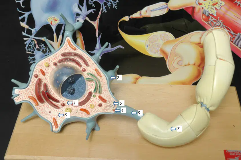
In the neuron model image, which structure is indicated by label 7?
Myelin sheath
Neurilemma
Axoplasm
Dendrite
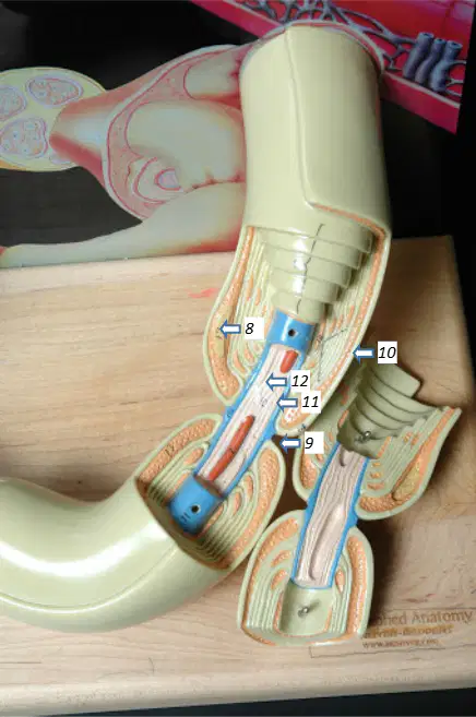
In the neuron model lower image, which structure is indicated by label 8?
Schwann cell
Node of Ranvier
Axolemma
Neurofibrils
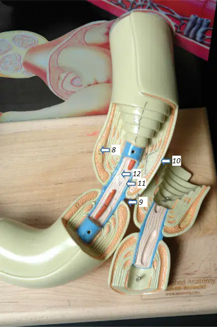
In the neuron model lower image, which structure is indicated by label 9?
Node of Ranvier
Neurilemma
Axoplasm
Soma
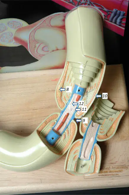
In the neuron model lower image, which structure is indicated by label 10?
Neurilemma
Axolemma
Myelin sheath
Dendrite
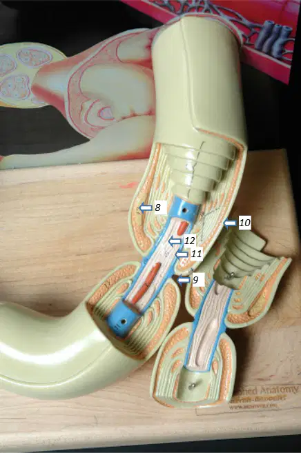
In the neuron model lower image, which structure is indicated by label 11?
Axolemma
Axoplasm
Soma
Schwann cell nucleus
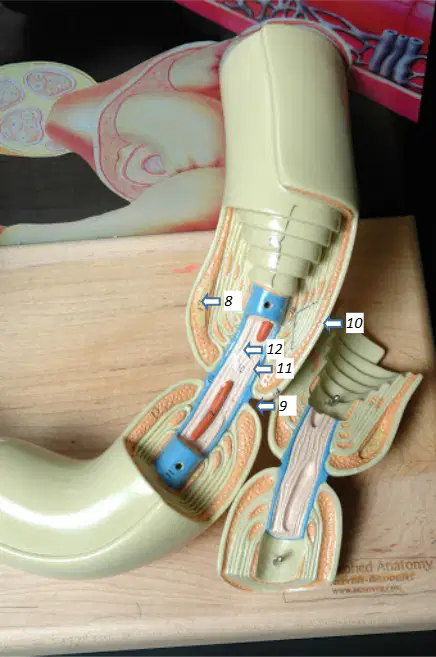
In the neuron model lower image, which structure is indicated by label 12?
Axoplasm
Axon hillock
Neurofibrils
Node of Ranvier
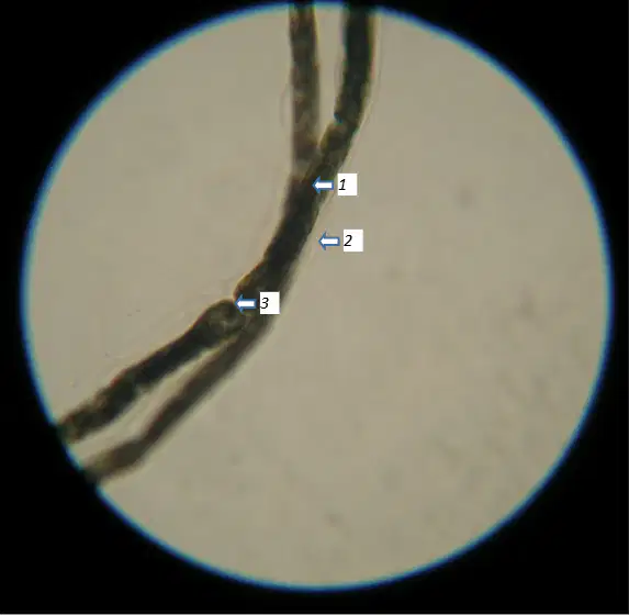
In the myelinated axon slide, identify the structure indicated by label 1.
Axon
Myelin sheath
Node of Ranvier
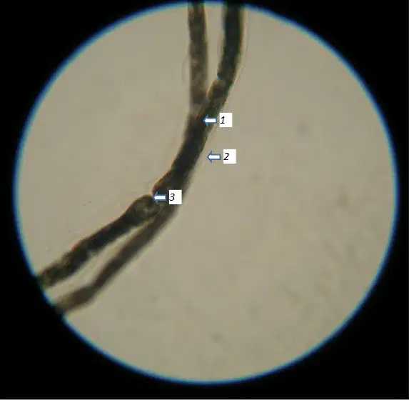
In the myelinated axon slide, identify the structure indicated by label 2.
Axon
Myelin sheath
Node of Ranvier
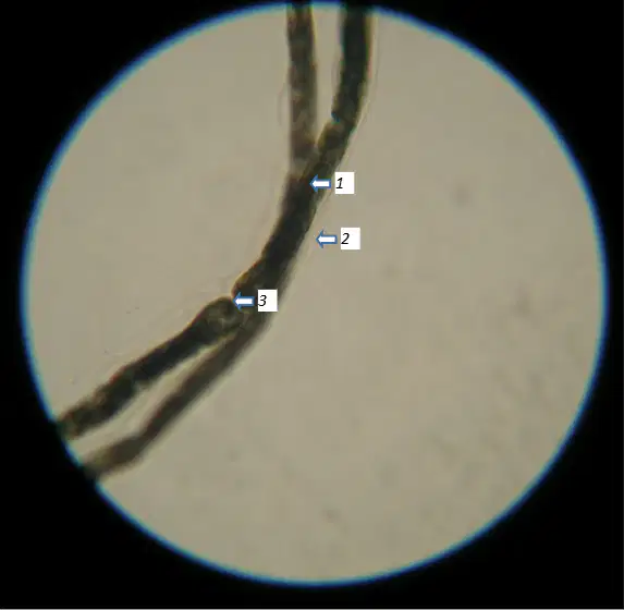
In the myelinated axon slide, identify the structure indicated by label 3.
Axon
Myelin sheath
Node of Ranvier
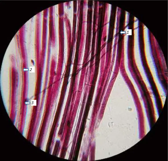
In the myoneural junction slide, identify the structure indicated by label 1.
Axon with synaptic terminals
Muscle fibers
Synapse or motor end plate
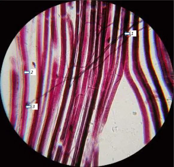
In the myoneural junction slide, identify the structure indicated by label 2.
Axon with synaptic terminals
Muscle fibers
Synapse or motor end plate
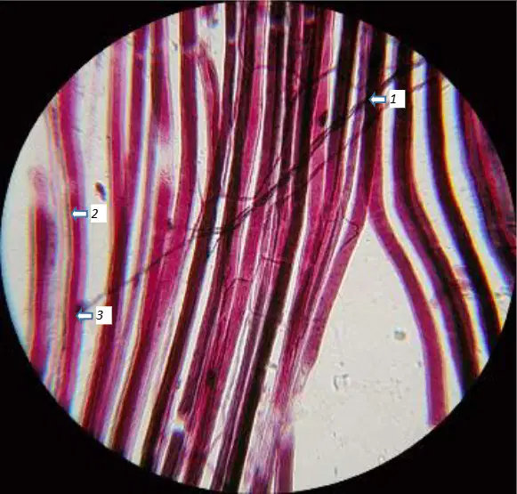
In the myoneural junction slide, identify the structure indicated by label 3.
Axon with synaptic terminals
Muscle fibers
Synapse or motor end plate

In the posterior view image of the sheep brain midbrain, identify the structure indicated as L. Superior Colliculus from the corpora quadrigemina.
Superior colliculus
Inferior colliculus
Pineal gland
Cerebellar hemisphere

In the same posterior view image of the sheep brain midbrain, identify the structure indicated as L. Inferior Colliculus from the corpora quadrigemina.
Inferior colliculus
Superior colliculus
Pineal gland
Vermis of cerebellum
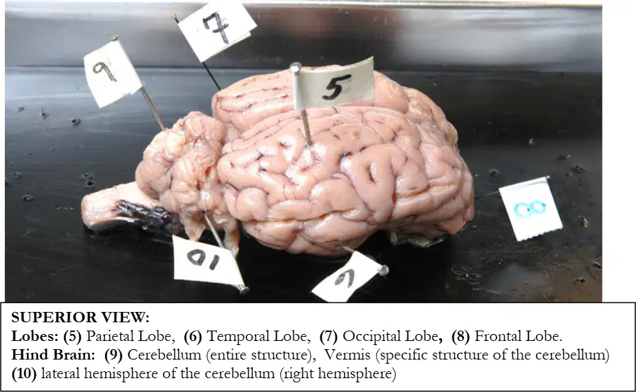
In the superior view of the cerebrum showing lobes, the flag labeled 5 indicates which lobe.
Parietal lobe
Temporal lobe
Occipital lobe
Frontal lobe
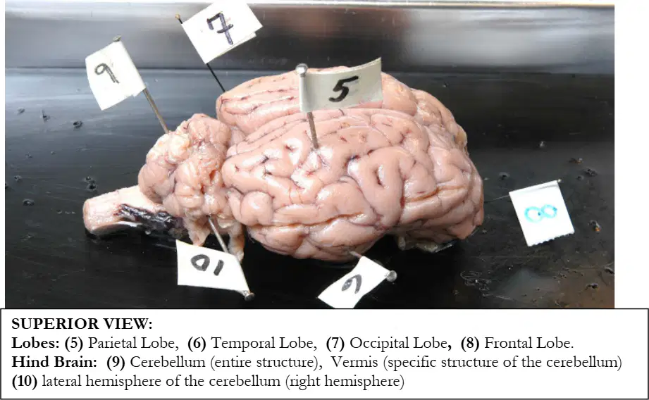
In the same superior view of the cerebrum showing lobes, the flag labeled 6 indicates which lobe.
Parietal lobe
Temporal lobe
Occipital lobe
Frontal lobe
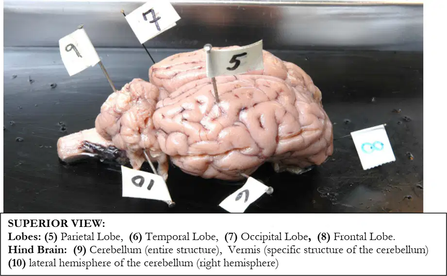
In the same superior view of the cerebrum showing lobes, the flag labeled 7 indicates which lobe.
Parietal lobe
Temporal lobe
Occipital lobe
Frontal lobe
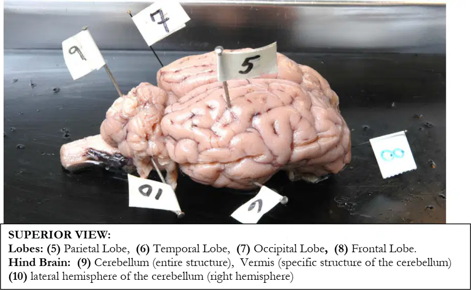
In the same superior view of the cerebrum showing lobes, the flag labeled 8 indicates which lobe.
Frontal lobe
Parietal lobe
Temporal lobe
Occipital lobe
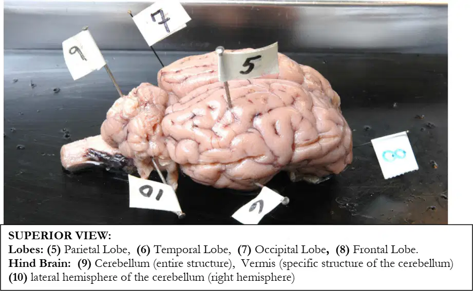
In the superior view focused on the hindbrain, the flag labeled 9 identifies which structure.
Cerebellum (entire structure)
Vermis (midline part of cerebellum)
Lateral hemisphere of cerebellum
Brainstem
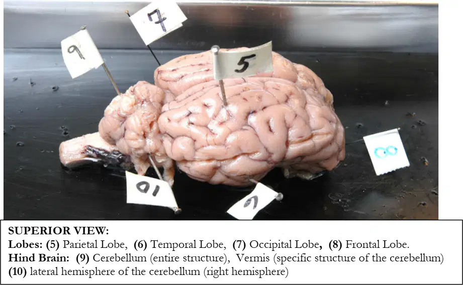
In the same superior view focused on the hindbrain, the flag labeled 10 identifies which structure.
Vermis (midline part of cerebellum)
Lateral hemisphere of cerebellum (right hemisphere)
Cerebellum (entire structure)
Pons
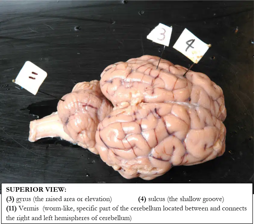
In the superior view highlighting surface features, the flag labeled 3 points to which feature.
Gyrus (elevation)
Sulcus (shallow groove)
Fissure (deep groove)
Vermis
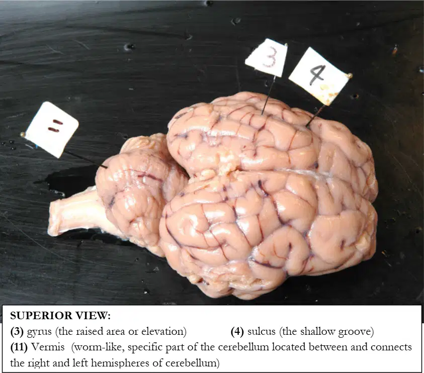
In the same superior view highlighting surface features, the flag labeled 4 points to which feature.
Gyrus (elevation)
Sulcus (shallow groove)
Longitudinal fissure
Arbor vitae
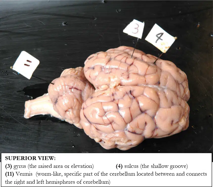
In the same superior view, the worm-like midline part of the cerebellum located between the right and left cerebellar hemispheres is identified by label 11. What is its name.
Vermis
Corpus callosum
Pons
Arbor vitae
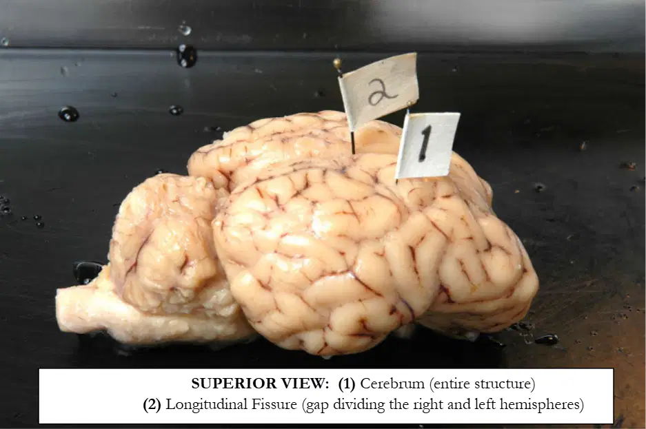
In the superior view of the brain, the flag labeled 1 identifies which structure.
Cerebrum (entire structure)
Cerebellum
Brainstem
Diencephalon
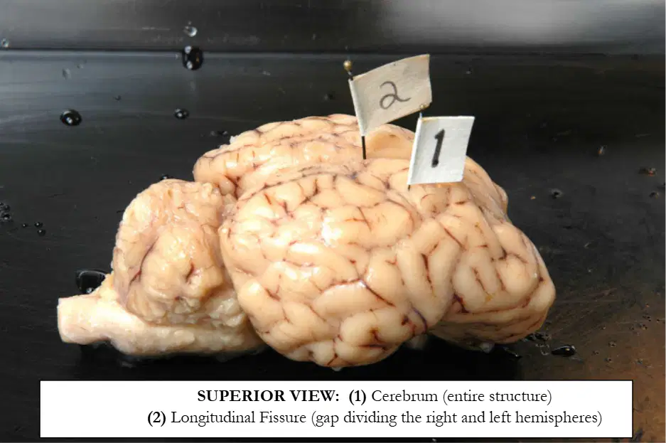
In the same superior view, the flag labeled 2 identifies which midline feature.
Longitudinal fissure
Central sulcus
Lateral sulcus
Parieto‑occipital sulcus
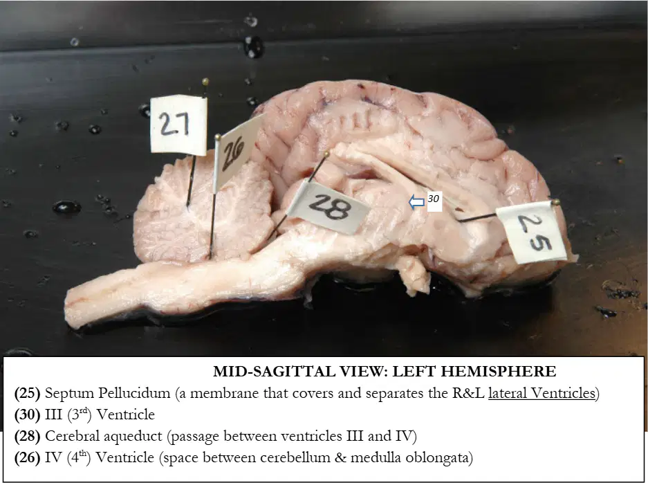
In the mid‑sagittal view of the left hemisphere, the flag labeled 25 identifies which structure.
Septum pellucidum
Thalamus
Pineal gland
Corpus callosum
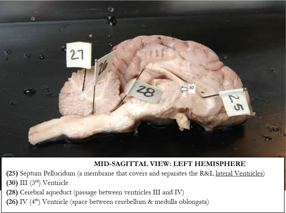
In the same mid‑sagittal view of the left hemisphere, the flag labeled 30 identifies which cavity.
III ventricle
Lateral ventricle
IV ventricle
Cerebral aqueduct
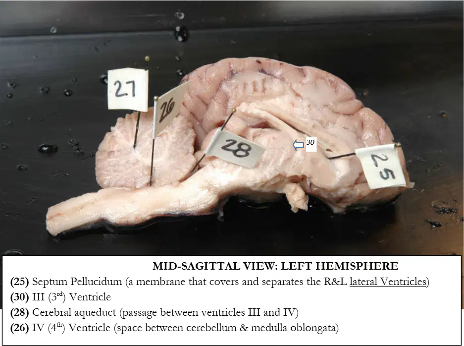
In the same mid‑sagittal view of the left hemisphere, the flag labeled 28 identifies which passage.
Cerebral aqueduct
Corpus callosum
Central canal
Longitudinal fissure
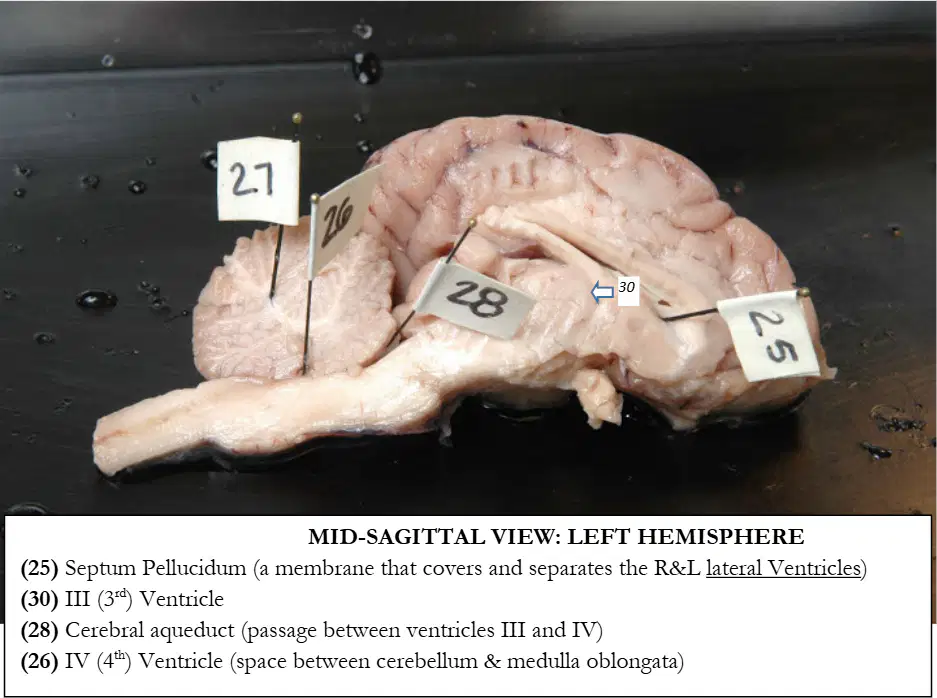
In the same mid‑sagittal view of the left hemisphere, the flag labeled 26 identifies which cavity.
IV ventricle
III ventricle
Lateral ventricle
Central canal
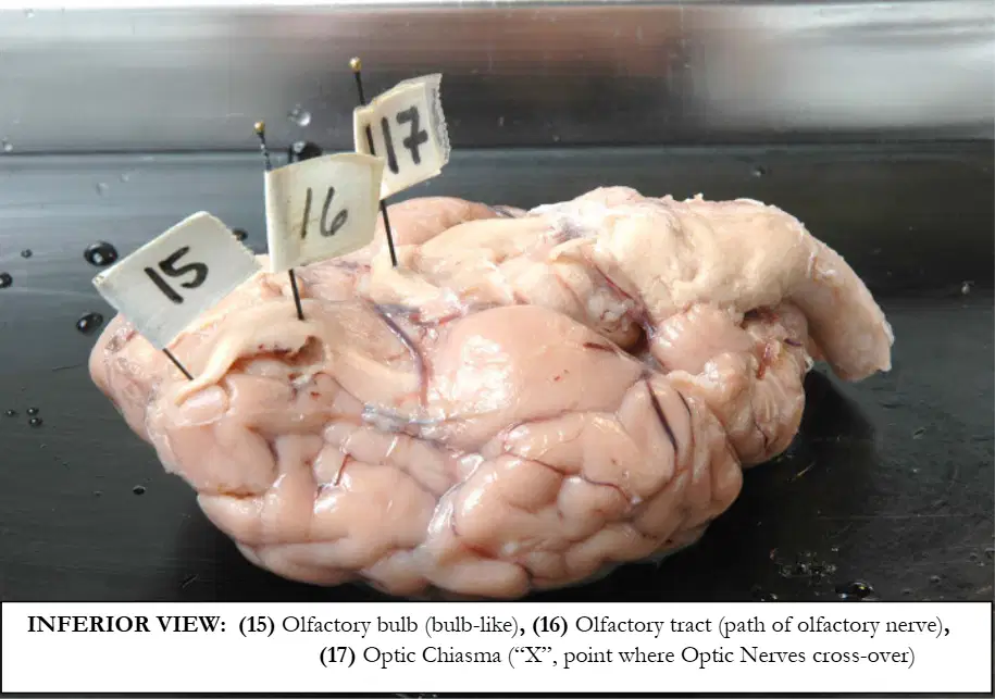
In the inferior view of the brain, the flag labeled 15 indicates which forebrain structure.
Olfactory bulb
Olfactory tract
Optic chiasma
Pituitary gland
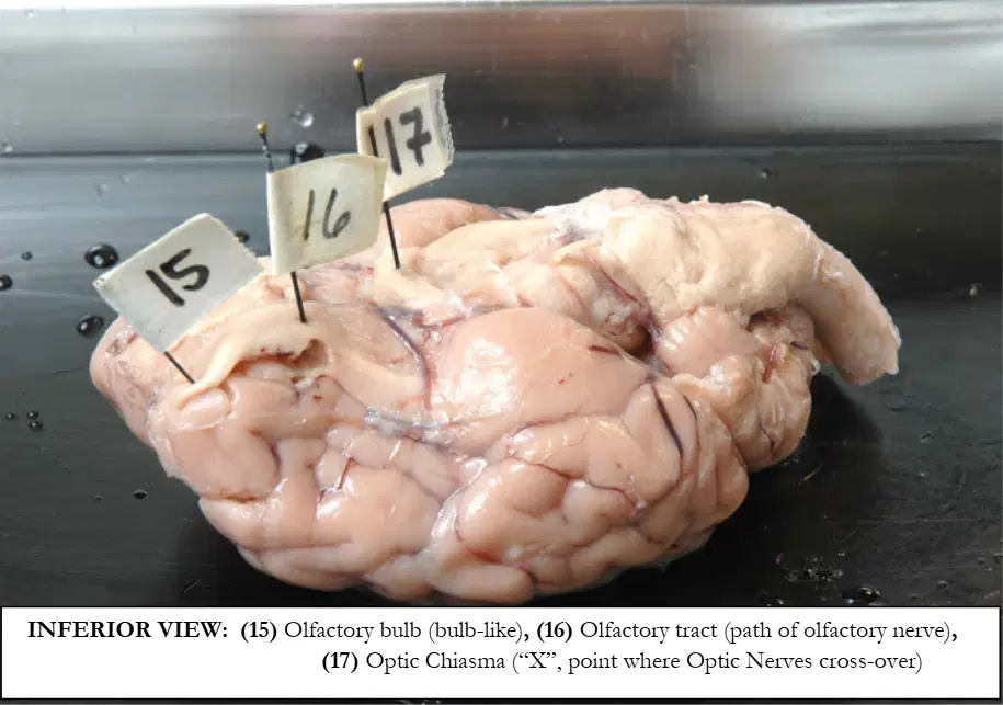
In the same inferior view, the flag labeled 16 indicates which pathway.
Olfactory tract
Optic nerve
Trigeminal nerve
Facial nerve
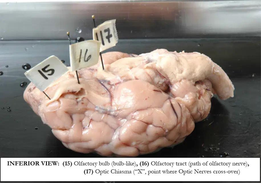
In the same inferior view, the flag labeled 17 indicates which crossing.
Optic chiasma
Olfactory bulb
Oculomotor nerve
Pons
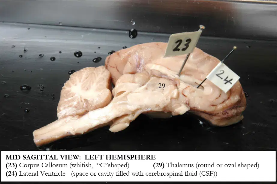
In the mid‑sagittal view of the left hemisphere, the flag labeled 23 identifies which structure.
Corpus callosum
Thalamus
Lateral ventricle
Pineal gland
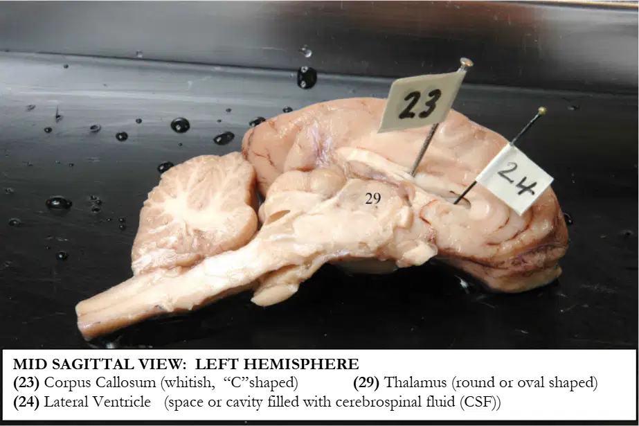
In the same mid‑sagittal view of the left hemisphere, the flag labeled 29 identifies which structure.
Thalamus
Corpus callosum
Pons
Hypothalamus
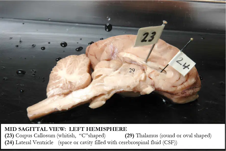
In the same mid‑sagittal view of the left hemisphere, the flag labeled 24 identifies which cavity.
Lateral ventricle
III ventricle
IV ventricle
Cerebral aqueduct

In the posterior view of the midbrain, the flag labeled 12 identifies which colliculus on the right side.
Right superior colliculus
Right inferior colliculus
Left superior colliculus
Left inferior colliculus
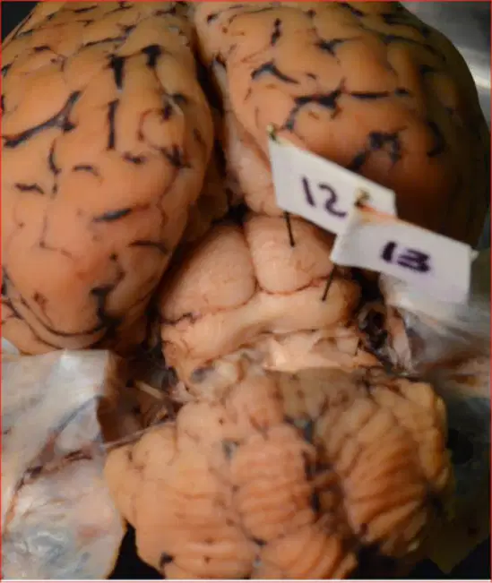
In the same posterior view of the midbrain, the flag labeled 13 identifies which colliculus on the right side.
Right inferior colliculus
Right superior colliculus
Left inferior colliculus
Left superior colliculus
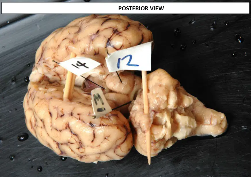
In the same posterior view, the flag labeled 14 identifies which midline structure.
Pineal gland
Pituitary gland
Thalamus
Mammillary body
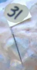
In the views showing meninges and endocrine structures, the flag labeled 31 identifies which outer meningeal layer.
Dura mater
Arachnoid mater
Pia mater
Falx cerebri

In the same set of views, the flag labeled 32 identifies which structure attached to the hypothalamus via the infundibulum.
Pituitary gland
Pineal gland
Thalamus
Mammillary body
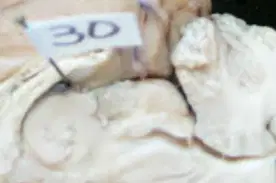
In the same set of views, the flag labeled 30 identifies which cavity surrounding the thalamus.
III ventricle
Lateral ventricle
IV ventricle
Cerebral aqueduct
In the sagittal human brain specimen, identify the numbered label that indicates the Cerebellum.
29
7
6
2
In the sagittal human brain specimen, identify the numbered label that indicates the Corpus Callosum.
7
29
10
6
In the sagittal human brain specimen, identify the numbered label that indicates the Lateral Ventricle.
6
10
2
7
In the sagittal human brain specimen, identify the numbered label that indicates the Falx Cerebri.
2
6
10
29
In the sagittal human brain specimen, identify the numbered label that indicates the Cerebral Aqueduct.
10
6
7
2
In the sagittal view with an endocrine organ shown hanging from the hypothalamus, which structure is suspended?
Pituitary gland
Pineal gland
Thymus
Adrenal cortex
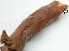
In the human spinal cord specimen, the structure labelled 30 is indicated by an arrow at a nerve exiting the cord laterally. Identify this labelled part.
Spinal nerve
Dorsal root ganglion
Cauda equina
Filum terminale
Dura mater
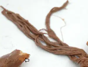
In the same specimen, the structure labelled 32 is the membranous covering surrounding the spinal cord at the superior end. Identify this labelled part.
Dura mater
Pia mater
Arachnoid mater
Spinal nerve
Conus medullaris
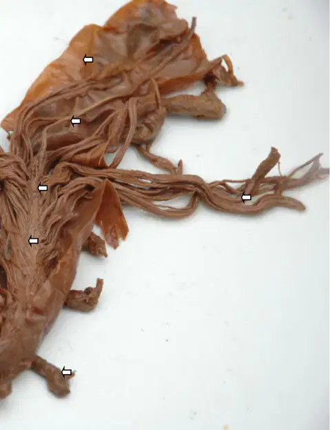
In the same specimen, the structure labelled 33 is a bundle of lumbar, sacral, and coccygeal nerve roots hanging inferiorly. Identify this labelled part.
Cauda equina
Conus medullaris
Filum terminale
Spinal nerve
Dorsal root
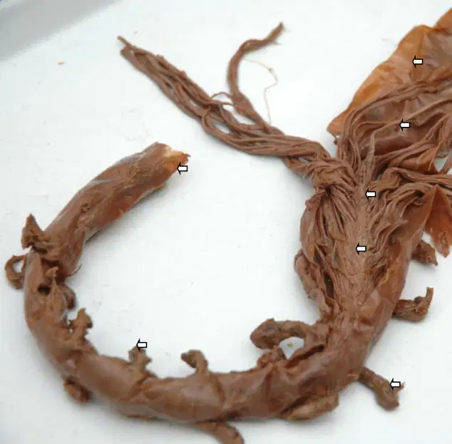
In the same specimen, the structure labelled 34 is the narrow, conical end of the spinal cord proper. Identify this labelled part.
Conus medullaris
Filum terminale
Cauda equina
Dorsal root ganglion
Posterior median sulcus
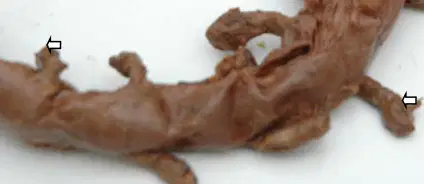
In the same specimen, the structure labelled 35 is the slender fibrous extension continuing from the conus to the coccyx. Identify this labelled part.
Filum terminale
Cauda equina
Conus medullaris
Dorsal root
Ventral root

In a spinal cord cross‑section with butterfly‑shaped gray matter and surrounding white columns, the tiny hole at the center is indicated by an arrow. Identify this labelled part.
Central canal
Gray commissure
Anterior median fissure
Posterior median sulcus
Lateral gray horn
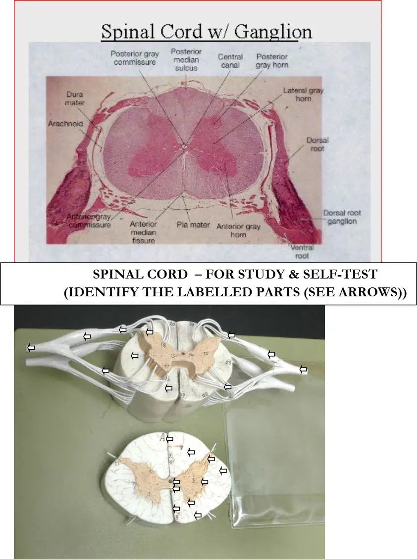
In the same cross‑section, the bar of gray matter that bridges the two sides around the central canal is indicated by an arrow. Identify this labelled part.
Gray commissure
Anterior median fissure
Posterior median sulcus
Dorsal root ganglion
Ventral root

In the same cross‑section, an arrow marks the deep midline groove on the anterior surface of the spinal cord. Identify this labelled part.
Anterior median fissure
Posterior median sulcus
Lateral gray horn
Anterior gray horn
Anterior white column
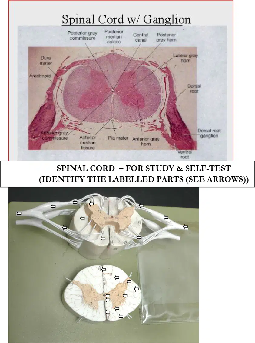
In the same cross‑section, an arrow marks the shallow midline groove on the posterior surface of the spinal cord. Identify this labelled part.
Posterior median sulcus
Anterior median fissure
Posterior gray horn
Posterior white column
Spinous process
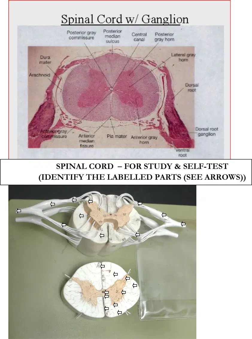
In the same model, an arrow indicates the swelling on a dorsal root just lateral to the cord. Identify this labelled part.
Dorsal root ganglion
Ventral root
Spinal nerve
Posterior gray horn
Lateral funiculus
In the ventral view note, all cranial nerves follow the sequence I to XII except which nerves? Select all that apply.
IV (Trochlear)
V (Trigeminal)
VI (Abducens)
VIII (Vestibulocochlear)
XI (Accessory, Spinal)
According to the mnemonic "Oh Once One Takes The Anatomy Final Very Good Vacation Ahead Ho Ho Ho," which word corresponds to cranial nerve VI?
Takes
Anatomy
Final
Vacation
Which nerve is cranial nerve I, as listed on the worksheet?
Olfactory
Optic
Oculomotor
Hypoglossal
Which nerve is cranial nerve VII, as listed on the worksheet?
Trigeminal
Facial
Vagus
Accessory, Spinal
According to the function column, what is the function of cranial nerve I (Olfactory)?
Smell
Vision
Eye movement
Taste and swallowing
According to the function column, what is the function of cranial nerve II (Optic)?
Hearing
Vision
Tongue movement
Shoulder movement
According to the function column, what is the function of cranial nerve VII (Facial)?
Facial expression
Major organs
Eye movement
Hearing
According to the function column, what is the function of cranial nerve XI (Accessory, Spinal)?
Tongue movement
Shoulder movement
Taste and swallowing
Smell
Based on the category mnemonic provided, what is the functional category of cranial nerve I (Olfactory)?
Sensory
Motor
Mixed
Based on the category mnemonic provided, what is the functional category of cranial nerve V (Trigeminal)?
Sensory
Motor
Mixed
Select all nerves that are classified as motor in the table:
III (Oculomotor)
IV (Trochlear)
VI (Abducens)
XI (Accessory, Spinal)
XII (Hypoglossal)
Select all nerves that are classified as sensory in the table:
I (Olfactory)
II (Optic)
V (Trigeminal)
VIII (Vestibulocochlear)
X (Vagus)
Which nerve is associated with hearing, according to the function column?
VI (Abducens)
VIII (Vestibulocochlear)
IX (Glossopharyngeal)
X (Vagus)
Which cranial nerve is labeled as "Accessory, Spinal" in the list?
IX
X
XI
XII

