
Font size
 Worksheets
Ch. 6 Skeletal System Study Guide
Total questions: 105
Worksheet time: 1hrs 15mins
The ends of a long bone are called the
diaphysis
epiphyses
periosteum
endosteum
The process by which bones increase in diameter:
ossification
hematopoiesis
appositional growth
longitudinal growth
Bone destroying cells:
osteocytes
osteoblasts
osteomasts
osteoclasts
Process of bone formation before birth:
ossification
hematopoiesis
appositional growth
atrophy
Dense bone tissue that appears smooth and homogeneous:
adipose tissue
compact bone
spongy bone
hyaline cartilage
Blood cell formation:
ossification
hemostasis
hematopoiesis
hemophilia
Remnant of the growth plate:
epiphyseal plate
spongy bone
epiphyseal line
lacunae
The long shaft of a bone:
epiphyses
diaphysis
periosteum
endosteum
Articular cartilage is found in what part of the long bone?
inside of the medullary cavity
on the outer surface of the diaphysis
the outer surface of the epiphyses
in the spaces of the spongy bone
What lines the medullary cavity and spaces of the spongy bone?
Periosteum
Epiphysis
Intramembranous
Endosteum
Type of bone found in the epiphysis
yellow marrow
endosteum
compact bone
spongy bone
Where is yellow marrow found?
compact bone
medullary cavity/canal
the diaphysis
the endosteium
The tough, fibrous outer layer of bone is called the _____.
epiphysis
endosteum
periosteum
red marrow
This is where two or more bones meet:
articulations
trabeculae
osteoblast
bone marrow
Growth ends when these ossify
deaphyses
epiphyseal plate
canaliculi
haversian canals
Type of marrow responsible for blood production
yellow
red
purple
blue
Which of the following lists the steps of fracture repair in the correct sequence?
hematoma, soft callus, bony callus, remodeling
soft callus, bony callus, hematoma, remodeling
remodeling, hematoma, bony callus, soft callus
hematoma, bony callus, soft callus, remodeling
The stem cell of our bone
Osteoblast
Osteogenic
Osteocyte
Chondrocyte
The function of an osteoblast
Destroys bone material
Exchanges nutrients & waste
Fights off bone diseases
Creates & lays down new bone material
The "new bone" material laid down by osteoblast is called the:
Osteocyte
Bone matrix
Articular cartilage
Osteon
Osteoblast will transform into this bone cell
Osteocyte
Osteoprogenitor
Osteoclast
Lacunae
This bone cell is responsible for exchanging nutrients/waste within the bone tissue
Osteoblat
Endosteum
Osteoclast
Osteocyte
Osteoclast develop from:
Osteoblast
Osteocytes
White blood cells
Cartilage
The axial skeleton is made up of.... (Select all that apply)
Bony Thorax
Limbs
Vertebral Column
Skull
Hip Girdle
How many cervical, thoracic, lumbar, and sacral vertebrae do humans have?
7, 12, 5, 5
12, 7, 5, 5
5, 12, 7, 5
5, 5, 12, 7
How many true ribs, false ribs, and floating ribs do humans have?
7, 5, 2
5, 7, 2
2, 5, 7
10, 2
How do true ribs differ from false ribs?
True ribs are attached directly to the sternum but the false ribs are not directly attached.
True ribs are not directly attached to the sternum but the false ribs are attached directly to the sternum
True ribs are not directly attached to the sternum but the false ribs are completely detached.
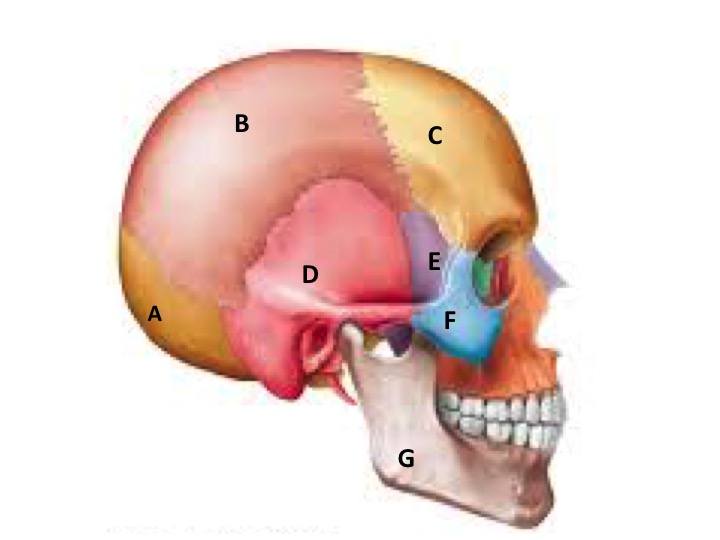
Which bone is labeled A
frontal
parietal
occipital
temporal
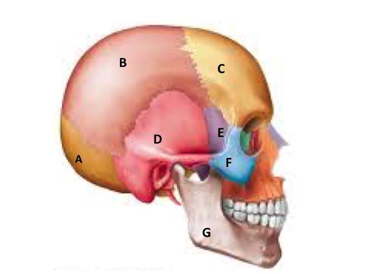
Which bone is labeled B
occipital
parietal
frontal
temporal
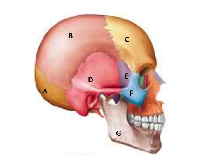
Which bone is labeled C
occipital
sphenoid
frontal
parietal
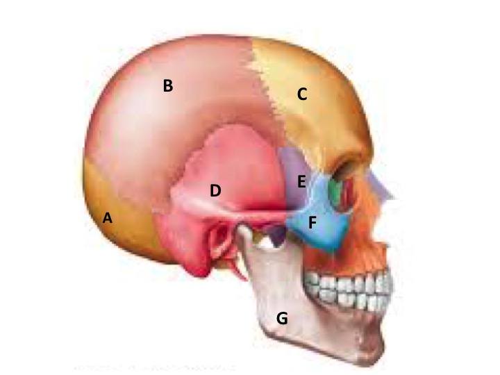
Which bone is labeled D
sphenoid
temporal
parietal
occipital
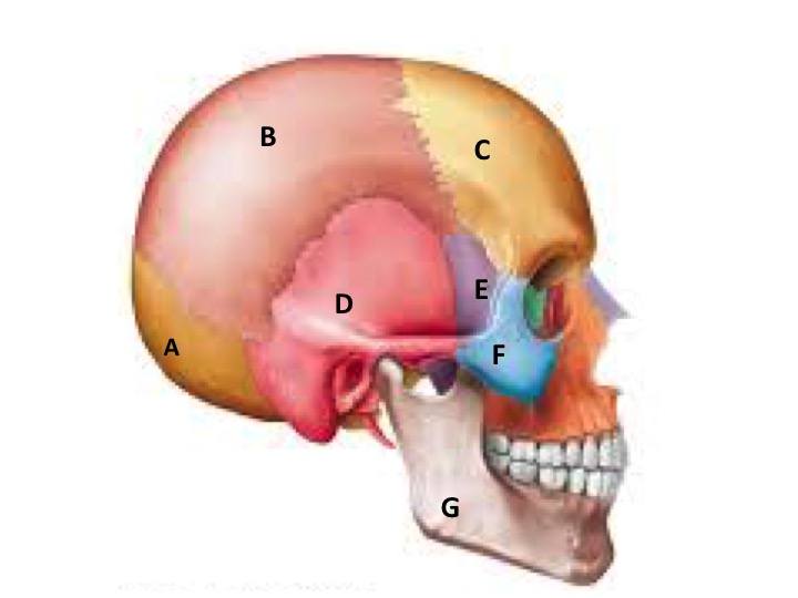
Which bone is labeled F
zygomatic
temporal
sphenoid
occipital
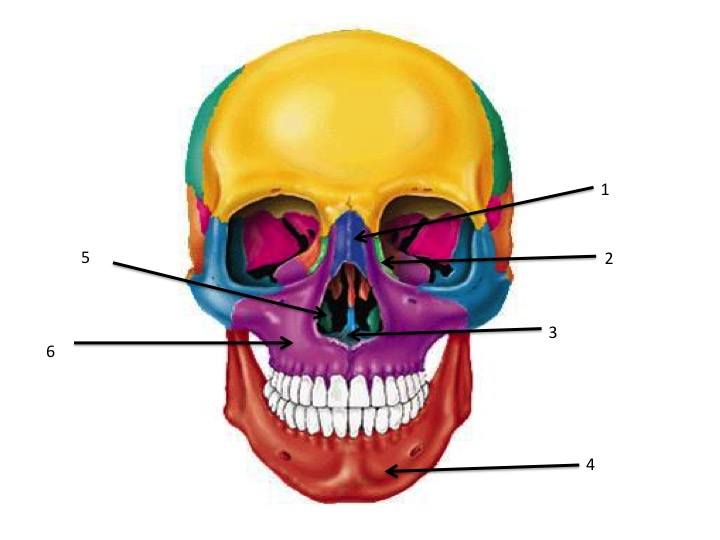
Which is bone 4
maxilla
mandible
zygomatic
nasal
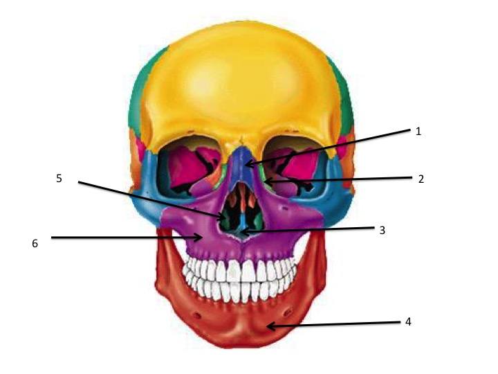
Which is bone 6
mandible
maxilla
sphenoid
nasal
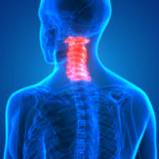
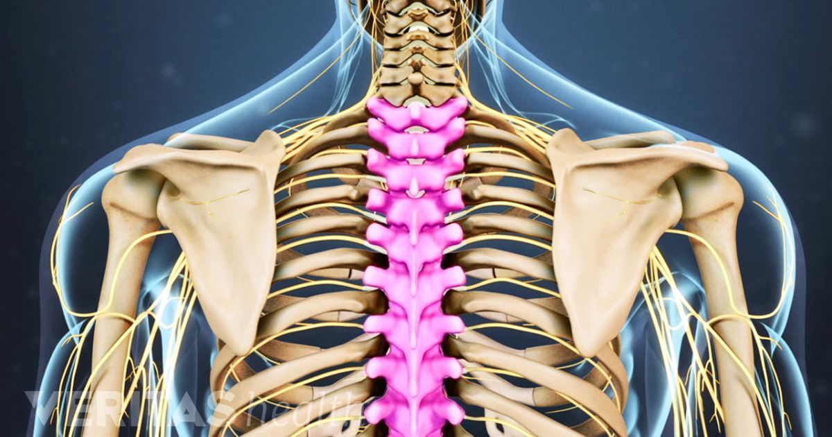
Which section of the vertebrae is highlighted
thoracic
cervical
lumbar
sacral
Which section of the vertebral column is highlighted
cervical
lumbar
thoracic
coccyx
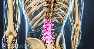
Which section of the vertebral column is shown?
cervical
lumbar
thoracic
sacral
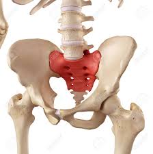
Which section of the vertebral column is highlighted
lumbar
cervical
coccyx
sacrum
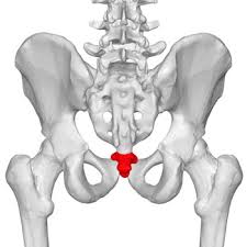
Which section of the vertebral column is highlighted
lumbar
sacrum
coccyx
cervical
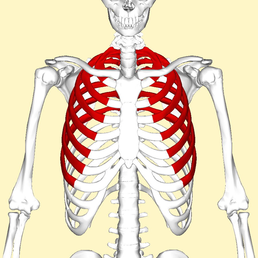
Which ribs are highlighted
true ribs
false ribs
floating ribs
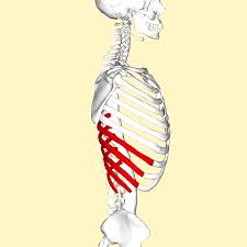
Which ribs are highlighted
true ribs
false ribs
floating ribs
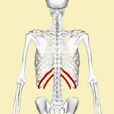
Which section of the rib cage is highlighted
true ribs
false ribs
floating ribs
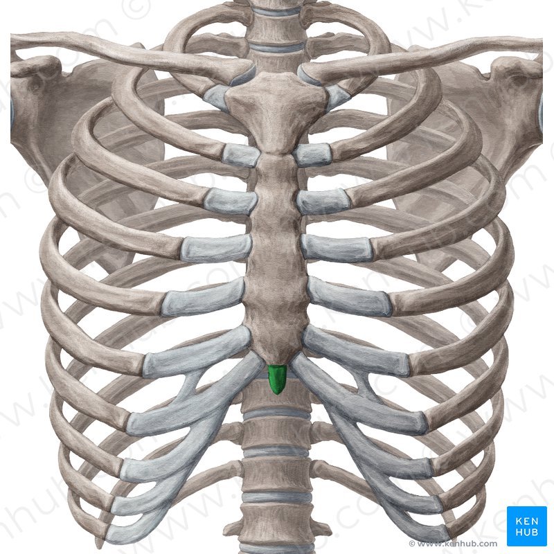
Which section of the sternum is highlighted in green
manubrium
body
xiphoid process
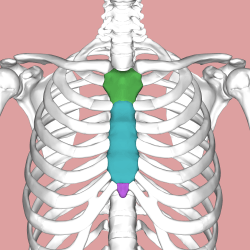
The entire bone is called...
Xiphoid Process
Manubrium
Body
Sternum
The clavicle attaches to the ___ medially.
tibia
scapula
sternum
carpal
The clavicle attaches to the ___ laterally.
tibia
scapula
sternum
carpal
The bones of the fingers and toes are called ___.
carpals
phalanges
tarsals
metacarpals
The bones in the palm area of the hands are called ___.
carpals
phalanges
tarsals
metacarpals
Which skeleton contains the upper and lower limbs and the shoulder and pelvic girdles?
Axial
Appendicular
The head of the humerus articulates with the
Tarsals
Acetabulum
Glenoid cavity
Trochlear notch
Forearm bone that is thumb side.
Radius
Ulna
Humerus
Metacarpal
Forearm bone that is pinky side.
Radius
Ulna
Humerus
Scapula
___ carpal bones are arranged in two irregular rows of four bones each wrist.
5
6
4
8

Ilium + Ischium + Pubis = ________
Coxal Bone
Pelvic Girdle
Bony Pelvis
Sacrum

Which bone is highlighted
humerus
femur
tibia
fibula
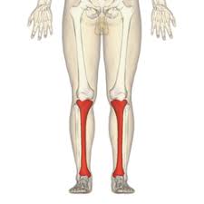
Which bone is highlighted
fibula
tibia
ulna
radius

Which bone is highlighted
ilium
ischium
pubic
sacrum

Which bone is highlighted
ilium
ischium
pubis
sacrum
coxal bone

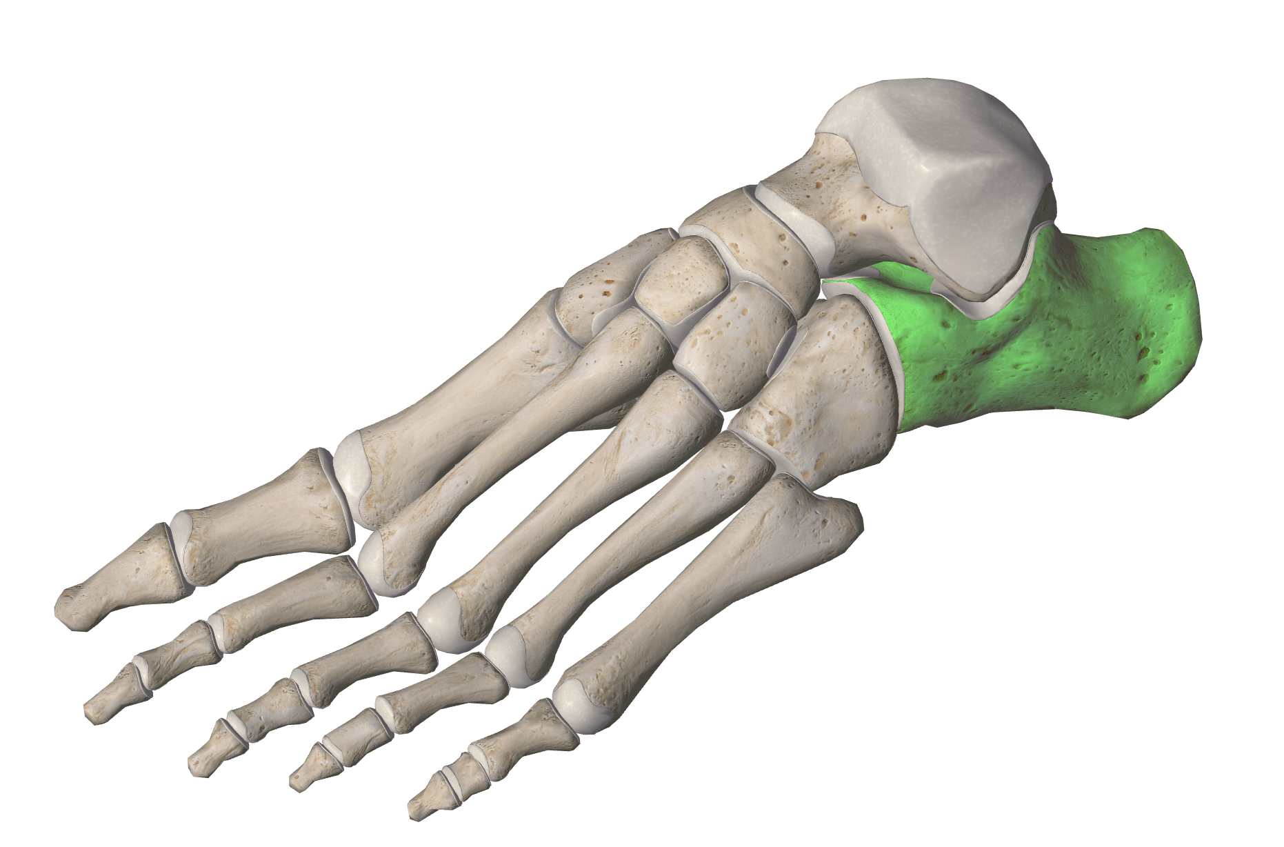
Which bone is highlighted
ilium
ischium
pubis
coccyx
sacrum

The arm is formed by a single bone, the ___ , which is a typical long bone.
humorous
femur
tibia
fibula




back of hand, lying on belly
depression
pronation
supination
extension

away from the mid-line
retraction
flexion
abduction
adduction

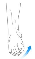
foot turned out
elevation
eversion
inversion
flexion

toes point upward
dorsi flexion
plantar flexion
supination
protraction

heels come up
elevation
abduction
plantar flexion
dorsi flexion



