
 Worksheets
A&P Spring Final Day 1 U2 & U3
Total questions: 98
Worksheet time: 53mins

Which tissue shown below is skeletal muscle tissue?
Muscle tissue 1
Muscle tissue 2
Muscle tissue 3
The heads of the myosin myofilaments are called ___ when they link the thick and thin filaments together.
Acetylcholine
ATP
Cross Bridges
Neuromuscular Junction
Skeletal muscle has 3 functions: mobility, posture & heat production. Posture is thanks to ___.
Muscle tone
You sitting up right
Muscles contracting & relaxing
Exercise
what types of tissues make up skeletal muscle?
muscle, connective, nervous
muscle, connective, nervous, epithelial
muscle, connective
muscle
Where will you find fascia connective tissue?
surrounding whole muscles
surrounding muscle fibers
surrounding fascicles
Fascia tapers off at the ends of skeletal muscles to form ___ which connects the skeletal muscle to a bone.
tendon
ligament
endomysium
perimysium
Between fascia & the skeletal muscle, you will find a layer of ___ connective tissue
epimysium
endomysium
perimysium
___ is a type of connective tissue which can be found wrapped around fascicles.
perimysium
endomysium
epimysium
Which of the following occurs when ATP binds to the myosin head?
Myosin head change to low energy configuration
Myosin head detach from actin
Myosin head undergoes power stroke
Myosin head forms cross bridge
Fascicles are bundles of ___.
muscle fibers (cells)
endomysium
myofilaments
myofibril
The outer cell membrane of a muscle cell/muscle fiber/myofiber is called a ___.
sarcolemma
sarcomere
sarcoplasm
sarcoplasmic reticulum

What is each #1 pointing to?
Acetylcholine
motor end plate
synaptic cleft
motor neuron
What is each #2 pointing to?
Acetylcholine
motor end plate
synaptic cleft
motor neuron
What is each #3 pointing to?
Acetylcholine
motor end plate
synaptic cleft
motor neuron
What is each #4 pointing to?
Acetylcholine
motor end plate
synaptic cleft
motor neuron
The gap between the motor neuron and the muscle fiber it supplies at the neuromuscular junction is called the .....
H band
Z zone
Synaptic Cleft
Sarcomere
Contractions are generated by
shortening of thick filaments
sliding of thin filaments past thick filaments
shortening of thin filaments
"accordian-like" folding of thick and thin filaments

Letter ___ would be found in a muscle like your latissimus dorsi, a large muscle on your back which makes imprecise movements.
A
B
C
Letter ___ would be found in a muscles like your fingers, allowing for precise movements.
A
B
C
which of the following occur during muscle relaxation
Release of calcium from sarcoplasmic reticulum
Acetyl choline released into synapse
ATP binds to myosin head
Tropomyosin covers myosin binding site
No cross bridge formation
Which of the following statements is true?
A motor neuron can attach to MANY muscle fibers
A motor neuron can only attach to ONE muscle fiber
A muscle fiber can attach to MANY motor neurons
Each muscle fiber has MANY motor end plates
Which of the following are the regulatory protein in muscle contraction?
Sarcoplasmic Reticulum
Troponin
Actin
Tropomyosin
Myosin

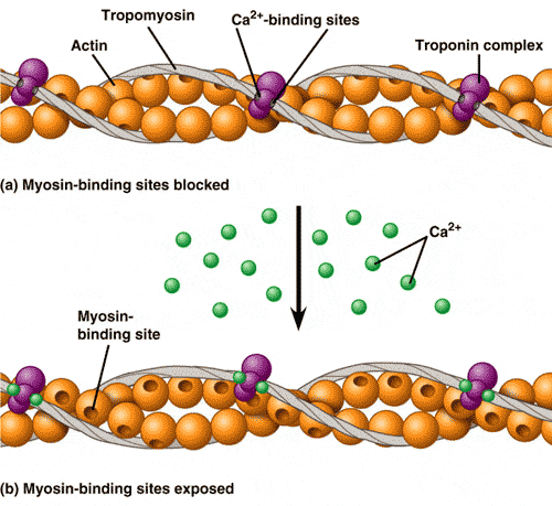
Use the image to determine the purpose of calcium:
causes ATP binding to actin
increases the action potential along the sarcolemma
binds to proteins on myosin
triggers the binding of actin and myosin
Which choice below covers the actin and prevents the myosin head to attach to the actin
Tropomyosin
Calcium and Oxygen
Myosin and Actin
Glycogen and Hemoglobin
A neurotransmitter that stimulates skeletal muscles
Acetylcholine
ATP
Cross Bridge
Neuromuscular junction
A neurotransmitter that stimulates skeletal muscles
Acetycholine
ATP
Cross bridge
Neuromuscular junction
Given the following events: 1. acetylcholine broken down 2. acetylcholine moves across the synaptic cleft 3. action potential reaches the presynaptic terminal of a motor neuron. 4. acetylcholine combines with a receptor molecule on a skeletal muscle. 5. action potential produced in a skeletal muscle cell.Choose the arrangement that lists the events in the order they occur at a neuromuscular junction.
2, 3, 4, 1, 5
3, 2, 4, 5, 1
2, 4, 3, 5, 1
4, 5, 2, 1, 3
The type of muscle contraction where the muscle tenses but does not shorten
Isometric
Contraction
Isotonic
Relaxation

During the upward phase (pressing) which muscle is undergoing concentric contraction?
biceps
triceps
abdominals
quads


Put steps of contraction in order.
rest, nerve action, ACh released, transmitted, Ca released
ACh released, Ca released, nerve action, transmitted, rest
rest, nerve action, Ca released, ACh released, transmitted
ACh released, Ca released, transmitted, nerve action, rest
Put steps of Contraction in order
Ca bound, activated, shift, cross bridge, repeat
Cross bridge, Ca bound, shift, activate, repeat
Activate, cross bridge, Ca bound, shift, repeat
Shift, activate, cross bridge, Ca bound, repeat
When the action reaches the muscle cell, it travels down the....
Sarcolemma
T Tubules
Sarcoplasmic Reticulum
Calcium Channel
What is the agonist and what is the antagonist in a bicep curl?
biceps are the agonist, triceps are the antagonist
triceps are the agonist, biceps are the antagonist
What is the opposing muscle group for the quadriceps?
biceps
lower back
hamstrings
obliques
The agonist and antagonist work in __________ to create movement
tandem
random
opposition
synchronization

Which type of bone is seen in the following image?
long bone
short bone
flat bone
irregular bone
none of these are correct
Which type of bone mostly serves as protection as it is platelike with broad surfaces (skull)?
long bone
short bone
flat bone
irregular bone
none of these
The hardest, most dense part of the bone tissue is the
periosteum
osteoblast
spongy bone
compact bone

True or False: The filled in portion of this image is accurately representing the axial skeleton.
True
False

Which correct # & term is pointing to the expanded end of long bone furthest from the point of attachment? (Check that the term is matched to the correct #)
#8: distal epiphysis
#8: proximal epiphysis
#9: diaphysis
#10: proximal epiphysis
#10: distal epiphysis

Identify the green highlighted area!
Mandible
Frontal
Parietal
Occipital

Identify the green highlighted area!
Zygamatic Arch
Temporal
Occipital
Maxilla

Identify the green highlighted area!
occipital
temporal
parietal
mandible

Maxilla
Mandible
Frontal
Zygomatic Arch

What is the yellow arrow pointing to?
Humerus
Olecranon Process
Foramen Magnum
Illium

Letter "B" identifies which of the following:
Axis
Atlas

Which part of the vertebral column is #4 referring?
cervical
lumbar
thoracic
sacral

Which part of the vertebral column is #6 referring?
Cervical
Lumbar
Thoracic
Sacrum

What section of the spine is highlighted?
Thoracic
Lumbar
Cervical
Sacrum

What section of the spine is circled?
Sacrum
Lumbar
Coccyx
Thoracic

What is #13 pointing to?
Malleus
Incus
Stapes

What is #10 pointing to?
Malleus
Incus
Stapes

What is #12 pointing to?
Malleus
Incus
Stapes

What is the #5 pointing towards?
Ulna
Radius
Tibia
Fibula

What is the #3 pointing towards?
Femur
Humerus
Radius
Ulna

What is the #4 pointing towards?
Humerus
Radius
Ulna
Tibia

What are the structures highlighted in red?
Ilium
Ischium
Clavicles
Humeri
How many pairs of true ribes do we have?
2
3
7
5
How many pairs of false ribs do we have?
2
3
7
5

What part of the skeleton is highlighted?
Sternum
Rib cage
Cervicall Spine

What part of the skeleton is highlighted?
Antecebulum
Olecranon Process
Manubrium
Xiphoid Process

What part of the skeleton is highlighted?
Antecebulum
Manubrium
Xiphoid Process
Olecranon Process
Name the "bones of the lower arm". Check all that apply
Femur
Ulna
Radius
Humerus
Tibia

What is the #1 pointing towards?
Ilium
Pubis
Ischium

What is #2 pointing towards?
Ilium
Pubis
Ischium

What is #3 pointing towards?
Ilium
Pubis
Ischium

What is #7 pointing towards?
Humerus
Femur
Tibia
Fibula

What is #14 pointing towards?
Radius
Tibia
Fibula
Ulna

What is #8 pointing towards?
cubital
scapula
patella
carpals

Which correct # & term is pointing to the place where fat is stored. (Check that the term is matched to the correct #)
#4: yellow marrow
#5: medullary cavity
#6: periosteum
#7: epiphyseal plates/disks
none of these are entirely correct
This type of bone is porous, allowing blood vessels to move through it and containing bone marrow
compact bone
spongy bone

What does this picture show?
osteon
hair follicle
hyaline cartilage
flat bone

Identify the structures circled in red
osteons
haversian canal
osteocyte is lacunae
medullary cavity

Identify the structures circled in red
osteon
haversian canal
osteocyte in lacuna
volkmann's canal

Identify the structures that are boxed
osteon
lamellae
volkmann's canal
haversian canal

Structure #2 identifying which part of the long bone
medullary cavity
epiphysis
periosteum
diaphysis
Osteoarthritis is defined as "A type of arthritis that occurs when flexible tissue at the ends of bones wears down.The wearing down of the protective tissue at the ends of bones occurs gradually and worsens over time. It causes symptoms such as joint pain in the hands, neck, lower back, knees, or hips"When a person has osteoarthritis, which part of a synovial joint is being worn down?
articular cartilage
ligaments
bursa
synovial fluid

You are conditioning for an iron man competition! To prepare, you build muscles in your arms by performing bicep curls as shown below. What joint connecting your humerus to your radius and ulna allows you to complete a bicep curl?
Ball and socket
saddle
hinge
gliding
condyloid
You are in watching a scary movie. At scariest part, you hear a noise outside. Sitting on your couch, you turn your head to look behind you out the window. What joint between the atlas and axis allows you to perform this motion?
Ball and socket
hinge
gliding
pivot
condyloid

You are doing the fitness challenge in PE and one requirement includes pull ups! To complete he pull up you must grasp on the pull up bar in order to complete the task. What joint connecting the last carpal bone in your hand to the first metacarpal bone in your thumb allows for the grasping of the pull up bar?
Ball and socket
Hinge
Saddle
Pivot

Identify the class of lever
first class lever
second class lever
third class lever

Identify the joint action being performed
flexion
hyperextension
eversion
circumduction

Identify the EFFORT in this lever system
A
B
C
Which of the following statements about osteoarthritis is/are true?
It occurs when the protective cartilage that cushions the ends of the bones wears down over time
Staying active and maintaining healthy weight might slow progression of the disease
Osteoarthritis symptoms can usually be managed, although the damage to joints can't be reversed
All are true
During a football game, a player's shoulder was forcibly displaced from the joint! Ouch! What condition is this?
Bursitis
Dislocation
Ruptured Disc
Spiral Fracture
Vitamin C aids in the production of which protein that accounts for about 30-35% of the dry weight of bone, and is responsible for providing flexibility and resilience to the bone.
Keratin
Collagen
Melanin
Calcium
In order to get this vitamin, you can: go outside and soak up some sun rays, consume eggs, and consume milk. Also, Mrs. G and other redheads actually make their own supply of this vitamin. Which vitamin are we describing
Vitamin B
Vitamin K
Vitamin C
Vitamin D

