
 Worksheets
A&P Semester 1 Review
Total questions: 103
Worksheet time: 1hrs 11mins
Closer to the head.
Anterior
Proximal
Superior
Medial
Away from the head.
Inferior
Lateral
Deep
Distal
Toward the front of the body.
Superficial
Anterior
Inferior
Proximal
Toward the back of the body.
Superficial
Distal
Superior
Posterior
Another term for superior.
Cranial
Caudal
Ventral
Dorsal
Another term for inferior.
Ventral
Superior
Caudal
Dorsal
Another term for anterior.
Dorsal
Distal
Lateral
Ventral
Another term for posterior.
Medial
Deep
Dorsal
Anterior
Toward the midline of the body
Inferior
Superficial
Proximal
Medial
Away from the midline of the body
Inferior
Distal
Lateral
Posterior
Closer to the trunk.
Proximal
Medial
Superior
Deep
Away from the trunk.
Superior
Distal
Lateral
Anterior
Toward the surface of the body.
Deep
Superficial
Inferior
Distal
Away from the surface of the body.
Medial
Inferior
Proximal
Deep
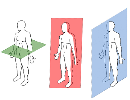
Divides the body into left and right portions.
Frontal
Transverse
Sagittal.
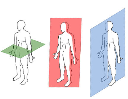
Divides the body into front and back portions.
Sagittal
Frontal
Transverse
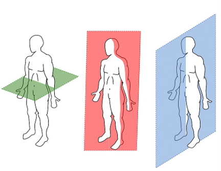
Divides the body into top and bottom portions.
Sagittal
Frontal
Transverse

Name this muscle
Pectoralis Major
Pectoralis Minor
Bicep
Hamstring

Name this muscle
Trapezius
Pectoralis Major
Deltoid
Tricep

Name this muscle
Gluteus Maximus
Deltoid
Tricep
Bicep

Name this muscle
Abdominal
Abdominals
Abs
Abbbs

Name this muscle
Hamstrings
Quads
Quadriceps
Groin

Deltoid
Latissimus Dorsi
Trapezius
Tricep

Name this muscle
Triceps
Biceps
Latissimus Dorsi
Deltoid

Name this muscle
Hamstring
Quadriceps
Booty
Gluteus Maximus

Name this muscle
Gastrocnemius
Achilles
Calves
Fibula

Name this muscle
Quadriceps
Quads
Calves
Hamstrings

Name this muscle
Deltoid
Trapezius
Latissimus Dorsi
Spinal Column
This type of muscle we have no voluntary control over
Cardiac
Smooth
Skeletal
Our intestines are an example of what type of muscle
Cardiac
Smooth
Skeletal
Our heart is this type of muscle
Cardiac
Smooth
Skeletal
These muscles are attached to our bones that allow us to move
Cardiac
Smooth
Skeletal

What skeletal muscle shape is this
Spindle
Flat
Triangular
Circular

What skeletal muscle shape is this
Spindle
Flat
Triangular
Circular

What skeletal muscle shape is this
Spindle
Flat
Triangular
Circular

Name this skeletal muscle shape
Spindle
Flat
Triangular
Circular

Name this skeletal muscle shape
Spindle
Flat
Triangular
Circular


Which of the following bones is NOT part of the axial skeleton?
clavicle
frontal
vertebrae
sternum
How many vertebrae are in the neck?
2
4
7
12

What bone is highlighted?
Radius
Ulna
Humerus
Carpals

What bone is highlighted?
Radius
Ulna
Humerus
Carpals

What bone is highlighted? (hint: inside of your leg)
Tibia
Fibula
Femur
Humerus

What bone is highlighted?
Skull (Mandible)
Clavicle
Mandible
Scapula
From wrist to fingertip, what is the order of bones?
Carpals, metacarpals, phalanges
Phalanges, metacarpals, carpals
Carpals, phalanges, metacarpals
Metacarpals, carpals, phalanges

Which of the following is not a function of bones?
Protection
Blood cell formation
Storage
Absorption


What must occur first before a muscle can contract?
An action potential must develop as energy/nutrients is gained from the digestive system
An action potential must travel to the muscle through the circulatory system
An action potential must enter the body through the respiratory system
An action potential must be sent through neurons in the nervous system
What neurotransmitter is used at the neuromuscular junction that sends a signal from the motor neuron to the skeletal muscle?
Glutamate
Acetylcholine
Norepinephrine
Dopamine
What happens when the action potential reaches the axon terminal of a motor neuron?
Voltage-gated channels open, releasing sodium
Voltage-gated channels open, releasing acetylcholinesterase
Voltage-gated channels open, releasing acetylcholine
Voltage-gated channels open, releasing calcium
What do calcium ions do once they are released?
Calcium enters the muscle tissue
Cause sodium ions to enter the muscle
Cause potassium ions to exit the muscle
Cause acetylcholine to be released
What is the function of acetylcholine?
ACh binds to receptors on the sarcolemma and opens ion channels
ACh binds to receptors on the sarcolemma and closes ion channels
ACh binds to ion channels and open them
ACh binds to ion channels and closes them
What ions are transported by the activated ion channel in muscle cells?
Sodium ions rush out of the cell, while potassium ions rush into the cell
Calcium ions rush out of the cell, while potassium ions rush into the cell
Sodium ions rush into the cell, while potassium ions rush out of the cell
Calcium ions rush into the cell, while potassium ions rush out of the cell
What is the purpose of the sodium potassium ion channel?
Create an action potential
Cause contraction
Create a membrane potential
Cause the release of ATP
The impulse/membrane potential caused by the ion channels affects what in the muscle cell?
The impulse releases more ACh
The impulse releases more calcium
The impulse releases ATP
The impulses releases ADP
Calcium inside the muscle cell binds to which filament, causing a shape change and revealing the active sites.
Actin
Myosin
Troponin
Tropomyosin
What binds to the active site on actin?
Actin
Myosin
Troponin
Tropomyosin
What is a cross-bridge?
When troponin and tropomyosin undergo a shape change
When the active site of actin is revealed
When the myosin heads bind to the actin active site
When the actin filaments slide past the myosin filaments
How do the actin filaments "slide"?
With the help of acetylcholine
With the help of ADP
With the help of ATP
With the help of calcium
When is ATP needed?
When actin slides
When myosin heads bind to actin
When myosin heads release from actin
When myosin slides
After the muscle is no longer stimulated, what occurs first
Acetylcholinesterase is released and removes ACh
The muscle continues to contract for a few more minutes
Enzymes break down calcium ions
The ion channels pump sodium back out of the cell and potassium back into the cell
Which ways do muscles get their energy from?
Creatine Phosphate
Anaerobic Respiration
Kreb Cycle
Aerobic Respiration
Which two bones are found in the leg?
Femur & Radius
Ulna and Fibula
Tibia and Humerus
Femur and Tibia


What's the name of this bone?
femur
ribs
hip
These types of bones are found in your ankles and wrists
Long
Sesamoid
Flat
Short
Irregular
What is the job of a long bone?
Support your organs during physical activity
Support the weight of your body during physical activity
To protect your vital organs during physical activity
To protect your muscles during physical activity
How many bones are in an adult human's body?
206
168
542
337
What connects muscle to bone?
Ligament
Tendon
Cartilage
Joint Capsule
What connects bone to bone?
Tendons
Ligaments
Joint Capsule
Bursae


What is the job of a flat bone?
Main bones used for gross motor movements
Support the weight of the body during physical exercise
Allow for fine motor movements
Protection for vital organs and anchor for muscles to attach
Can you name correct term for the bone protecting your brain?
Craniul
Skull
Cranium
Where on your body would you find the Patella bone?
Knee
Elbow
Shoulder
Ankle
Your Radius, Ulna, Femure, Tibia, Fibula and Humerus are all what type of bone?
Short
Long
Flat
Irregular
Your heart is in your chest cavity (thorax).
True
False


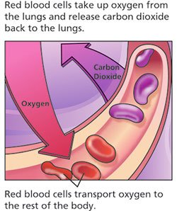

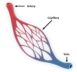

The proteins found on the surface of RBCs that allow us to type blood as A, B, O or AB are called
antigens
antibodies
aggluntates
Rh factors
Tyler has blood type AB+, he can donate blood to a person that has blood type?
AB+
AB-
O-
O+
Laney is the universal donor, what blood type does she have?
O-
O+
AB+
AB-

